. 2018 Dec:82:184-196.
doi: 10.1016/j.actbio.2018.10.018. Epub 2018 Oct 14.

Bioactive effects of silica nanoparticles on bone cells are size, surface, and composition dependent

Affiliations

Bioactive effects of silica nanoparticles on bone cells are size, surface, and composition dependent

Shin-Woo Ha et al. Acta Biomater. 2018 Dec.

Abstract

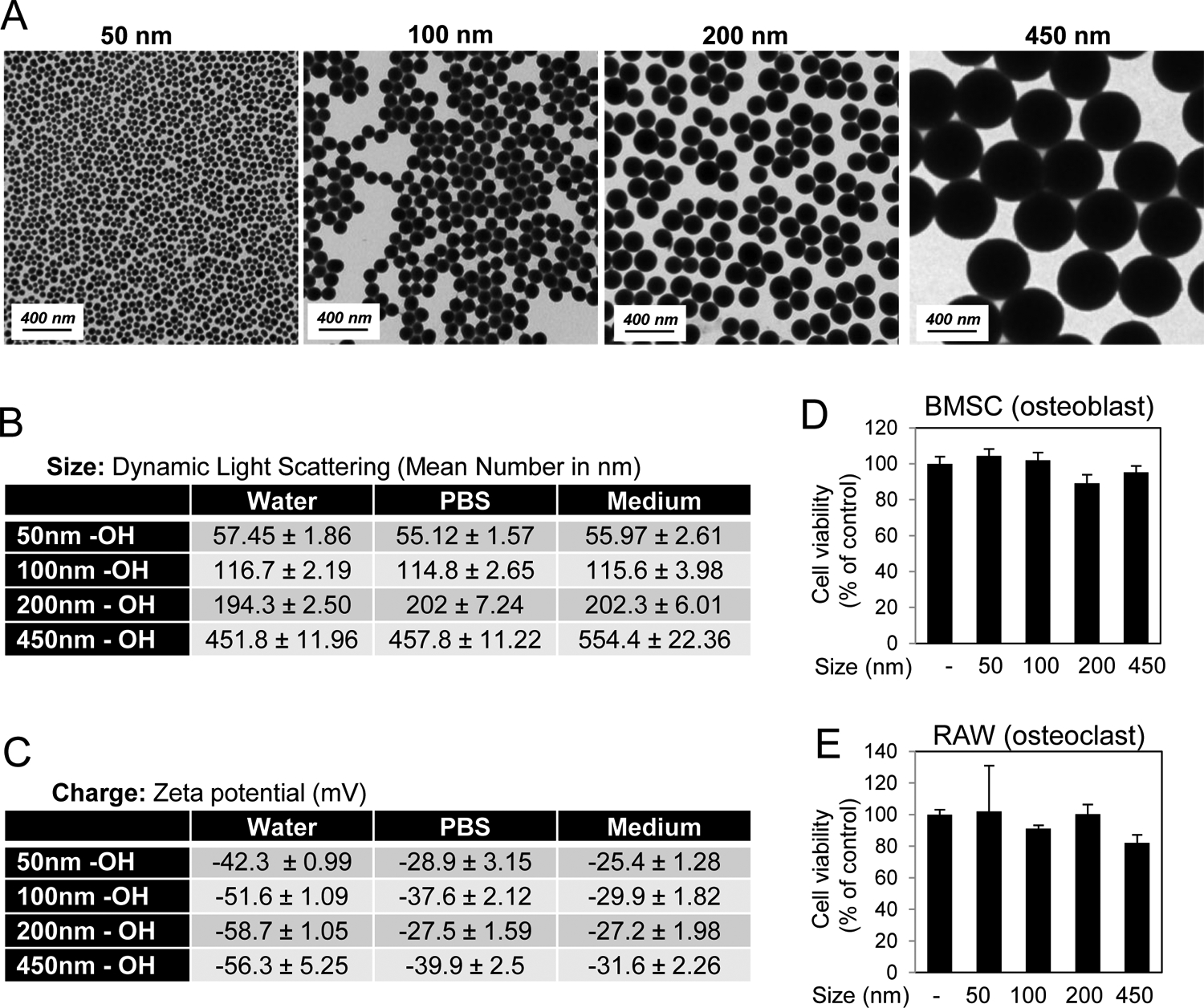
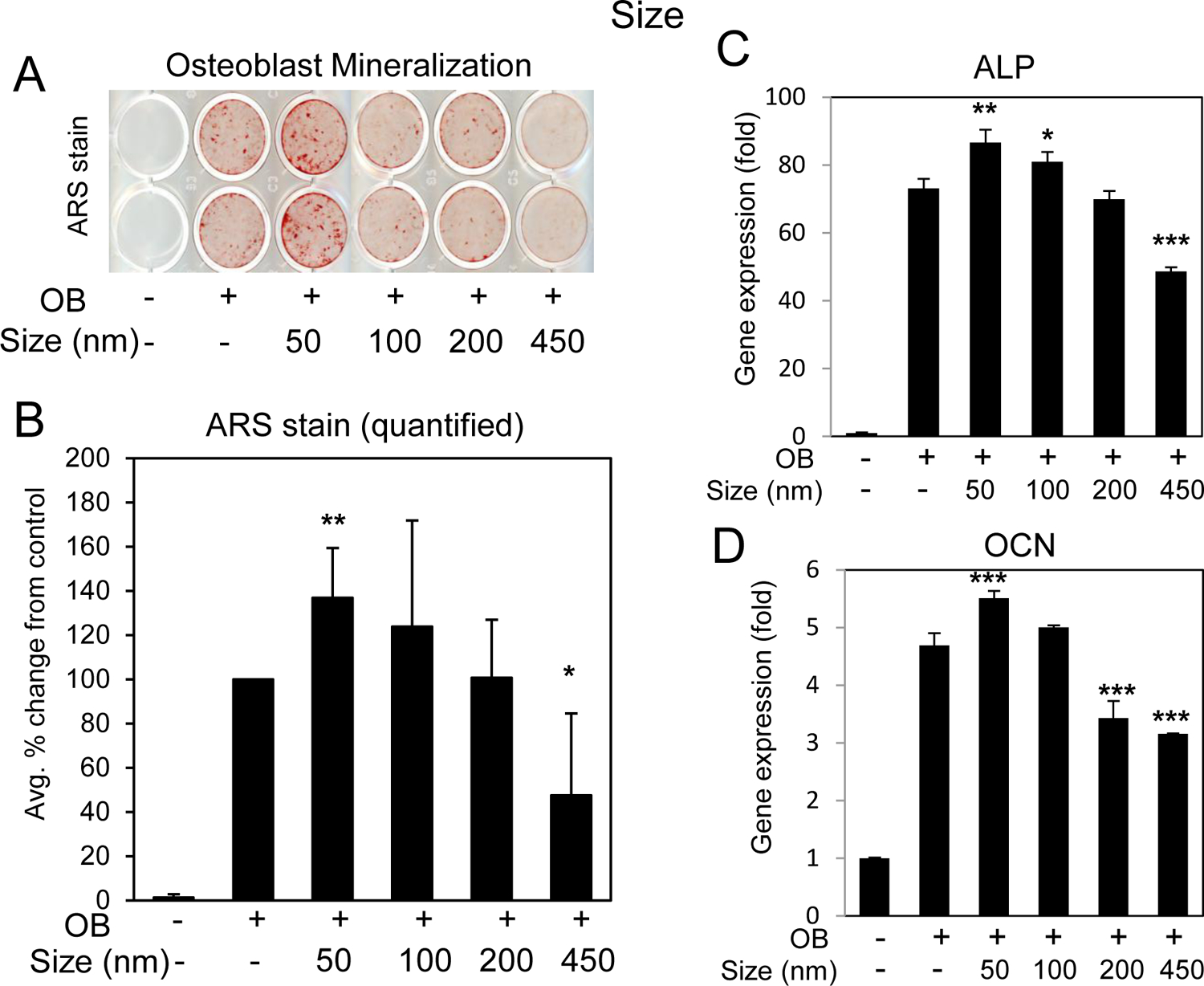
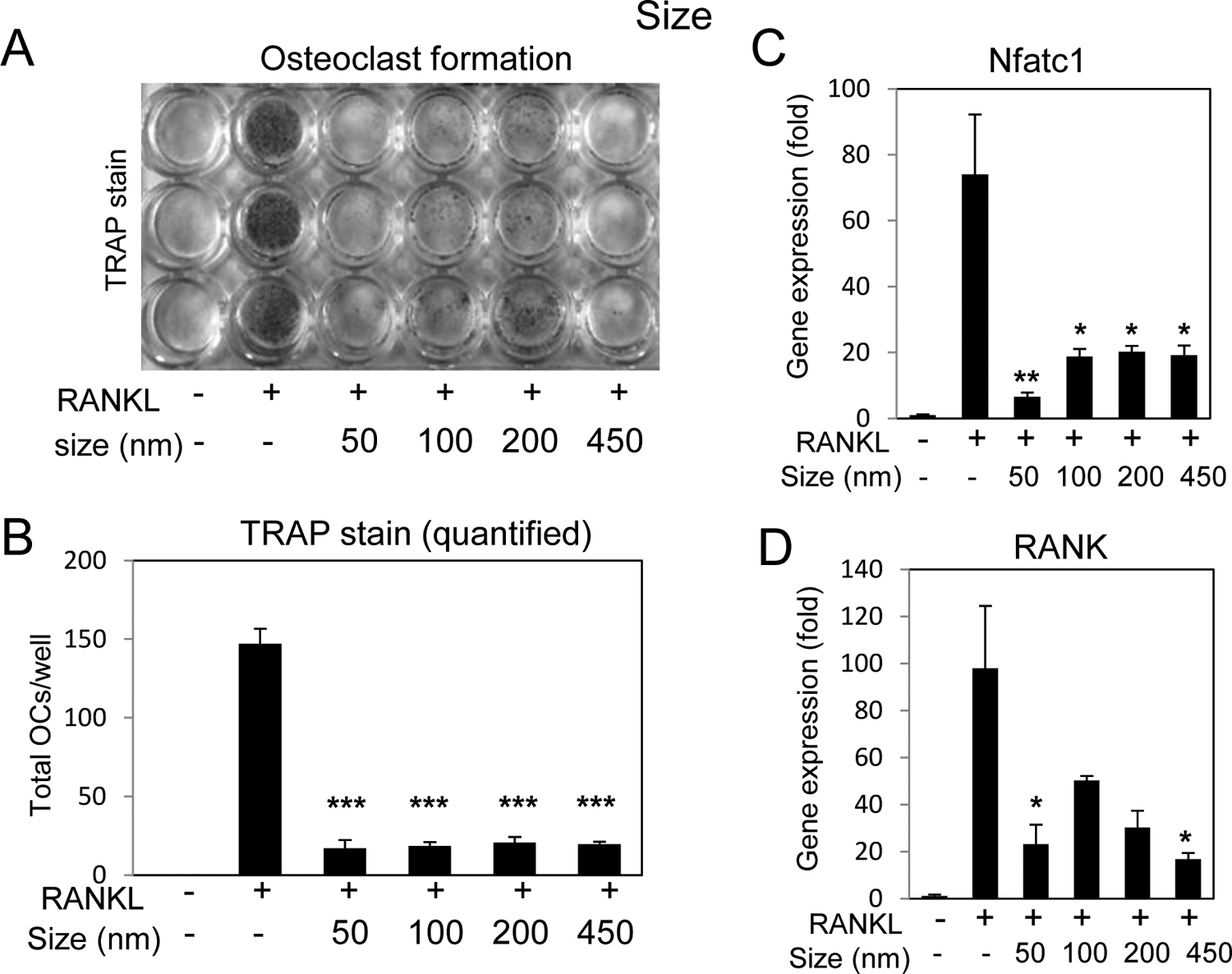
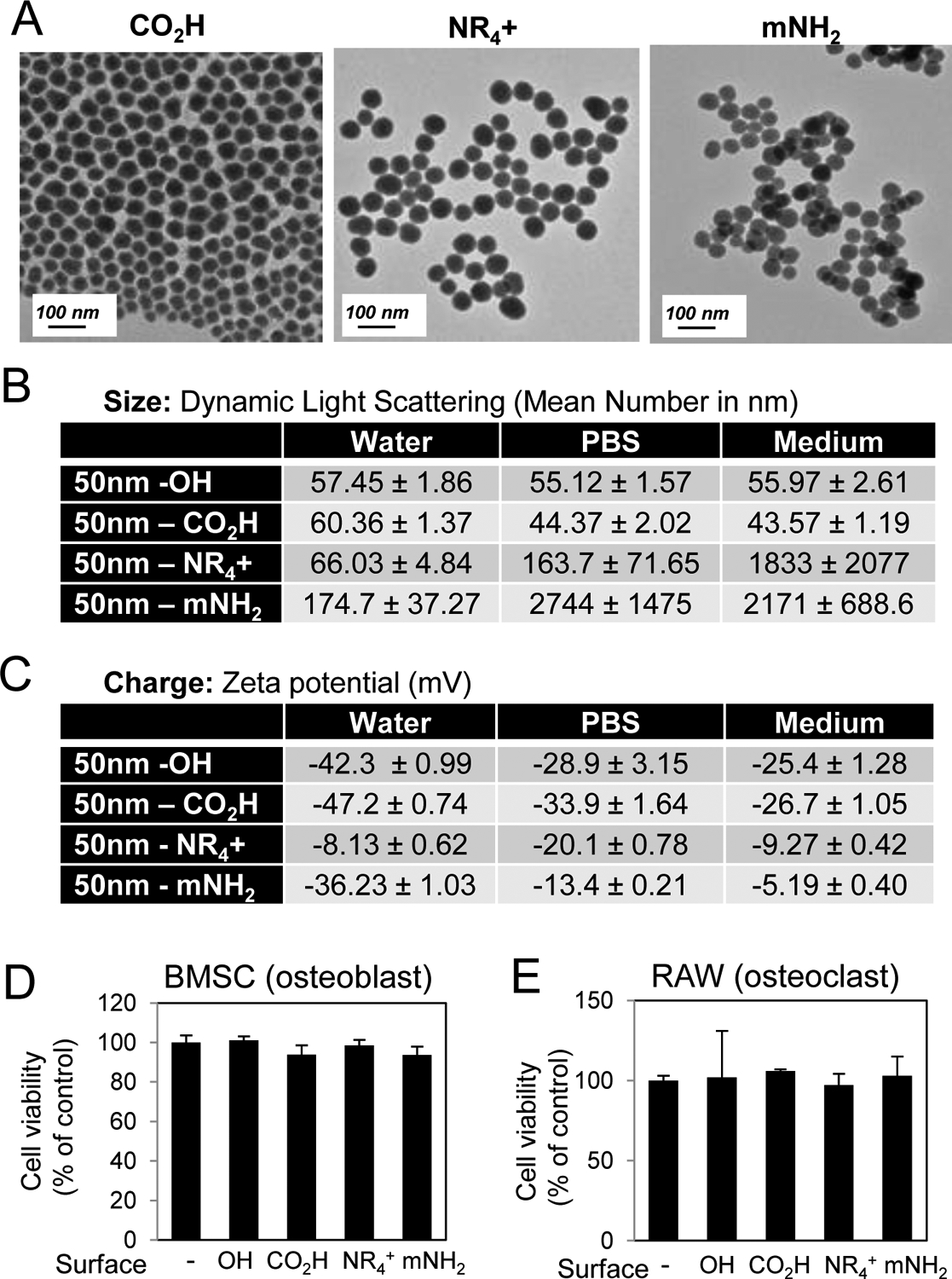
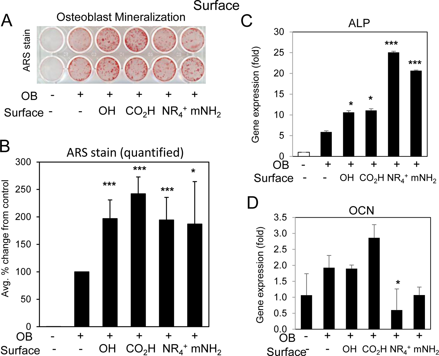
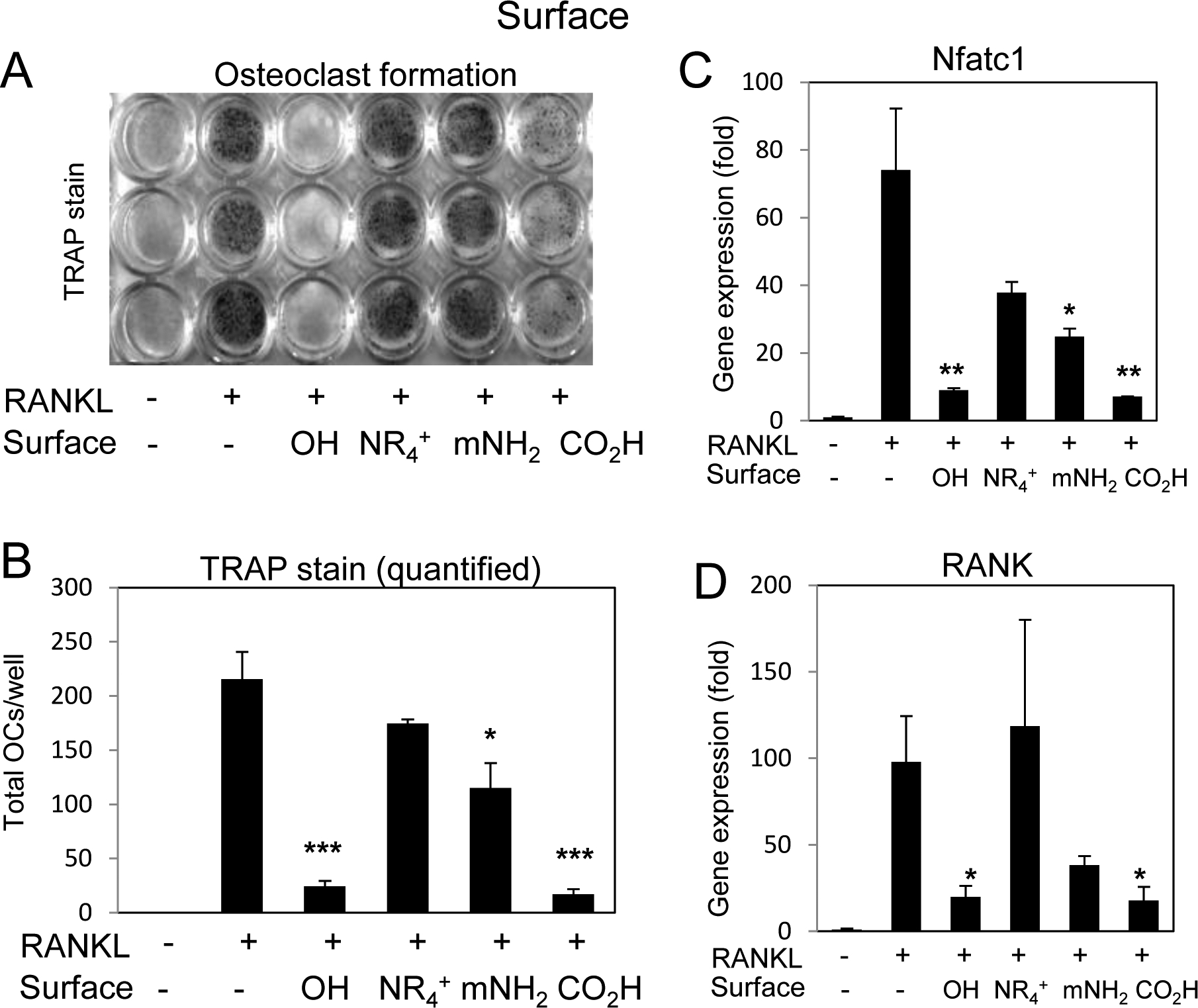
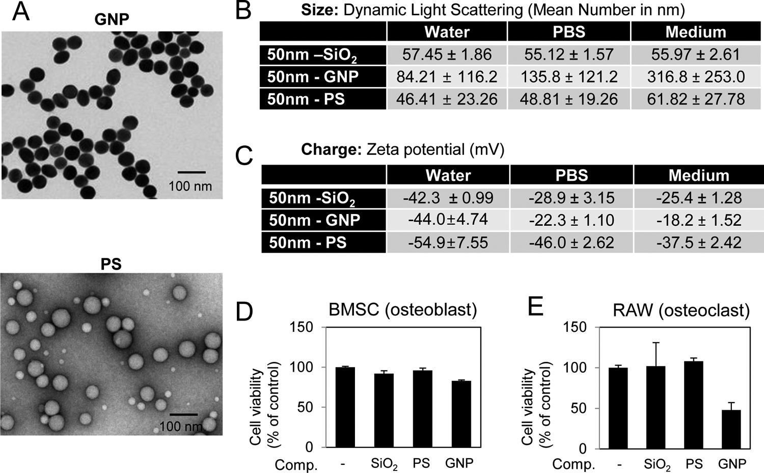
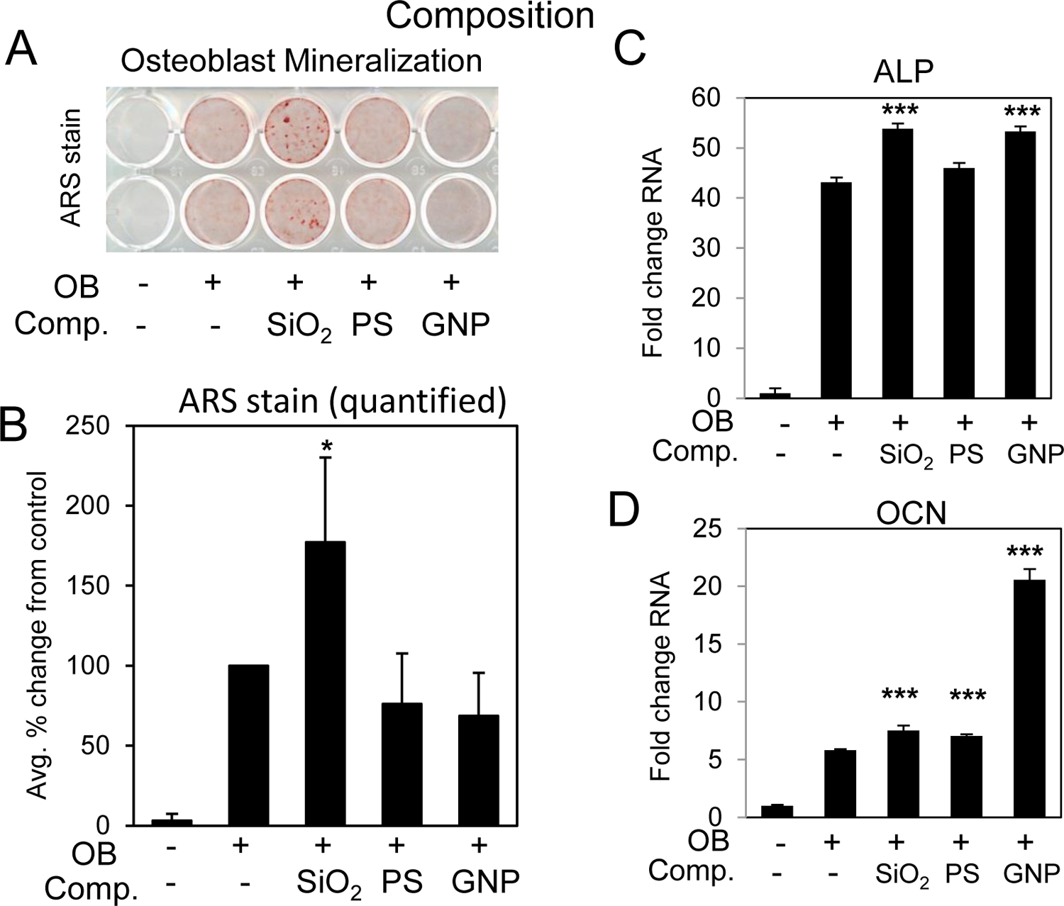
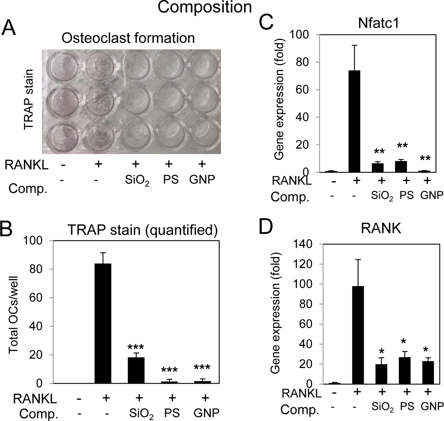
Silica based nanoparticles have been demonstrated to have intrinsic biologic activity towards the skeleton and to function by promoting the differentiation of bone forming osteoblasts while inhibiting the differentiation of bone resorbing osteoclasts. The excitement surrounding nanomedicine in part revolves around the almost unlimited possibilities for varying the physicochemical properties including size, composition, and surface charge. To date few studies have attempted to manipulate these characteristics in concert to optimize a complex biologic outcome. Towards this end, spherical silica nanoparticles of various sizes (50-450 nm), of different surface properties (OH, CO2H, NR4+, mNH2), and of different composition (silica, gold, and polystyrene) were synthesized and evaluated for biological activity toward skeletal cells. Osteoblast activity was most influenced by composition and size variables, whereas osteoclasts were most affected by surface property variation. The study also establishes nanoparticle mediated suppression of Nfatc1, a key transcriptional regulator for osteoclast differentiation, identifying a novel mechanism of action. Collectively, the study highlights how during the design of bioactive nanoparticles, it is vital to consider not only the myriad of physical properties that can be manipulated, but also that the characteristics of the target cell plays an equally integral role in determining biological outcome. STATEMENT OF SIGNIFICANCE: Silica nanomaterials represent a promising biomaterial for beneficial effects on bone mass and quality as well as regenerative tissue engineering and are currently being investigated for intrinsic bioactivity towards the primary cells responsible for skeletal homeostasis; osteoblasts and osteoclasts. The goal of the current study was to assess the physical properties of silica nanoparticles that impart intrinsic bioactivity by evaluating size, surface charge, and composition. Results reveal differential influences of the physical properties of nanoparticles towards osteoblasts and osteoclasts. This study provides new insights into the design of nanoparticles to specifically target different aspects of bone metabolism and highlights the opportunities provided by nanotechnology to modulate a range of cell specific biological responses for therapeutic benefit.

Keywords: Bone cells; Composition; Silica nanoparticles; Size; Surface charge.


Conflict of interest statement

Conflict of Interest

The authors declare no conflict of interest.

Figures

Fig. 1.
Fig. 1.
Characterization of varying sized silica nanoparticles. A) Shape was assessed by Transmission Electron Microscopy. B) Size of nanoparticles was characterized by Dynamic Light Scattering in water, phosphate buffered saline (PBS), and medium (Avg. of 3 readings). C) Zeta Potential of nanoparticles was measured in water, DPBS, and cell culture medium (Avg. of 3 readings). Cell viability of D) BMSCs in response to 72 hr treatment with 10 μg/ml nanoparticles and E) RAW264.7 cells in response to 72 hr treatment with 25 μg/ml nanoparticles as indicated (N=3–6). Avg.±Stdev.
Fig. 2.
Fig. 2.. Silica nanoparticle size and enhancement of osteoblast differentiation.
BMSCs were cultured in growth medium or medium supplemented with ascorbic acid and beta glycerophosphate (OB) and treated with 10 μg/ml silica nanoparticles of varying size as indicated. Cells were stained after 11–14 days for A) mineralization with Alizarin Red S staining and B) the average of six independent experiments quantified by image J. Data are expressed and percent change from the positive control (+OB) ±Stdev. Gene expression of C) alkaline phosphatase (ALP) and D) osteocalcin (OCN) are expressed as fold change (±Stdev) relative to untreated. Statistical analysis compares nanoparticle treated relative to positive control (+OB) by Student’s t test. *P< 0.05, **P< 0.01, **P< 0.005.
Fig. 3.
Fig. 3.. Silica nanoparticle size and inhibition of osteoclast differentiation.
A) RAW264.7 cells were cultured in growth medium or medium supplemented with RANKL (15 ng/ml) and treated with silica nanoparticles of varying size as indicated and after 3–4 days the cultures were stained for tartrate resistant alkaline phosphatase (TRAP). B) Multinucleated (≥3 nuclei) TRAP positive cells were quantified were quantified from plates in (A); average of 3 wells ±Stdev and representative of 3 independent experiments. Cells were treated with RANKL (15 ng/ml) for 16 hours and harvested for RNA analysis of C) Nfatc1 or D) RANK by qRT-PCR expressed as fold change relative to untreated control Avg.±Stdev. *P< 0.05, **P< 0.01, and ***P< 0.005 relative to RANKL treated Students t test (n=3).
Fig. 4.
Fig. 4.. Characterization of nanoparticles with varied surface properties.
A) The shape of nanoparticles with varied surfaces was characterized by TEM and B) size was assessed by Dynamic Light Scattering in water, PBS, and medium (Avg. of 3 readings). C) Zeta Potential of nanoparticles was measured in water, DPBS, and cell culture medium (Avg. of 3 readings). Cell viability of D) BMSCs in response to 72 hr treatment with 10 μg/ml nanoparticles and E) RAW264.7 cells in response to 72 hr treatment with 25 μg/ml nanoparticles as indicated (N=3–6). Avg.±Stdev.
Fig. 5.
Fig. 5.. Nanoparticle surface properties and enhancement of osteoblast differentiation.
BMSCs or MC3T3-E1 cells were cultured in growth medium or medium supplemented with ascorbic acid and beta glycerophosphate (OB) and treated with 10 μg/ml silica nanoparticles of varying surface properties as indicated. Cells were stained after 11–14 days for A) mineralization with Alizarin Red S staining and B) the average of six independent experiments quantified by image J. Data are expressed and percent change from the positive control (+OB) ±Stdev. Gene expression of C) alkaline phosphatase (ALP) and D) osteocalcin (OCN) are expressed as fold change (±Stdev) relative to untreated. Statistical analysis compares nanoparticle treated relative to positive control (+OB) by Student’s t test. *P< 0.05, **P< 0.01, **P< 0.005.
Fig. 6.
Fig. 6.. Silica nanoparticle surface properties and enhancement of osteoclast differentiation.
A) RAW264.7 cells were cultured in growth medium or medium supplemented with RANKL (15 ng/ml) and treated with silica nanoparticles with varying surfaces as indicated and after 3–4 days the cultures were stained for tartrate resistant alkaline phosphatase (TRAP). B) Multinucleated (≥3 nuclei) TRAP positive cells were quantified were quantified from plates in (A); average of 3 wells ±Stdev and representative of 3 independent experiments. Cells were treated with RANKL (15 ng/ml) for 16 hours and harvested for RNA analysis of C) Nfatc1 or D) RANK by qRT-PCR expressed as fold change relative to untreated control Avg.±Stdev. *P< 0.05, **P< 0.01, and ***P< 0.005 relative to RANKL treated Students t test (n=3).
Fig. 7.
Fig. 7.. Characterization of spherical nanoparticles of varying composition.
A) The shape of the gold (GNP) and polystyrene (PS) nanoparticles was characterized by TEM and B) size by Dynamic Light Scattering in water, PBS, and medium (Avg. of 3 readings). C) Zeta Potential of nanoparticles was measured in water, DPBS, and cell culture medium (Avg. of 3 readings). Cell viability of D) BMSCs in response to 72 hr treatment with 10 μg/ml nanoparticles and E) RAW264.7 cells in response to 72 hr treatment with 25 μg/ml nanoparticles as indicated (N=3–6). Avg.±Stdev.
Fig. 8.
Fig. 8.. Nanoparticle composition and enhancement of osteoblast differentiation.
BMSCs were cultured in growth medium or medium supplemented with ascorbic acid and beta glycerophosphate (OB) and treated with 10 μg/ml of 50 nm nanoparticles composed of SiO2:silica nanoparticles, GNP:gold nanoparticles and PS:polystyrene. Cells were stained after 11–14 days for A) mineralization with Alizarin Red S staining and B) the average of four independent experiments quantified by image J. Data are expressed and percent change from the positive control (+OB) ±Stdev. Gene expression of C) alkaline phosphatase (ALP) and D) osteocalcin (OCN) are expressed as fold change (±Stdev) relative to untreated. Statistical analysis compares nanoparticle treated relative to positive control (+OB) by Student’s t test. *P< 0.05, **P< 0.01, **P< 0.005.
Fig. 9.
Fig. 9.. Nanoparticle composition and inhibition of osteoclast differentiation.
A) RAW264.7 cells were cultured in growth medium or medium supplemented RANKL (15 ng/ml) and treated with spherical 50nm nanoparticles of varying compositions; SiO2:silica nanoparticles, GNP:gold nanoparticles and PS:polystyrene as indicated and after 3–4 days the cultures were stained for tartrate resistant alkaline phosphatase (TRAP). B) Multinucleated (≥3 nuclei) TRAP positive cells were quantified were quantified from plates in (A); average of 3 wells ±Stdev and representative of 3 independent experiments. Cells were treated with RANKL (15 ng/ml) for 16 hours and harvested for RNA analysis of C) Nfatc1 or D) RANK by qRT-PCR expressed as fold change relative to untreated control Avg.±Stdev. *P< 0.05, **P< 0.01, and ***P< 0.005 relative to RANKL treated Students t test (n=3).

References

    1. Shang L, Nienhaus K, Nienhaus GU. Engineered nanoparticles interacting with cells: size matters. J Nanobiotechnology. 12 (2014) 5. - PMC - PubMed
    1. Frohlich E. The role of surface charge in cellular uptake and cytotoxicity of medical nanoparticles. Int J Nanomedicine. 7 (2012) 5577–5591. - PMC - PubMed
    1. Verma A, Stellacci F. Effect of surface properties on nanoparticle-cell interactions. Small. 6 (2010) 12–21. - PubMed
    1. Liu Y, Tan J, Thomas A, Ou-Yang D, Muzykantov VR. The shape of things to come: importance of design in nanotechnology for drug delivery. Ther Deliv. 3 (2012) 181–194. - PMC - PubMed
    1. Albanese A, Tang PS, Chan WC. The Effect of Nanoparticle Size, Shape, and Surface Chemistry on Biological Systems. Annu Rev Biomed Eng. (2012). - PubMed

Publication types

Substances