Review
. 2018 Oct 15;10(10):560.
doi: 10.3390/v10100560.

Temperature Sensitive Mutations in Influenza A Viral Ribonucleoprotein Complex Responsible for the Attenuation of the Live Attenuated Influenza Vaccine

Affiliations
Review

Temperature Sensitive Mutations in Influenza A Viral Ribonucleoprotein Complex Responsible for the Attenuation of the Live Attenuated Influenza Vaccine

Luis Martínez-Sobrido et al. Viruses. .

Abstract

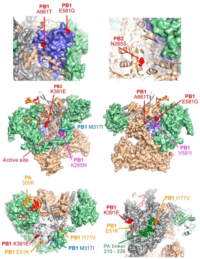
Live attenuated influenza vaccines (LAIV) have prevented morbidity and mortality associated with influenza viral infections for many years and represent the best therapeutic option to protect against influenza viral infections in humans. However, the development of LAIV has traditionally relied on empirical methods, such as the adaptation of viruses to replicate at low temperatures. These approaches require an extensive investment of time and resources before identifying potential vaccine candidates that can be safely implemented as LAIV to protect humans. In addition, the mechanism of attenuation of these vaccines is poorly understood in some cases. Importantly, LAIV are more efficacious than inactivated vaccines because their ability to mount efficient innate and adaptive humoral and cellular immune responses. Therefore, the design of potential LAIV based on known properties of viral proteins appears to be a highly appropriate option for the treatment of influenza viral infections. For that, the viral RNA synthesis machinery has been a research focus to identify key amino acid substitutions that can lead to viral attenuation and their use in safe, immunogenic, and protective LAIV. In this review, we discuss the potential to manipulate the influenza viral RNA-dependent RNA polymerase (RdRp) complex to generate attenuated forms of the virus that can be used as LAIV for the treatment of influenza viral infections, one of the current and most effective prophylactic options for the control of influenza in humans.

Keywords: attenuated; cold-adapted; influenza vaccine; influenza virus; live-attenuated influenza virus; nucleoprotein; recombinant influenza virus; temperature-sensitive; viral polymerase complex.


Conflict of interest statement

The authors declare no conflict of interest.

Figures

Figure 1
Figure 1
Influenza A virion structure, genome organization, and viral replication and transcription. (A) Virion structure: IAV are surrounded by a lipid bilayer containing the two viral glycoproteins hemagglutinin (HA) and neuraminidase (NA). Also, in the virion membrane is the ion channel matrix 2 (M2) protein. Under the viral lipid bilayer is a protein layer composed of the matrix 1 (M1) protein and the nuclear export protein (NEP). Inside the virion are the eight viral (v)RNA segments that are encapsidated by the viral nucleoprotein (NP) as viral ribonucleoprotein (vRNP) complexes. Associated with each vRNP is the viral RNA-dependent RNA polymerase (RdRp) complex made of the three polymerase subunits PB2, PB1, and PA that, together with the viral NP are the minimal components required for viral replication and transcription. Viral proteins in the virion particle (top) and in the vRNP (bottom) are indicated. The viral proteins showed in the illustration of the viral particle do not maintain the stoichiometric that is found in a mature virion. (B) Genome organization: IAV contain eight single-stranded, negative-sense, viral RNA segments (PB2, PB1, PA, HA, NP, NA, M, and NS). Each viral segment contains non-coding regions (NCR) at the 3′ and 5′ ends (black lines). Also, at the 3′ and 5′ end of the viral RNAs are the packaging signals, responsible for the efficient encapsidation into nascent virions (white boxes). Numbers represent the nucleotide length of the 3′ or 5′ packaging signals in each of the viral RNA segments. (C) Viral genome replication, transcription and translation: IAV NP and the viral polymerase complex PB2, PB1, and PA associated with the viral RNA form the vRNP complexes that are responsible for viral replication and transcription. vRNP complexes initiate transcription from the viral promoters located within the NCR at the 3′ termini of each of the vRNAs. Transcription results in the synthesis of IAV mRNA that are translated to proteins. IAV polymerase complex is also involved in the replication of the vRNA into a complementary (c)RNA that serve as template for the amplification of vRNAs. Newly synthesized vRNPs, together with the structural viral proteins result in the formation of new IAV.
Figure 2
Figure 2
Structure of the IAV polymerase complex. The IAV polymerase complex structure is shown in three different views (left to right) rotated by ≈90° between each, and the same coloring is retained in all images (PDB code 4WSB). A dynamic version of this figure where the molecules can be rotated in real time is available online as Figure S1. Panel A shows the PB1 RdRp subunit in grey with its thumb domain colored blue, the active site within conserved motif C shown in magenta, and the 5′ and 3′ RNAs shown in red and orange, respectively. Panel B adds the PB2 subunit in wheat color with its two major domains (RNA and cap binding) indicated. PB2 associates with a large surface on PB1, effectively forming one large globular structure from which the PB2 cap binding and RNA binding domains protrude. Panel C illustrates how the RdRp PA subunit wraps around the PB2-PB1 complex, with its N-terminal endonuclease domain on one side of the complex, the PB1 binding domain forming the major exterior surface of the complex on the other side, and the linker sequence being extended across the PB1 surface. Panel D shows the to-scale structure of the poliovirus polymerase elongation complex, among the smallest single subunit viral polymerases, for size comparison and in the same orientations (PDB code 3OL6).
Figure 3
Figure 3
Schematic representation of the IAV virus polymerase complex protein subunits. Graphical illustration of IAV PB1 (A), PB2 (B), PA (C), and NP (D), showing the location of the conserved functional domains. Amino acid substitutions responsible for the temperature sensitive (ts), attenuated (att), and cold-adapted (ca) phenotype of the MDV LAIV A/Ann Arbor/6/60 (PB1: K391E, E581G, and A661T; PB2: N265S; NP D34G), Len/17 (PB1: K265N and V591I; PB2: V478L), and Len/47 (Len/47 substitutions and additional amino acid changes in PB1: M317I; PB2: S490R; NP: L341I) are indicated in red, magenta, or blue, respectively. NLS, nuclear localization signal. Figure not to scale.
Figure 4
Figure 4
Generation of LAIV: (A) Isolation of the A/Ann Arbor/6/60 H2N2 LAIV MDV: A/Ann Arbor/6/60 H2N2 wild-type (WT) was serially passage 100 times (P1 to P100) in chicken kidney tissue culture (CKTC) cells under gradually reduced temperatures (36 °C to 24 °C). The obtained A/Ann Arbor/6/60 LAIV contained several mutations and the ones responsible for the att, ca, and ts has been mapped to a single residue in PB2 (N265S), three residues in PB1 (K391E, E581G, and A661T) and a single residue (D34G) in NP (Figure 2). The A/Ann Arbor/6/60 H2N2 LAIV is used as master donor virus (MDV) to produce the seasonal LAIV. (B) Generation of the A/Leningrad/134/17/57 H2N2 LAIV MDV: A/Leningrad/134/17/57 H2N2 WT was serially passage 20 times (P1 to P20) in embryonated chicken eggs at optimal temperature of 32 °C. Then, the A/Leningrad/134/17/57 H2N2 LAIV MDV was obtained after 17 additional passages at 25 °C. (C) Schematic representation to produce the seasonal LAIV: The traditional method for generating reassortant virus is based on the co-infection with two influenza viruses in eggs. Both the WHO candidate IAV (left) and the A/Ann Arbor/6/60 H2N2 LAIV MDV (right) are inoculated in eggs followed by the selection of appropriate seed viruses by amplification in the presence of antibodies against the HA and NA of A/Ann Arbor/6/60 H2N2. The resulting virus containing the HA (red) and NA (blue) segments from the WHO-recommended IAV strain and the six inteRNAl vRNAs of the MDV A/Ann Arbor/6/60 H2N2 LAIV is amplified and used for vaccine production and used as a seasonal LAIV.
Figure 5
Figure 5
Attenuation and reversion mutations in the IAV polymerase complex. Attenuation mutations in the IAV polymerase: The locations of various IAV mutations found in the MDV LAIV and in revertant strains are shown as CPK spheres on the background of the polymerase complex colored as in Figure 2, with some subunits being shown as surfaces and others as cartoons in various orientations as needed for clarity. A dynamic version of this figure where the molecules can be rotated in real time is available on-lines as Figure S2. Mutations in the US LAIV MDV A/Ann Arbor/6/60 H2N2 are shown in red. Mutations present in the Russian LAIV MDV Len/17 or Len/47 (A/Leningrad/134/57 H2N2) strain are shown in magenta or blue, respectively. PB2 mutations V478L (Len/17) and S490R (Len/47) are not indicated in the structure. For more information see Figure 2 and Table 1. A set of engineered mutations in the PA subunit linker segment that wraps around PB1 are shown in green. Reversion mutations in PB1 (E51K, I171V) and PA (N350K) that restore virulence in the US LAIV MDV A/Ann Arbor/6/60 H2N2 (Table 2, green) are shown in orange.

References

    1. Chiapponi C., Faccini S., De Mattia A., Baioni L., Barbieri I., Rosignoli C., Nigrelli A., Foni E. Detection of influenza D virus among swine and cattle, Italy. Emerg. Infect. Dis. 2016;22:352–354. doi: 10.3201/eid2202.151439. - DOI - PMC - PubMed
    1. Foni E., Chiapponi C., Baioni L., Zanni I., Merenda M., Rosignoli C., Kyriakis C.S., Luini M.V., Mandola M.L., Bolzoni L., et al. Influenza D in Italy: Towards a better understanding of an emerging viral infection in swine. Sci. Rep. 2017;7:11660. doi: 10.1038/s41598-017-12012-3. - DOI - PMC - PubMed
    1. Su S., Fu X., Li G., Kerlin F., Veit M. Novel influenza D virus: Epidemiology, pathology, evolution and biological characteristics. Virulence. 2017;8:1580–1591. doi: 10.1080/21505594.2017.1365216. - DOI - PMC - PubMed
    1. Baker S.F., Nogales A., Finch C., Tuffy K.M., Domm W., Perez D.R., Topham D.J., Martinez-Sobrido L. Influenza A and B virus intertypic reassortment through compatible viral packaging signals. J. Virol. 2014;88:10778–10791. doi: 10.1128/JVI.01440-14. - DOI - PMC - PubMed
    1. Xu X., Lindstrom S.E., Shaw M.W., Smith C.B., Hall H.E., Mungall B.A., Subbarao K., Cox N.J., Klimov A. Reassortment and evolution of current human Influenza A and B viruses. Virus. Res. 2004;103:55–60. doi: 10.1016/j.virusres.2004.02.013. - DOI - PubMed

Publication types

MeSH terms