. 2018 Oct 15;7(10):170.
doi: 10.3390/cells7100170.

Samp1 Mislocalization in Emery-Dreifuss Muscular Dystrophy

Affiliations

Samp1 Mislocalization in Emery-Dreifuss Muscular Dystrophy

Elisabetta Mattioli et al. Cells. .

Abstract

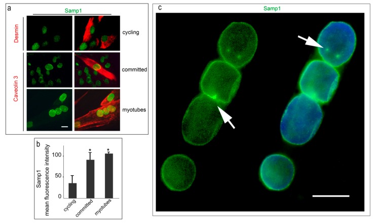
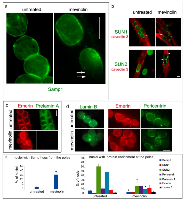
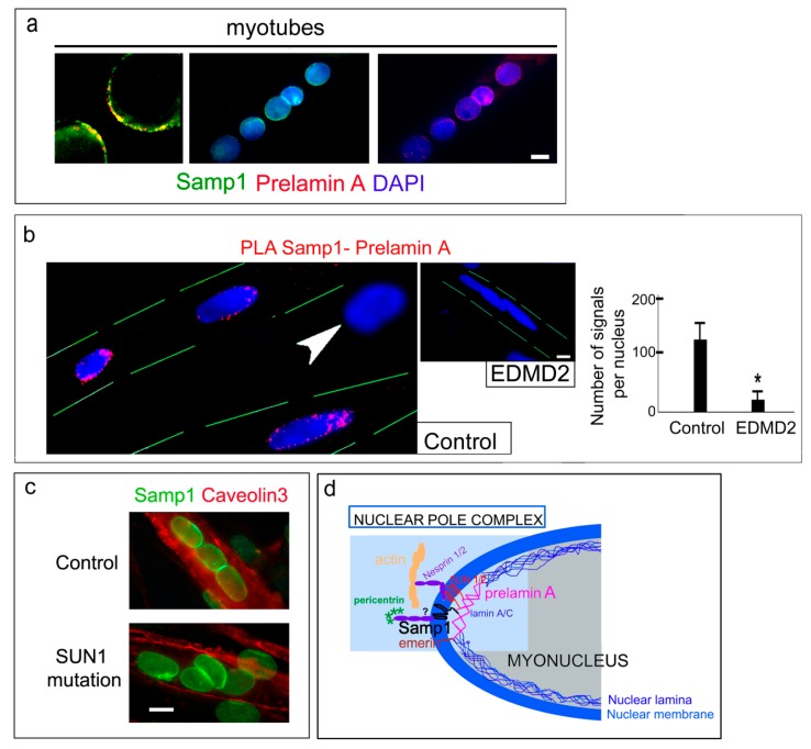
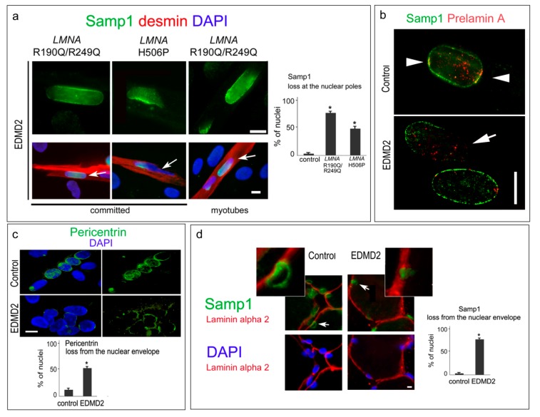
LMNA linked-Emery-Dreifuss muscular dystrophy (EDMD2) is a rare disease characterized by muscle weakness, muscle wasting, and cardiomyopathy with conduction defects. The mutated protein lamin A/C binds several nuclear envelope components including the Linker of Nucleoskeleton and Cytoskeleton (LINC) complex and the inner nuclear membrane protein Samp1 (Spindle Associated Membrane Protein 1). Considering that Samp1 is upregulated during muscle cell differentiation and it is involved in nuclear movement, we hypothesized that it could be part of the protein platform formed by LINC proteins and prelamin A at the myotube nuclear envelope and, as previously demonstrated for those proteins, could be affected in EDMD2. Our results show that Samp1 is uniformly distributed at the nuclear periphery of normal human myotubes and committed myoblasts, but its anchorage at the nuclear poles is related to the presence of farnesylated prelamin A and it is disrupted by the loss of prelamin A farnesylation. Moreover, Samp1 is absent from the nuclear poles in EDMD2 myotubes, which shows that LMNA mutations associated with muscular dystrophy, due to reduced prelamin A levels in muscle cell nuclei, impair Samp1 anchorage. Conversely, SUN1 pathogenetic mutations do not alter Samp1 localization in myotubes, which suggests that Samp1 lies upstream of SUN1 in nuclear envelope protein complexes. The hypothesis that Samp1 is part of the protein platform that regulates microtubule nucleation from the myotube nuclear envelope in concert with pericentrin and LINC components warrants future investigation. As a whole, our data identify Samp1 as a new contributor to EDMD2 pathogenesis and our data are relevant to the understanding of nuclear clustering occurring in laminopathic muscle.

Keywords: Emery-Dreifuss Muscular Dystrophy type 2 (EDMD2); LINC complex; Samp1 (NET5); myonuclear positioning; prelamin A.


Conflict of interest statement

The authors declare no conflict of interest.

Figures

Figure 1
Figure 1
Samp1 is recruited to the nuclear periphery in differentiating human control myoblasts. (a) Samp1 (green) and desmin (red) were co-stained in cycling human myoblasts (cycling). Samp1 and caveolin 3 (red) were co-stained in committed myoblasts (committed) and myotubes (myotubes). Nuclear envelope localization of Samp1 is observed in all caveolin 3-positive cells. (b) Quantitative analysis of mean fluorescence intensity is reported in the graphs. (c) A myotube labeled with Samp1 antibody and counterstained with DAPI shows a few sites of accumulation of Samp1 in areas possibly corresponding to centrosome remnants (arrows). Statistically significant differences by the Mann Whitney test are indicated by asterisks (*). Scale bars, 10 µm.
Figure 2
Figure 2
Loss of Samp1 from the nuclear poles in the absence of farnesylated prelamin A. Human control myotubes left untreated (untreated) or treated with mevinolin (mevinolin) are shown. (a) Samp1 staining in myotubes. Arrows show a nuclear pole devoid of Samp1 in a myotube subjected to mevinolin. (b) Co-staining of SUN1 or SUN2 with caveolin 3. SUN1 and SUN2 loss from the nuclear poles is observed in mevinolin-treated myotubes. The arrow points to a nuclear pole connected to a nucleus of a fusing myoblast. Arrowheads show protein loss from a nuclear pole. (c) Human myotubes co-stained for emerin and farnesylated prelamin A (Diatheva 1188-2 antibody). The antibody used to detect prelamin A is specific for farnesylated prelamin A, which is not detectable in mevinolin treated cells. (d) Staining of lamin B (left panel) and co-staining of emerin and pericentrin (right panel) in myotubes. (e) Quantitative analysis of the percentage of nuclei showing loss of Samp1 staining at the nuclear pole(s) (left panel) and protein accumulation at the nuclear poles (right panel) in untreated or mevinolin-treated cells. Mean values of three counts are reported in the graphs. Statistical significance of differences (p < 0.05) is indicated by an asterisk (*). Scale bars, 10 µm.
Figure 3
Figure 3
Samp1 co-localizes with farnesylated prelamin A at the nuclear poles of human myotubes. (a) Co-staining of Samp1 and prelamin A in human control myotubes (myotubes). Left panel, high magnification of a double stained nuclear pole showing protein colocalization. Single fluorescence of Samp1 and prelamin A are shown along with DAPI staining of nuclei. (b) PLA of prelamin A and Samp1 (PLA Prelamin A-Samp1, red dots) in control myotubes. The arrowhead indicates a mononucleated myoblast that is negative for PLA. An EDMD2 myotube negative for PLA is shown in the inset. Graphs show quantitative analysis of PLA signals reporting the average number of spots in nuclei (80 nuclei per sample were counted). (c) Immunostaining of Samp1 and Caveolin 3 in control myotubes (control) and myotubes from a muscular dystrophy caused by SUN1 mutation (SUN1 mutation, see methods for details) [17]. (d) Schematic representation of the proposed protein platform (myo-Nuclear Pole Complex) at the nuclear envelope of differentiated human myoblasts. Prelamin A interacts with Samp1, SUN1/2, and emerin. SUN1/2 link the complex to nesprins and the actin cytoskeleton. The hypothesis (indicated by “?”) that Samp1 binds nesprin 2G and pericentrin warrants future investigation. Statistically significant differences by the Mann Whitney test are indicated by asterisks (*). Nuclei are counterstained with DAPI (blue). Scale of bars, 10 µm.
Figure 4
Figure 4
Mis-localization of Samp1 in the EDMD2 nuclei. (a) Samp1 staining in EDMD2 myoblasts committed to differentiation (committed) and myotubes (myotubes). Myotubes are double stained for desmin (red). Upper panel, high magnification of nuclei indicated by arrows. The percentage of nuclei showing Samp1 loss at the nuclear poles (at least one pole) is reported in the graph. (b) Double staining of prelamin A and Samp1 in differentiated control and EDMD2 myoblasts. Arrowheads indicate co-localization, the arrow indicates a nuclear pole devoid of Samp1 and prelamin A fluorescence. (c) Pericentrin staining in the control and the EDMD2 myotubes. Percentage of nuclei showing pericentrin loss at the nuclear periphery (negative nuclei) is reported in the graph. (d) Samp1 and laminin alpha 2 were co-stained in muscle tissue from control (control) and EDMD2 (EDMD2). Nuclei inside the basal lamina outlined by laminin alpha 2 are myonuclei. Percentage of nuclei showing Samp1 loss from the nuclear envelope is reported in the graph. Magnification of selected nuclei (arrows) is shown in the insets. Nuclei in (a,c,d) are counterstained with DAPI. Statistically significant differences by the Mann Whitney test are indicated by asterisks (*). Scale of bars, 10 µm.

References

    1. Burke B., Stewart C.L. The nuclear lamins: Flexibility in function. Nat. Rev. Mol. Cell Biol. 2013;14:13–24. doi: 10.1038/nrm3488. - DOI - PubMed
    1. Chang W., Worman H.J., Gundersen G.G. Accessorizing and anchoring the linc complex for multifunctionality. J. Cell Biol. 2015;208:11–22. doi: 10.1083/jcb.201409047. - DOI - PMC - PubMed
    1. Schwartz C., Fischer M., Mamchaoui K., Bigot A., Lok T., Verdier C., Duperray A., Michel R., Holt I., Voit T., et al. Lamins and nesprin-1 mediate inside-out mechanical coupling in muscle cell precursors through fhod1. Sci. Rep. 2017;7:1253. doi: 10.1038/s41598-017-01324-z. - DOI - PMC - PubMed
    1. Lee Y.L., Burke B. Linc complexes and nuclear positioning. Semin. Cell Dev. Biol. 2018;82:67–76. doi: 10.1016/j.semcdb.2017.11.008. - DOI - PubMed
    1. Zhang X., Lei K., Yuan X., Wu X., Zhuang Y., Xu T., Xu R., Han M. Sun1/2 and syne/nesprin-1/2 complexes connect centrosome to the nucleus during neurogenesis and neuronal migration in mice. Neuron. 2009;64:173–187. doi: 10.1016/j.neuron.2009.08.018. - DOI - PMC - PubMed