. 2018 Dec 14;293(50):19344-19353.
doi: 10.1074/jbc.RA118.005850. Epub 2018 Oct 16.

Binding of a physiological substrate causes large-scale conformational reorganization in cytochrome P450 51

Affiliations

Binding of a physiological substrate causes large-scale conformational reorganization in cytochrome P450 51

Tatiana Y Hargrove et al. J Biol Chem. .

Abstract

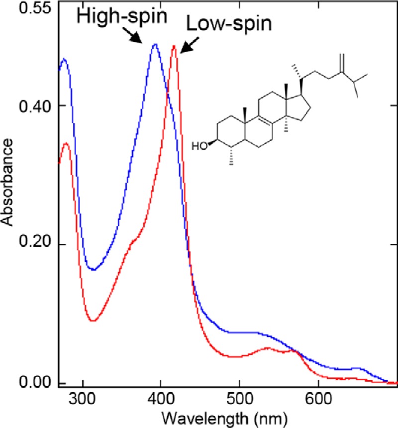
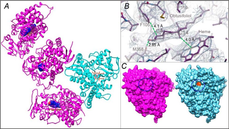
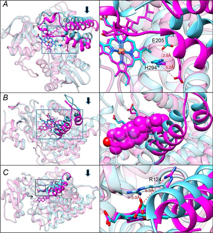
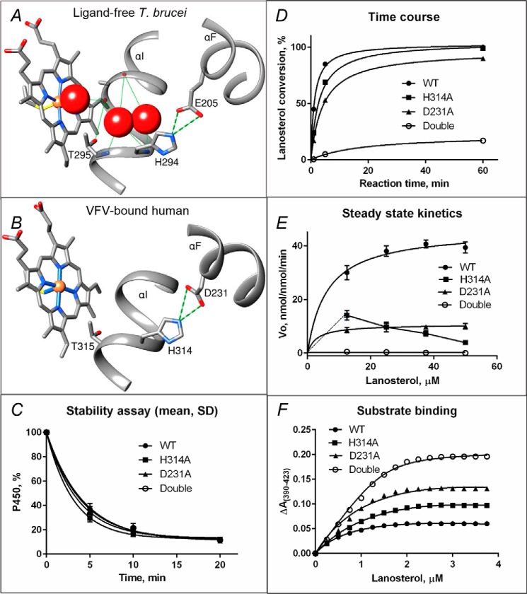
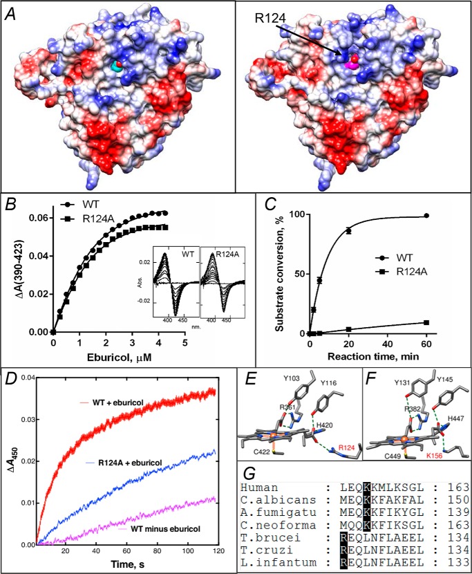
Sterol 14α-demethylases (CYP51s) are phylogenetically the most conserved cytochromes P450, and their three-step reaction is crucial for biosynthesis of sterols and serves as a leading target for clinical and agricultural antifungal agents. The structures of several (bacterial, protozoan, fungal, and human) CYP51 orthologs, in both the ligand-free and inhibitor-bound forms, have been determined and have revealed striking similarity at the secondary and tertiary structural levels, despite having low sequence identity. Moreover, in contrast to many of the substrate-promiscuous, drug-metabolizing P450s, CYP51 structures do not display substantial rearrangements in their backbones upon binding of various inhibitory ligands, essentially representing a snapshot of the ligand-free sterol 14α-demethylase. Here, using the obtusifoliol-bound I105F variant of Trypanosoma cruzi CYP51, we report that formation of the catalytically competent complex with the physiological substrate triggers a large-scale conformational switch, dramatically reshaping the enzyme active site (3.5-6.0 Å movements in the FG arm, HI arm, and helix C) in the direction of catalysis. Notably, our X-ray structural analyses revealed that the substrate channel closes, the proton delivery route opens, and the topology and electrostatic potential of the proximal surface reorganize to favor interaction with the electron-donating flavoprotein partner, NADPH-cytochrome P450 reductase. Site-directed mutagenesis of the amino acid residues involved in these events revealed a key role of active-site salt bridges in contributing to the structural dynamics that accompanies CYP51 function. Comparative analysis of apo-CYP51 and its sterol-bound complex provided key conceptual insights into the molecular mechanisms of CYP51 catalysis, functional conservation, lineage-specific substrate complementarity, and druggability differences.

Keywords: X-ray crystallography; catalysis; conformational change; cytochrome P450; drug target; druggability; sterol 14alpha-demethylase (CYP51); sterol biosynthesis; structure-function; substrate binding.


Conflict of interest statement

The authors declare that they have no conflicts of interest with the contents of this article

Figures

Figure 1.
Figure 1.
Spectral changes induced in the I105F mutant of T. cruzi CYP51 by obtusifoliol. Red, low-spin water-bound ferric P450; Soret band maximum, 417 nm. Blue, >85% high-spin substrate bound ferric P450; Soret band maximum, 393 nm. The increase in the absorbance at 280 nm is due to HPCD, which was used to dissolve the sterol.
Figure 2.
Figure 2.
X-ray structure of CYP51 in the substrate-bound (magenta) and ligand-free (cyan) state. A, the asymmetric unit. The carbon atoms of the substrate (obtusifoliol) molecule are seen as blue spheres, and the C3 OH oxygen is red. The heme is shown as a stick model, and the carbon atoms are orange. B, the 2FoFc electron density map of the obtusifoliol area is shown as a mesh contoured at 1.2 σ, prepared in Coot. The distances between the C14α-methyl group and the heme iron, the C3-OH and the carbonyl oxygen of Met-358, and the C4 atom and Phe-105 are marked with dashed green lines. C, surface representation of the substrate-bound and ligand-free molecules, distal P450 view. The substrate entry area (closed in the substrate-bound and open in the ligand-free structure) is circled. The heme is seen as orange spheres (see also Fig. S2C).
Figure 3.
Figure 3.
Conformational switch in the CYP51 structure upon binding of the substrate. The three segments of the active site that experience large-scale conformational rearrangements are shown in cyan (ligand-free structure) and magenta (substrate-bound structure). Arrows show the direction of movement. The other portions of the molecules are presented as semitransparent ribbons of the corresponding colors. Left panels, the overall view of the superimposed structures (see also Fig. S6); right panels, the enlarged view of the squared fragments. The structures were superimposed in LSQKAB (CCP4 Suite). A, the FG arm and the conserved histidine-acid salt-bridge opening (activation of proton relay machinery). B, the HI segment. In the enlarged fragment, obtusifoliol is presented as van der Waals spheres to show that without the movement it would collide with the (cyan) I helix. C, helix C. The 3.5–4 Å movement of this helix reshapes the CYP51 proximal surface. The inset shows rearrangements in the position of Arg-124, whose guanidino group now protrudes above the CYP51 proximal surface, bringing additional positive charge to its electrostatic potential (shown in Fig. 5).
Figure 4.
Figure 4.
Proposed proton delivery route in the CYP51 family. A, ligand-free water-coordinated protozoan CYP51 (PDB code 3G1Q, 1.8 Å resolution). The water molecules are presented as red spheres. B, inhibitor-bound human CYP51 (PDB code 4UHI, 2.0 Å resolution). C–F, mutants of the His-acid salt bridge in human CYP51. C, stability at 42 °C monitored as the decrease in the P450 content, the initial P450 concentration was 2 μm (see also Fig. S7). D, time course of substrate conversion, 0.5 μm P450, 25 μm lanosterol. E, Michaelis–Menten plots using 0.25 μm P450, 1 min reaction; points are shown as means of three determinations ± S.D. F, spectral response to lanosterol; 2 μm P450 (fit to Morrison equation, nonlinear regression). The estimated low-to-high spin transition was 30, 50, 68, and 94% in the WT, H314A, D231A, and the H314A/D231A double mutant proteins, respectively (see also Table 2).
Figure 5.
Figure 5.
In protozoan CYP51, Arg-124 must be involved into the electron transfer. A, proximal surface of the ligand-free (left panel) and substrate-bound (right panel) I105F T. cruzi CYP51 molecules colored by electrostatic potential (red for negative and blue for positive charge). The heme is seen in cyan and magenta, respectively. In the substrate-bound structure repositioning of helix C and exposure of the guanidino group of Arg-124 (marked with an arrow) adds positive charge to the surface, whereas in the substrate-free structure the guanidino group of Arg- 124 forms the H-bond with the heme ring D propionate. B, spectral response of the R124A mutant to the substrate binding, 2 μm P450; Kd = 0.6 μm; the high-spin form content 36% (fit to Morrison equation nonlinear regression). The response of the WT protein (Kd 0.5 μm, 39% high-spin) is shown as a comparison. C, time course of substrate conversion by T. cruzi CYP51, WT, and the R124A mutant. The reaction mixture contained 1 μm P450, 2 μm T. brucei CPR, and 50 μm eburicol. D, enzymatic reduction. Rates of reduction of (ferric) T. cruzi WT and R124A were measured under anaerobic conditions in the presence of CO at 23 °C, using final concentrations of 1 μm P450, 2 μm CPR, 150 μm DLPC, and 5 μm eburicol and estimated by global fitting of the 380–520-nm data points (from >4 individual reductions, ± S.D.) to first-order plots in the OLIS software: WT enzyme, 1.56 ± 0.24 min−1; WT minus substrate, 0.20 ± 0.02 min−1; R124A enzyme (with substrate), 0.65 ± 0.02 min−1. E, heme support in protozoan CYP51, ligand-free T. brucei, PDB code 3G1Q. F, heme support in VFV-bound human CYP51, PDB code 4UHI. Arg-124 and Lys-156 are labeled in red. G, fragment of CYP51 sequence alignment showing the Arg and Lys residues that form the salt bridge with the heme.

References

    1. Lepesheva G. I., and Waterman M. R. (2007) Sterol 14α-demethylase cytochrome P450 (CYP51), a P450 in all biological kingdoms. Biochim. Biophys. Acta 1770, 467–477 10.1016/j.bbagen.2006.07.018 - DOI - PMC - PubMed
    1. Guengerich F. P. (2007) Mechanisms of cytochrome P450 substrate oxidation: MiniReview. J. Biochem. Mol. Toxicol. 21, 163–168 10.1002/jbt.20174 - DOI - PubMed
    1. Hargrove T. Y., Wawrzak Z., Liu J., Waterman M. R., Nes W. D., and Lepesheva G. I. (2012) Structural complex of sterol 14α-demethylase (CYP51) with 14α-methylenecyclopropyl-Δ7–24,25-dihydrolanosterol. J. Lipid Res. 53, 311–320 10.1194/jlr.M021865 - DOI - PMC - PubMed
    1. Rittle J., and Green M. T. (2010) Cytochrome P450 compound I: capture, characterization, and C–H bond activation kinetics. Science 330, 933–937 10.1126/science.1193478 - DOI - PubMed
    1. Lepesheva G. I., Friggeri L., and Waterman M. R. (2018) CYP51 as drug targets for fungi and protozoan parasites: past, present and future. Parasitology 145, 1820–1836 10.1017/S0031182018000562 - DOI - PMC - PubMed

Publication types

LinkOut - more resources