. 2018 Dec 7;293(49):19127-19135.
doi: 10.1074/jbc.RA118.005367. Epub 2018 Oct 16.

Comprehensive elucidation of the structural and functional roles of engineered disulfide bonds in antibody Fc fragment

Affiliations

Comprehensive elucidation of the structural and functional roles of engineered disulfide bonds in antibody Fc fragment

Fang Zeng et al. J Biol Chem. .

Abstract

Therapeutic monoclonal antibodies and Fc-fusion proteins containing antibody Fc fragment may tend to destabilize (e.g. unfold and aggregate), which leads to loss of functions and increase of adverse risks. Although engineering of an additional disulfide bond has been performed in Fc or Fc domains for optimization, the relationships between introduced disulfide bond and alteration of the stability, aggregation propensity and function were still unclear and should be addressed for achievement of better therapeutic outcome. Here, we constructed three human IgG1 Fc mutants including FcCH2-s-s- (one engineered disulfide bond in CH2 domain), FcCH3-s-s- (one engineered disulfide bond in CH3 domain), and FcCH3-s-s-CH2-s-s- (two engineered disulfide bonds in CH2 and CH3 domains, respectively) for evaluation. As expected, each mutated domain shows obviously increased stability during thermo-induced unfolding, and FcCH3-s-s-CH2-s-s- is most thermo-stable among wildtype Fc (wtFc) and three mutants. The order of overall stability against denaturant is FcCH3-s-s-CH2-s-s- > FcCH2-s-s- > FcCH3-s-s- > wtFc. Then the aggregation propensity was compared among these four proteins. Under conditions of incubation at 60 °C, their aggregation resistance is in the order of FcCH3-s-s-CH2-s-s- > FcCH2-s-s- > FcCH3-s-s- ≈ wtFc. In contrast, the order is FcCH3-s-s-CH2-s-s- > FcCH3-s-s- > FcCH2-s-s- ≈ wtFc under acidic conditions. In addition, the Fc-mediated functions are not obviously affected by engineered disulfide bond. Our results give a comprehensive elucidation of structural and functional effects caused by additional disulfide bonds in the Fc fragment, which is important for Fc engineering toward the desired clinical performance.

Keywords: Fc engineering; Fc fragment; Fc receptor; aggregation; circular dichroism (CD); disulfide bond; function; fusion protein; monoclonal antibody; stability.


Conflict of interest statement

The authors declare that they have no conflicts of interest with the contents of this article

Figures

Figure 1.
Figure 1.
Design of Fc mutants with additional disulfide bond. A, the structure of wtFc obtained from an intact IgG1 with solved crystal structure (Protein Data Bank code 1HZH (12)) presented by PyMOL (PyMOL Molecular Graphics System, version 1.2r 1 Schrödinger, LLC). The CH2 and CH3 domains are colored cyan and green, respectively. The native cysteines that form disulfide are colored yellow. The distances of two α-carbon atoms in CH2 and CH3 are 6.6 and 6.5 Å, respectively. Asn297 colored purple is the glycosylation site. Residues Leu242 and Lys334 colored red in CH2 could be mutated to two cysteines to introduce the additional disulfide bond (the distance of two α-carbon atoms is 6.7 Å), while residues Pro343 and Ala431 colored orange in CH3 could be replaced by two cysteines to get the engineered disulfide bond (the distance of two α-carbon atoms is 5.7 Å). B, sequence alignment of wtFc and designed mutants FcCH2-s-s-, FcCH3-s-s-, and FcCH3-s-s-CH2-s-s-.
Figure 2.
Figure 2.
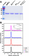
Molecular mass of wtFc, FcCH2-s-s-, FcCH3-s-s-, and FcCH3-s-s-CH2-s-s-. A, SDS-PAGE of purified wtFc, FcCH2-s-s-, FcCH3-s-s-, and FcCH2-s-s CH3-s-s-. B, SEC analysis of purified wtFc, FcCH2-s-s-, FcCH3-s-s-, and FcCH3-s-s-CH2-s-s-. The standard curve is used for calculation of molecular mass.
Figure 3.
Figure 3.
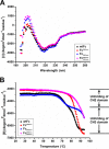
Thermostability of wtFc, FcCH2-s-s-, FcCH3-s-s-, and FcCH3-s-s-CH2-s-s-. A, secondary structure at 25 °C measured by CD. A major negative peak at 216 nm in each protein indicates typical β-sheet structure. B, thermo-induced unfolding curve measured by CD. The CD signal was recorded at 216 nm in the temperature range from 25–94 °C with heating rate of 0.5 °C/min. Experiments were repeated at least twice, and the results from one representative experiment are presented.
Figure 4.
Figure 4.
Urea-induced unfolding of wtFc, FcCH2-s-s-, FcCH3-s-s-, and FcCH3-s-s-CH2-s-s-. The fraction folded (ff) of the protein was calculated as ff = ([θ] − [θU])/([θF] − [θU]). [θF] and [θU] were the mean residue ellipticities at 216 nm of folded state (F) in the absence of urea and unfolded state (U) in the presence of urea concentration of 10 m. The urea concentrations at half of unfolding course of wtFc, FcCH2-s-s-, FcCH3-s-s-, and FcCH3-s-s-CH2-s-s- are 7.3, 8.7, 7.8, and 9.0 m, respectively. Experiments were repeated at least twice, and the results from one representative experiment are presented.
Figure 5.
Figure 5.
Aggregation resistance of wtFc, FcCH2-s-s-, FcCH3-s-s-, and FcCH3-s-s-CH2-s-s- measured by turbidity assay and size-exclusion chromatography. A, turbidity assay of wtFc, FcCH2-s-s-, FcCH3-s-s-, and FcCH3-s-s-CH2-s-s- after 60 °C incubation at different time points. B, evaluation of aggregation formation by SEC. In addition to Peak 1, there is an obvious aggregation peak (Peak 2) in wtFc and FcCH3-s-s-, respectively, at the end of 60 °C incubation. FcCH3-s-s-CH2-s-s- and FcCH2-s-s- are more resistant to aggregation. However, compared with FcCH3-s-s-CH2-s-s-, a very small Peak 2 could be observed in FcCH2-s-s-. The inset is a standard curve as used above. Experiments were repeated twice, and the results from one representative experiment are presented.
Figure 6.
Figure 6.
The aggregation tendency of wtFc, FcCH2-s-s-, FcCH3-s-s-, and FcCH3-s-s-CH2-s-s- in acidic conditions. Sample aliquots were taken at predetermined time intervals and analyzed by recording the absorbance at 320 nm. Experiments were repeated twice, and the results from one representative experiment are presented.
Figure 7.
Figure 7.
Flow cytometry for measurement of binding of wtFc, FcCH2-s-s-, FcCH3-s-s-, and FcCH3-s-s-CH2-s-s- to FcγRI. A, FcγRI-positive U937 cells were incubated with 40 nm wtFc, FcCH2-s-s-, FcCH3-s-s-, and FcCH3-s-s-CH2-s-s- proteins. The obvious fluorescence intensity shift could be observed. In the case of FcγRI-negative HeLa cells, the shift is minor. B, fluorescence intensity shifts at different protein concentrations from 0 to 1,000 nm. All four proteins bind to U937 cells in a dose-dependent manner. MFI, mean fluorescence intensity.
Figure 8.
Figure 8.
Determination of binding of wtFc, FcCH2-s-s-, FcCH3-s-s-, and FcCH3-s-s-CH2-s-s- to shFcRn at pH 6.0 by BLI. In general, all four proteins bind to shFcRn with high similarity. The experiments were repeated twice, and the results from one representative experiment are presented.

References

    1. Yang C., Gao X., and Gong R. (2017) Engineering of Fc Fragments with optimized physicochemical properties implying improvement of clinical potentials for Fc-based therapeutics. Front. Immunol. 8, 1860 - PMC - PubMed
    1. Edelman G. M., Cunningham B. A., Gall W. E., Gottlieb P. D., Rutishauser U., and Waxdal M. J. (1969) The covalent structure of an entire γG immunoglobulin molecule. Proc. Natl. Acad. Sci. U.S.A. 63, 78–85 10.1073/pnas.63.1.78 - DOI - PMC - PubMed
    1. Thies M. J., Talamo F., Mayer M., Bell S., Ruoppolo M., Marino G., and Buchner J. (2002) Folding and oxidation of the antibody domain C(H)3. J. Mol. Biol. 319, 1267–1277 10.1016/S0022-2836(02)00375-3 - DOI - PubMed
    1. McAuley A., Jacob J., Kolvenbach C. G., Westland K., Lee H. J., Brych S. R., Rehder D., Kleemann G. R., Brems D. N., and Matsumura M. (2008) Contributions of a disulfide bond to the structure, stability, and dimerization of human IgG1 antibody CH3 domain. Protein Sci. 17, 95–106 - PMC - PubMed
    1. Sakurai K., Nakahata R., Lee Y. H., Kardos J., Ikegami T., and Goto Y. (2015) Effects of a reduced disulfide bond on aggregation properties of the human IgG1 CH3 domain. Biochim. Biophys. Acta 1854, 1526–1535 10.1016/j.bbapap.2015.02.020 - DOI - PubMed

Publication types

Associated data