. 2019 Jun;16(6):602-610.
doi: 10.1038/s41423-018-0164-2. Epub 2018 Oct 16.

High levels of circulating GM-CSF+CD4+ T cells are predictive of poor outcomes in sepsis patients: a prospective cohort study

Affiliations

High levels of circulating GM-CSF+CD4+ T cells are predictive of poor outcomes in sepsis patients: a prospective cohort study

Huihuang Huang et al. Cell Mol Immunol. 2019 Jun.

Abstract

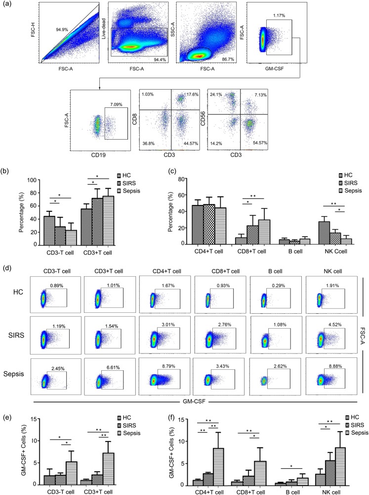
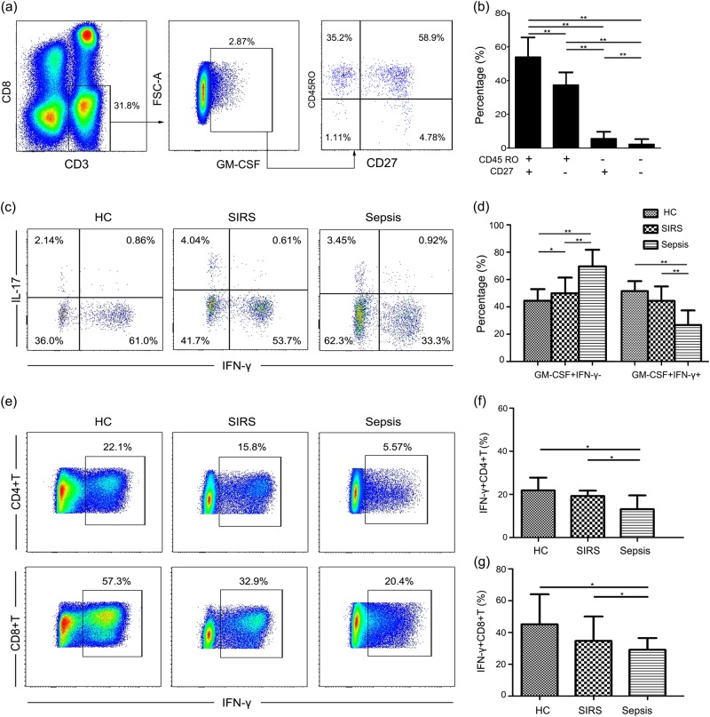
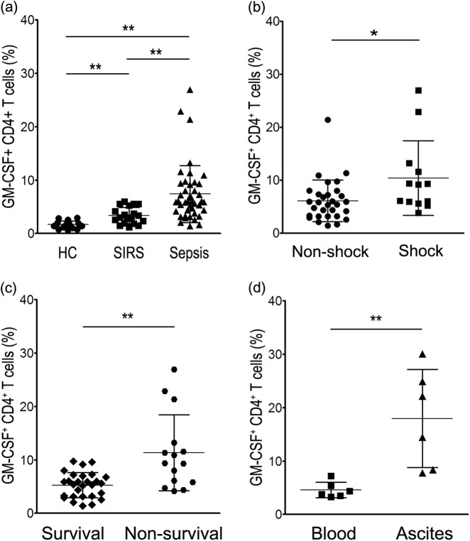
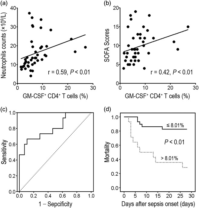
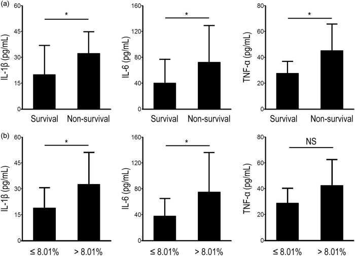
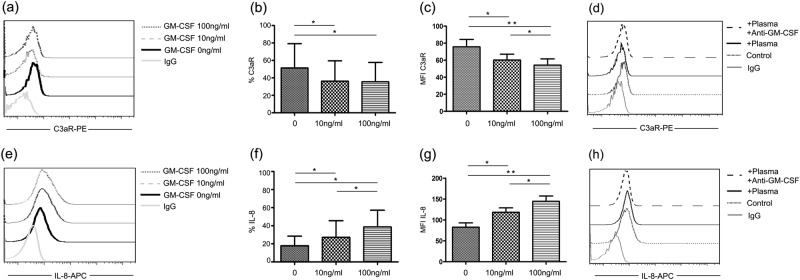
Granulocyte colony-stimulating factor (GM-CSF), produced by CD4+ T cells, has recently been implicated in the pathogenesis of inflammatory diseases, such as multiple sclerosis and juvenile arthritis. However, the role of GM-CSF-producing CD4+ T cells in sepsis remains unknown. This study reports peripheral changes in GM-CSF-producing CD4+ T cells in septic patients and the possible underlying mechanism by which GM-CSF influences the outcome of sepsis. Forty-three septic patients, 20 SIRS patients, and 20 healthy controls were enrolled in this study and followed for 28 days to assess mortality. We measured the peripheral frequency of GM-CSF+CD4+ T cells and recorded their associated relationship with disease progression. Our data demonstrated that peripheral GM-CSF-producing CD4+ T cells were significantly higher in septic patients than in both SIRS patients and healthy controls. These cells exhibit a memory phenotype and impaired IFN-γ-secreting capacity in sepsis patients. Using a receiver operating curve analysis with 8.01% as a cut-off point, the percentage of GM-CSF+CD4+ T cells could predict the outcome of septic patients. Combined with the increase in GM-CSF-producing CD4+ T cells, inflammatory cytokines IL-1β and IL-6 were also upregulated. Using an in vitro neutrophil model, we found that GM-CSF inhibited C3aR expression, while inducing IL-8 production. Furthermore, this effect was transferrable in plasma from sepsis patients and was attenuated by inhibition of GM-CSF using an anti-GM-CSF antibody. These results indicate that GM-CSF-producing CD4+ T cells may serve as a marker of sepsis severity. Thus, targeting GM-CSF overproduction may benefit sepsis patients.

Keywords: CD4+ T cells; GM-CSF; SIRS; sepsis.


Conflict of interest statement

The authors declare no competing interests.

Figures

Fig. 1
Fig. 1
GM-CSF is primarily produced by CD4+ T cells. a Representative dot plots illustrating the source of GM-CSF in lymphocyte population. b Pooled data showing the proportion of CD3+ and CD3 T cells from GM-CSF-positive cells. c Pooled data showing the proportion of CD4+ T cells, CD8+ T cells, NK cells, and B cells from GM-CSF-positive cells. d Representative dot plots showing expression of GM-CSF in different lymphocyte populations. e Pooled data illustrating expression of GM-CSF among CD3+ T cells and CD3 T cells in different groups. f Pooled data showing expression of GM-CSF in CD4+ T cells, CD8+ T cells, NK cells, and B cells in different groups. Each group included eight subjects for pooled data collection. *P < 0.05; **P < 0.01
Fig. 2
Fig. 2
Phenotype and cytokine production of GM-CSF+CD4+ T cells. a Representative dot plots showing expression of CD45RO and CD27 on GM-CSF+CD4+ T cells. b Pooled data showing expression of CD45RO and CD27 on GM-CSF+CD4+ T cells. c Representative dot plots showing expression of IFN-γ and IL-17A in the population of GM-CSF+CD4+ T cells. d Pooled data showing expression of IFN-γ in GM-CSF+ CD4+ T cells in different groups. e Representative dot plots showing expression of IFN-γ in CD4+ T cells and CD8+ T cells. f Pooled data showing expression of IFN-γ in CD4+ T cells in different groups.  g Pooled data showing expression of IFN-γ in CD8+ T cells in different groups. Each group included eight subjects for pooled data collection. *P < 0.05; **P < 0.01
Fig. 3
Fig. 3
Dynamics of GM-CSF+ CD4+ T-cell expression in sepsis patients. a Pooled data showing expression of GM-CSF+ CD4+ T cells in healthy controls and patients with SIRS and sepsis. b, c Increasing GM-CSF+ CD4+ T-cell expression is associated with sepsis severity. d Differential expression of GM-CSF+ CD4+ T cells in blood and ascites from patients with sepsis caused by intra-abdominal infection. *P < 0.05; **P < 0.01
Fig. 4
Fig. 4
Increase of GM-CSF+CD4+ T-cell expression is positively associated with sepsis severity. a, b GM-CSF+CD4+ T-cell levels are positively correlated with neutrophil counts and SOFA scores in sepsis patients. c Receiving operating curve of GM-CSF+ CD4+ T cells on Day 0 for predicting mortality. d Kaplan–Meier survival curves of septic patients showing that patients with high levels of GM-CSF+CD4+ T cells ( > 8.01%) have increased mortality compared with patients with low levels of GM-CSF+ CD4+ T cells ( ≤ 8.01%). Follow-up period: 28 days. P < 0.01
Fig. 5
Fig. 5
Pro-inflammatory cytokine production in sepsis patients is associated with increased GM-CSF+ CD4+ T cells. a Pooled data showing that production of pro-inflammatory cytokines, including IL-1β, IL-6, and TNF-α, is associated with sepsis outcome. b Sepsis patients with high proportions of GM-CSF+CD4+ T cells ( > 8.01%) have increased expression of IL-1β and IL-6 compared with patients with low proportions of GM-CSF+ CD4+ T cells ( ≤ 8.01%). *P < 0.05; NS no significance
Fig. 6
Fig. 6
Increased GM-CSF production is associated with decreased C3aR expression and increased IL-8 production in neutrophils. ac Representative dot plots and pooled data showing expression of C3aR on neutrophils following incubation with different doses of GM-CSF. d C3aR on healthy neutrophils incubated with sepsis patient plasma before and after blocking with an anti-GM-CSF antibody. eg Representative dot plots and pooled data showing production of IL-8 in neutrophils following incubation with different doses of GM-CSF. h IL-8 in healthy neutrophils incubated with sepsis patient plasma before and after blocking with anti-GM-CSF antibody. *P < 0.05; **P < 0.01

References

    1. Levy MM, et al. SCCM/ESICM/ACCP/ATS/SI:2001SCCM/ESICM/ACCP/ATS/SIS International Sepsis Definitions Conference. Crit. Care Med. 2003;31:1250–1256. doi: 10.1097/01.CCM.0000050454.01978.3B. - DOI - PubMed
    1. Balk RA. Severe sepsis and septic shock. Crit. Care Clin. 2000;16:179–192. doi: 10.1016/S0749-0704(05)70106-8. - DOI - PubMed
    1. Van Nieuwenhuijze A, et al. GM-CSF as a therapeutic target in inflammatory diseases. Mol. Immunol. 2013;56:675–682. doi: 10.1016/j.molimm.2013.05.002. - DOI - PubMed
    1. Codarri L, et al. RORγt derives production of the cytokine GM-CSF in helper T cells, which is essential for the effector phase of autoimmune neuroinflammation. Nat. Immunol. 2011;12:560–567. doi: 10.1038/ni.2027. - DOI - PubMed
    1. EI-Behi M, et al. The encephalitogenicity of TH17 cells is dependent on IL-1- and IL-23-induced production of the cytokine GM-CSF. Nat. Immunol. 2011;12:568–575. doi: 10.1038/ni.2031. - DOI - PMC - PubMed

Publication types

Substances