Review
. 2019 Feb;76(3):405-419.
doi: 10.1007/s00018-018-2938-1. Epub 2018 Oct 16.

Pathological processes activated by herpes simplex virus-1 (HSV-1) infection in the cornea

Affiliations
Review

Pathological processes activated by herpes simplex virus-1 (HSV-1) infection in the cornea

Lulia Koujah et al. Cell Mol Life Sci. 2019 Feb.

Abstract

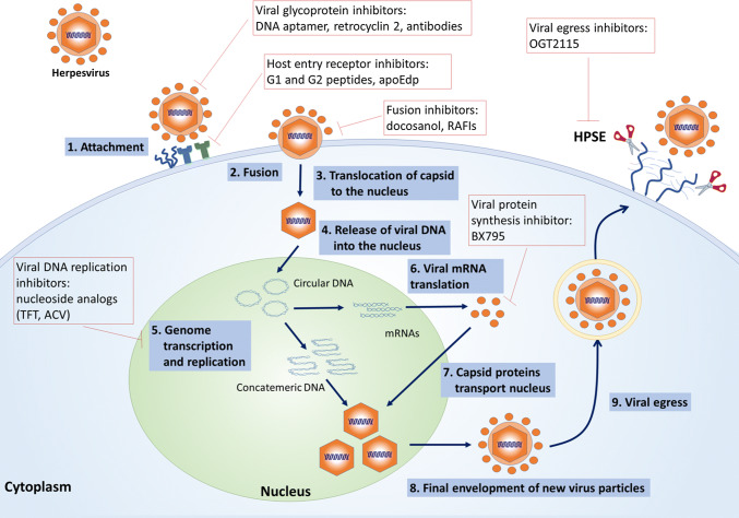
Herpes simplex virus type-1 (HSV-1) is a ubiquitous pathogen that infects a large majority of the human population worldwide. It is also a leading cause of infection-related blindness in the developed world. HSV-1 infection of the cornea begins with viral entry into resident cells via a multistep process that involves interaction of viral glycoproteins and host cell surface receptors. Once inside, HSV-1 infection induces a chronic immune-inflammatory response resulting in corneal scarring, thinning and neovascularization. This leads to development of various ocular diseases such as herpes stromal keratitis, resulting in visual impairment and eventual blindness. HSV-1 can also invade the central nervous system and lead to encephalitis, a relatively common cause of sporadic fetal encephalitis worldwide. In this review, we discuss the pathological processes activated by corneal HSV-1 infection and existing antiviral therapies as well as novel therapeutic options currently under development.

Keywords: Anti-viral therapy; Herpes simplex virus; Immune response; Ocular herpes; Pathological process.


Figures

Fig. 1
Fig. 1
Herpes simplex virus-1 structure. The glycoproteins essential for viral entry into host cells are listed
Fig. 2
Fig. 2
HSV-1 entry glycoproteins and their known receptors. Initial interactions of HSV envelope glycoproteins gB and/or gC with heparan sulfate proteoglycans (HSPG) occur on the surface of a host cell, which facilitate viral attachment and surfing on membrane projections to reach the plasma membrane. This is followed by specific interactions of HSV gD and gB with their corresponding receptors leading to membrane fusion and delivery of the viral nucleocapsid and tegument proteins into the cytoplasm
Fig. 3
Fig. 3
TLR2, TLR3 and TLR9 regulate a robust immune response following HSV-1 infection. TLR2 resides on the cell surface and recognizes viral proteins. TLR2 signals through a myeloid differentiation factor 88 (MyD88) or Toll-interleukin 1 receptor domain containing adaptor protein (TIRAP)-dependent cascade, resulting in the recruitment of IR-1R-associated protein kinase (IRAK1) and TNF receptor-associated factor 6 (TRAF6), both of which are upstream of the IKK complex and NF-κB. TLR9 is expressed in the endosome and recognizes the virus by rich CpG sequences and signals through MyD88-dependent pathway that induces NF-κB activation. TLR3 is also expressed in the endosome and recognizes dsRNA. TLR3 signals through MyD88-dependent pathway, activating NF-κB, as well as TIR-domain-containing adapter-inducing interferon-β (TRIF), which is upstream of TBK-1, and leads to the activation of IRF-3
Fig. 4
Fig. 4
Induction of corneal neovascularization and inflammation by HSV-1 infection: HSV-1 infection results in the increased expression of heparanase (HPSE) which causes heparan sulfate proteoglycan (e.g., syndecan) shedding. HPSE-degraded heparan sulfate releases VEGF and FGF-2, which signal ERK phosphorylation and cause pro-inflammatory cytokine induction. Production of pro-inflammatory factors (IL-6, MMP-9, FGF-2, HGF) then contributes to inflammation and infiltration of macrophages, T cells, and Nk cells in the cornea. In addition, HSV-1-infected corneas predominantly produce VEGF-A that leads to angiogenic sprouting
Fig. 5
Fig. 5
Virus replication cycle and the steps which are targeted by antiviral drugs currently in use or under investigation

References

    1. Agelidis AM, Hadigal SR, Jaishankar D, Shukla D. Viral activation of heparanase drives pathogenesis of herpes simplex virus-1. Cell Rep. 2017;20:439–450. doi: 10.1016/j.celrep.2017.06.041. - DOI - PMC - PubMed
    1. Ahmad R, El Bassam S, Cordeiro P, Menezes J. Requirement of TLR2-mediated signaling for the induction of IL-15 gene expression in human monocytic cells by HSV-1. Blood. 2008;112:2360–2368. doi: 10.1182/blood-2008-02-137711. - DOI - PubMed
    1. Akhtar J, Tiwari V, Oh MJ, Kovacs M, Jani A, Kovacs SK, Valyi-Nagy T, Shukla D. HVEM and nectin-1 are the major mediators of herpes simplex virus 1 (HSV-1) entry into human conjunctival epithelium. Invest Ophthalmol Vis Sci. 2008;49:4026–4035. doi: 10.1167/iovs.08-1807. - DOI - PMC - PubMed
    1. Akira S, Takeda K. Toll-like receptor signalling. Nat Rev Immunol. 2004;4:499–511. doi: 10.1038/nri1391. - DOI - PubMed
    1. Akira S, Takeda K, Kaisho T. Toll-like receptors: critical proteins linking innate and acquired immunity. Nat Immunol. 2001;2:675–680. doi: 10.1038/90609. - DOI - PubMed