. 2018 Oct 17;8(1):18.
doi: 10.1186/s12348-018-0160-9.

Acute multifocal retinitis: a retrospective review of 35 cases

Affiliations

Acute multifocal retinitis: a retrospective review of 35 cases

Sana Khochtali et al. J Ophthalmic Inflamm Infect. .

Abstract

Background: Acute multifocal retinitis is a rare condition that has been considered to be often idiopathic. The purpose of this study was to analyze clinical features and causes of acute multifocal retinitis.

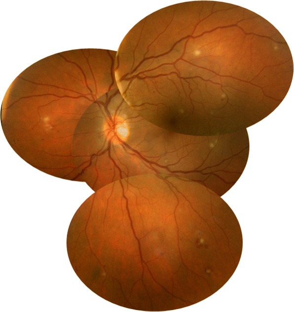
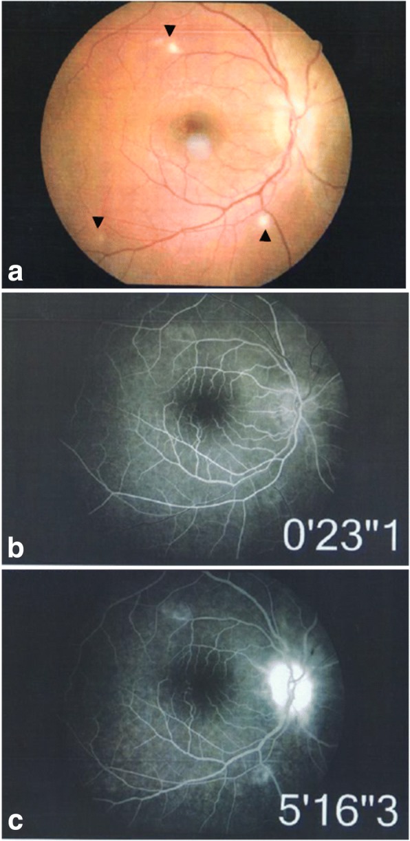
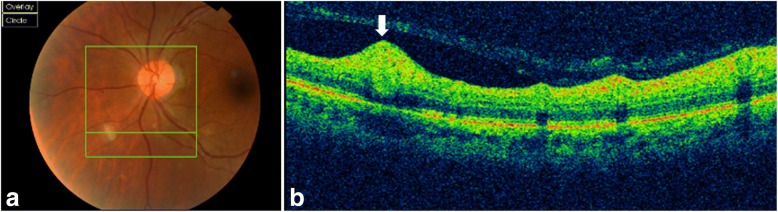
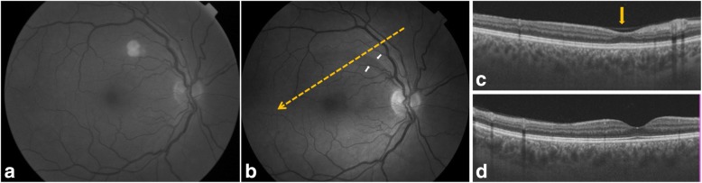
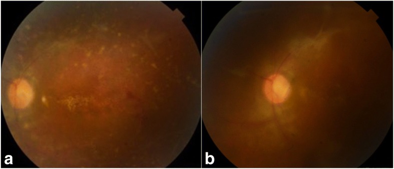
Results: This study is a retrospective review of the charts of 35 patients with acute multifocal retinitis. Patients with three or more retinal lesions in at least one eye, with at least one lesion of less than 500 μm in size were included. All patients had complete ophthalmological examination, fundus photography, and fluorescein angiography. Twelve patients (34.3%) had optical coherence tomography. An extensive work-up was performed including a detailed comprehensive medical history, examination by an internist and an infectious disease specialist, a chest X-ray, Mantoux test, and laboratory tests for syphilis, human immunodeficiency virus, Bartonella, and Rickettsia. Of the 35 patients, 25 (71.4%) had bilateral involvement and 10 (28.6%) had unilateral involvement (total number of eyes: 60). Mean best-corrected visual acuity (BCVA) was 20/25 (range, 20/1000-20/20). Retinal lesions ranged from 3 to more than 20 in number in at least 1 eye, and from 150 to 1500 μm in size. Associated findings included mild anterior chamber inflammation in 5 eyes (8.3%), mild vitritis in 46 eyes (76.7%), optic disc swelling in 9 eyes (15%), macular star in 4 eyes (6.7%), exudative retinal detachment in 6 eyes (10%), and branch retinal artery occlusion in 6 eyes (10%). Acute multifocal retinitis was found to be caused by Rickettsia conorii infection in 20 patients (57.1%), Rickettsia typhi infection in 4 patients (11.4%), cat-scratch disease in 8 patients (22.9%), and syphilis in 1 patient (2.9%). It was idiopathic in two patients (5.7%). Retinal lesions resolved without scarring in 3 to 12 weeks in all but three eyes (5%), in which residual retinal pigment epithelial changes were noted.

Conclusion: Rickettsial disease was the most common cause of acute multifocal retinitis. Other identified causes included cat-scratch disease and syphilis, and a very small subset of patients was diagnosed with idiopathic multifocal retinitis.

Keywords: Branch retinal artery occlusion; Cat-scratch disease; Posterior uveitis; Retinitis; Rickettsial disease.


Conflict of interest statement

Ethics approval and consent to participate

This work adheres to the guidelines and principles by the Declaration of Helsinki. It was approved by the ethics committee of our institution.

Consent for publication

Not applicable.

Competing interests

The authors declare that they have no competing interests.

Publisher’s Note

Springer Nature remains neutral with regard to jurisdictional claims in published maps and institutional affiliations.

Figures

Fig. 1
Fig. 1
A 42-year-old patient complained of floaters. He reported a 2-week history of fever and malaise. Composite fundus photograph of the left eye shows multiple small white retinal lesions in the posterior pole and the periphery, with a few retinal hemorrhages. Laboratory testing was positive for Mediterranean spotted fever (Rickettsia Conorii infection)
Fig. 2
Fig. 2
A 38-year-old male patient presented with acute scotoma in the left eye. Color fundus photograph shows three retinal infiltrates with a triangle-shaped area of retinal ischemic whitening adjacent to a retinal infiltrate (arrow) (a). Fluorescein angiography confirms the presence of branch retinal artery occlusion corresponding to the area of retinal ischemic whitening (arrow). It also shows early hypofluorescence of the larger infiltrates and isofluorescence of the smaller infiltrate (b) and late staining of all three retinal infiltrates, retinal vascular leakage, and optic disc hyperfluorescence (c). Laboratory testing was positive for Mediterranean spotted fever (Rickettsia Conorii infection)
Fig. 3
Fig. 3
A 32-year-old female patient presented with vision blurring in the right eye. The patient reported having been recently scratched by a kitten. There was a right relative afferent pupillary defect. Color fundus photograph shows retinal infiltrates (arrowheads) (a). Fluorescein angiography shows mild peripheral staining of retinal lesions (b, c) and late optic disc hyperfluorescence (c). Serology confirmed a diagnosis of cat-scratch disease
Fig. 4
Fig. 4
A 38-year-old female patient complained of floaters OU. Color fundus photograph shows three white retinal infiltrates, inferior to the optic disc (a). Spectral-domain OCT image section passing through the largest lesion reveals a focal area of retinal thickening with hyperreflectivity extending from the retinal nerve fiber layer to the outer nuclear layer (white arrow), sparing of the retinal pigment epithelium and a mild posterior shadowing (b). Laboratory testing was positive for murine typhus (Rickettsia typhi infection)
Fig. 5
Fig. 5
A 43-year-old patient complained of floaters, OU. Red-free fundus photograph of the right eye shows a mid-sized white retinal infiltrate in the posterior pole (a). Serology confirmed Mediterranean spotted fever. Red-free fundus photograph, taken 3 months after presentation, shows resolution of the retinal infiltrate with development of a retinal nerve fiber layer defect demarcated by the white arrows (b). Spectral-domain OCT image section passing through the clinically visible localized retinal nerve fiber layer defect (yellow dashed arrow in b) shows an indentation (arrow) corresponding to a thinning of the retinal nerve fiber layer (c). Spectral-domain OCT image section passing through the foveola shows normal findings (d)
Fig. 6
Fig. 6
A 23-year-old female patient presented with vision blurring in the left eye. Color fundus photograph shows numerous white retinal lesions, associated with diffuse venous sheathing and a few retinal hemorrhages (a). Serology was positive for syphilis. Color fundus photograph, 2 months after presentation, shows residual retinal pigment epithelial changes and venous sheathing (b)

References

    1. Goldstein BG, Pavan PR. Retinal infiltrates in six patients with an associated viral syndrome. Retina. 1985;5:144–150. doi: 10.1097/00006982-198500530-00002. - DOI - PubMed
    1. Foster RE, Gutman FA, Meyers SM, Lowder CY. Acute multifocal inner retinitis. Am J Ophthalmol. 1991;111(6):673–681. doi: 10.1016/S0002-9394(14)76768-6. - DOI - PubMed
    1. Cunningham ET, Jr, Schatz H, McDonald HR, Johnson RN. Acute multifocal retinitis. Am J Ophthalmol. 1997;123(3):347–357. doi: 10.1016/S0002-9394(14)70130-8. - DOI - PubMed
    1. Giuliari GP, Chang PY, Muci-Mendoza R, Cortez RT. A case of acute multifocal inner retinitis associated with nonspecific viral illness. Retin Cases Brief Rep. 2012;6(1):40–42. doi: 10.1097/ICB.0b013e3182051d06. - DOI - PubMed
    1. Yew YH, Subrayan V. Spectral-domain optical coherence tomographic appearance of AMR. JAMA Ophthalmol. 2013;131(8):1097–1099. doi: 10.1001/jamaophthalmol.2013.2489. - DOI - PubMed

LinkOut - more resources