

doi: 10.1002/ajh.25321.
Epub 2018 Nov 9.
Platelet sequestration and consumption in the placental intervillous space contribute to lower platelet counts during pregnancy
Affiliations
- PMID: 30328633
- PMCID: PMC6447301
- DOI: 10.1002/ajh.25321
Item in Clipboard
Platelet sequestration and consumption in the placental intervillous space contribute to lower platelet counts during pregnancy
Am J Hematol.
2019 Jan.
No abstract available
Conflict of interest statement
Figures
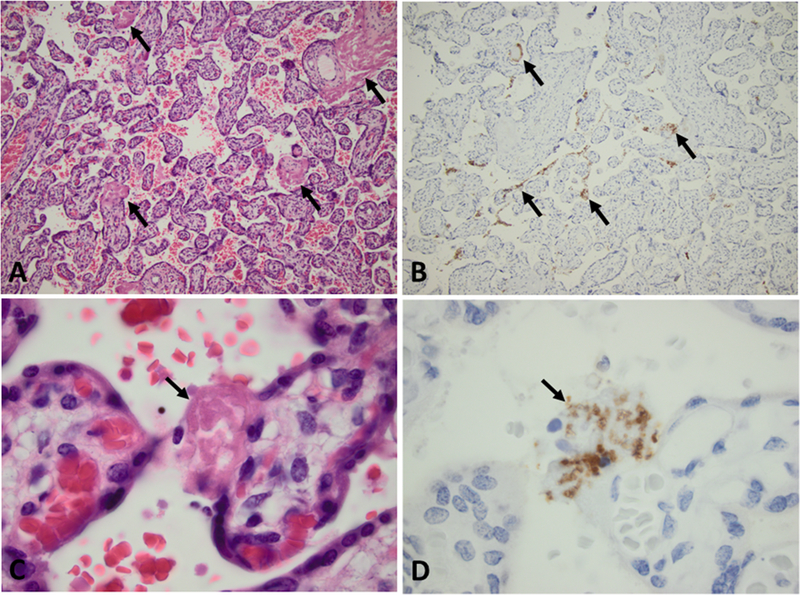
Placental morphology and immunohistochemical staining of sections of a placenta obtained at delivery and included in this study. Placental sections were stained with hematoxylin and eosin (H&E) to demonstrate the structure of the villi and perivillous fibrinoid, and also with a rabbit anti-CD61 (platelet membrane glycoprotein IIIa) monoclonal antibody by ProPath referral pathology service, Dallas, TX. A. Lowmagnification view of the placental parenchyma illustrating mature terminal villi. Perivillous fibrinoid filling gaps in the syncytiotrophoblast surface of the villi are identified by the homogeneous pink stain. Representative examples of the perivillous fibrinoid are indicated by the arrows. (H&E, 10X objective) B. An adjacent location in the same placental section stained with anti-CD61 (brown color) demonstrating platelets within the perivillous fibrinoid, in a linear pattern along the villous surfaces throughout this section. Representative examples of platelet staining are indicated by the arrows. (CD61 immunostaining, 10X objective) C. High-magnification of the placental parenchyma illustrates a mature terminal villus. The arrow indicates perivillous fibrinoid sealing a gap in the syncytiotrophoblast surface. (H&E, 100X objective) D. An adjacent location stained with anti-CD61 (brown color) demonstrating platelets within the perivillous fibrinoid, indicated by the arrow. (CD61 immunostaining, 100X objective)
References
-
- Jonsson V, Bock JE, Nielsen JB. Significance of plasma skimming and plasma volume expansion. J Appl Physiol 1992;72:2047–51. - PubMed
-
- Kaufmann P, Huppertz B, Frank H-G. The fibrinoids of the human placenta: origin, composition and functional relevance. Ann Anatomy 1996;178:485–501. - PubMed
-
- Benirschke K, Burton GJ, Baergen RA. Pathology of the Human Placenta Heidelberg, Germany: Springer-Verlag; 2012.
Publication types
MeSH terms
Substances
Grants and funding
LinkOut - more resources
Full Text Sources
Medical