. 2018 Dec 15;29(26):3155-3167.
doi: 10.1091/mbc.E18-05-0277. Epub 2018 Oct 17.

Emerin induces nuclear breakage in Xenopus extract and early embryos

Affiliations

Emerin induces nuclear breakage in Xenopus extract and early embryos

Matthew R Dilsaver et al. Mol Biol Cell. .

Abstract

Emerin is an inner nuclear membrane protein often mutated in Emery-Dreifuss muscular dystrophy. Because emerin has diverse roles in nuclear mechanics, cytoskeletal organization, and gene expression, it has been difficult to elucidate its contribution to nuclear structure and disease pathology. In this study, we investigated emerin's impact on nuclei assembled in Xenopus laevis egg extract, a simplified biochemical system that lacks potentially confounding cellular factors and activities. Notably, these extracts are transcriptionally inert and lack endogenous emerin and filamentous actin. Strikingly, emerin caused rupture of egg extract nuclei, dependent on the application of shear force. In egg extract, emerin localized to nonnuclear cytoplasmic membranes, and nuclear rupture was rescued by targeting emerin to the nucleus, disrupting its membrane association, or assembling nuclei with lamin A. Furthermore, emerin induced breakage of nuclei in early-stage X. laevis embryo extracts, and embryos microinjected with emerin were inviable, with ruptured nuclei. We propose that cytoplasmic membrane localization of emerin leads to rupture of nuclei that are more sensitive to mechanical perturbation, findings that may be relevant to early development and certain laminopathies.


Figures
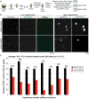
FIGURE 1:
FIGURE 1:
Emerin induces breakage of X. laevis egg extract nuclei. (A) Experimental approach. See Materials and Methods for further details. (B) Representative images of nuclei treated with dialysis buffer or 5 nM recombinant emerin. (C) Quantification of the nuclear breakage phenotype. The intact GFP+ nuclei were counted for buffer- or emerin-treated nuclei in eight different X. laevis egg extracts. Above each pair of bars is the percentage of intact emerin-treated nuclei compared with the buffer control. Above the graph is the average of all data.
FIGURE 2:
FIGURE 2:
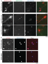
TRC40 rescues emerin’s nuclear localization. (A) Experiments were performed as in Figure 1, except that the extract was supplemented with 1 µM DiI and 0.4 µM SNAP-emerin conjugated to Alexa Fluor 488. Representative images of small and large emerin puncta are shown. (B) Nuclei assembled in X. laevis egg extract as shown in Figure 1 were supplemented with 8 nM TRC40-EMD or an equivalent volume of dialysis buffer. After a 30-min incubation, nuclei were fixed, spun down onto coverslips, and stained with an anti-emerin antibody and Hoechst. Emerin images were acquired with the same exposure time. Representative images are shown.
FIGURE 3:
FIGURE 3:
Rescue of nuclear breakage. Experiments and quantification were performed as in Figure 1. Data are plotted as the mean and SD from multiple independent experiments. The wild-type emerin data are the same as presented in Figure 1C (n = 8 extracts). TRC40-emerin was added at 5 nM (n = 6 extracts). For the “emerin + lamin A experiment,” recombinant lamin A was added during nuclear assembly at 1 nM before the addition of 5 nM emerin (n = 5 extracts). The emerin-ΔTM protein (deletion of transmembrane domain amino acids 228–242) was added at 5 nM (n = 4 extracts). The Sec61β protein was added at 5 nM (n = 2 extracts), and 50 nM Sec61β also did not induce nuclear breakage (unpublished data). Across all experiments, the average number of intact nuclei for buffer controls was 240. Statistical analysis was performed relative to the buffer control, which was normalized to 100% intact nuclei (bold horizontal line). ***p < 0.005; NS, not significant.
FIGURE 4:
FIGURE 4:
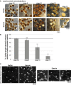
Early stage X. laevis embryonic nuclei are susceptible to emerin-induced breakage. (A) Embryo extracts containing endogenous embryonic nuclei were prepared from different-stage X. laevis embryos. At least 30 embryos were used per extract. Extracts were supplemented with 5 nM emerin or an equivalent volume of dialysis buffer as a control and incubated at room temperature for 30 min. Nuclei were stained with Hoechst and visualized as in Figure 1. Representative images from one experiment out of three are shown. Average nuclear cross-sectional area at each developmental stage is indicated in µm2 (Jevtic and Levy, 2015). (B) Experiments were performed with X. laevis egg extract as in Figure 1, except that the lengths of nuclear assembly and emerin incubation were varied as indicated, so that the nuclei were different sizes when emerin was added. Representative images from one experiment out of three are shown. Average nuclear cross-sectional area for each condition is indicated in µm2.
FIGURE 5:
FIGURE 5:
Microinjected emerin protein induces nuclear breakage and death in X. laevis embryos. (A) One-cell embryos were microinjected with emerin to achieve a final concentration of 0.5 nM within the embryo or an equivalent volume of dialysis buffer and allowed to develop at room temperature. Representative images at different developmental stages are shown. (B) At the indicated developmental stages, the viable embryos were counted. Inviable embryos were those that had stopped dividing or exhibited a puffy white appearance indicative of apoptosis (Johnson et al., 2010; Du Pasquier et al., 2011; Tokmakov et al., 2011; Gillespie et al., 2012; Willis et al., 2012; Iguchi et al., 2013; Broadus et al., 2015). The number of viable embryos was normalized to the buffer control. From 30 to 60 embryos were analyzed per condition per experiment. Averages from three independent experiments are shown. Error bars represent SD. Statistical analysis was performed relative to the stage 5–6 embryos. ***p < 0.005; NS, not significant. (C) Nuclei in buffer- or emerin-microinjected embryos were visualized with Hoechst at stage 12. Representative images from one experiment out of three are shown.
FIGURE 6:
FIGURE 6:
Microinjected emerin mRNA induces loss of nuclear integrity and death in X. laevis embryos. (A) One-cell embryos were microinjected with 1500 pg of emerin mRNA (dissolved in water) or an equivalent volume of water as a control and allowed to develop. Representative images at different developmental stages are shown. (B) One-cell embryos were microinjected with the indicated amounts of emerin mRNA or an equivalent volume of water as a control and allowed to develop. At the indicated developmental stages, the viable embryos were counted. Inviable embryos were those that had stopped dividing or exhibited a puffy white appearance indicative of apoptosis (Johnson et al., 2010; Du Pasquier et al., 2011; Tokmakov et al., 2011; Gillespie et al., 2012; Willis et al., 2012; Iguchi et al., 2013; Broadus et al., 2015). The number of viable embryos was normalized to water-microinjected controls. From 11 to 89 embryos were analyzed per condition per experiment (43 embryos on the average). Averages from three independent experiments are shown. Error bars represent SD. Statistical analysis was performed relative to the water-microinjected control embryos. ***p < 0.005; NS, not significant. Injection with 100 pg emerin mRNA had no effect on embryo viability, and 2000 pg emerin mRNA exerted an effect similar to 1500 pg (unpublished data). (C) One-cell embryos were microinjected with 1000 pg GFP-NLS mRNA with or without 1500 pg emerin mRNA and allowed to develop. When control embryos reached stage 12, nuclei in microinjected embryos were visualized with Hoechst and GFP-NLS. Representative images from one experiment out of two are shown. (D) When control embryos reached stage 12, extracts were prepared from equivalent numbers of microinjected embryos described in C. Equivalent volumes of extract were supplemented with Hoechst, applied to a slide, overlaid with a coverslip, and incubated for 15 min. Images were acquired and the intact GFP+ nuclei per ∼660 × 660 µm field were counted. Nuclei from at least six fields were counted per experiment and condition. Averages from two independent experiments are shown. Error bars represent SD. ***p < 0.005.

Similar articles

Cited by

References

    1. Barton LJ, Soshnev AA, Geyer PK. (2015). Networking in the nucleus: a spotlight on LEM-domain proteins. Curr Opin Cell Biol , 1–8. - PMC - PubMed
    1. Beckhelling C, Chang P, Chevalier S, Ford C, Houliston E. (2003). Pre-M phase-promoting factor associates with annulate lamellae in Xenopus oocytes and egg extracts. Mol Biol Cell , 1125–1137. - PMC - PubMed
    1. Bengtsson L, Wilson KL. (2006). Barrier-to-autointegration factor phosphorylation on Ser-4 regulates emerin binding to lamin A in vitro and emerin localization in vivo. Mol Biol Cell , 1154–1163. - PMC - PubMed
    1. Berk JM, Simon DN, Jenkins-Houk CR, Westerbeck JW, Gronning-Wang LM, Carlson CR, Wilson KL. (2014). The molecular basis of emerin-emerin and emerin-BAF interactions. J Cell Sci , 3956–3969. - PMC - PubMed
    1. Berk JM, Tifft KE, Wilson KL. (2013). The nuclear envelope LEM-domain protein emerin. Nucleus , 298–314. - PMC - PubMed

Publication types

MeSH terms

LinkOut - more resources