. 2018 Dec 15;29(26):3168-3182.
doi: 10.1091/mbc.E18-06-0353. Epub 2018 Oct 17.

Genetic and epigenetic determinants establish a continuum of Hsf1 occupancy and activity across the yeast genome

Affiliations

Genetic and epigenetic determinants establish a continuum of Hsf1 occupancy and activity across the yeast genome

David Pincus et al. Mol Biol Cell. .

Abstract

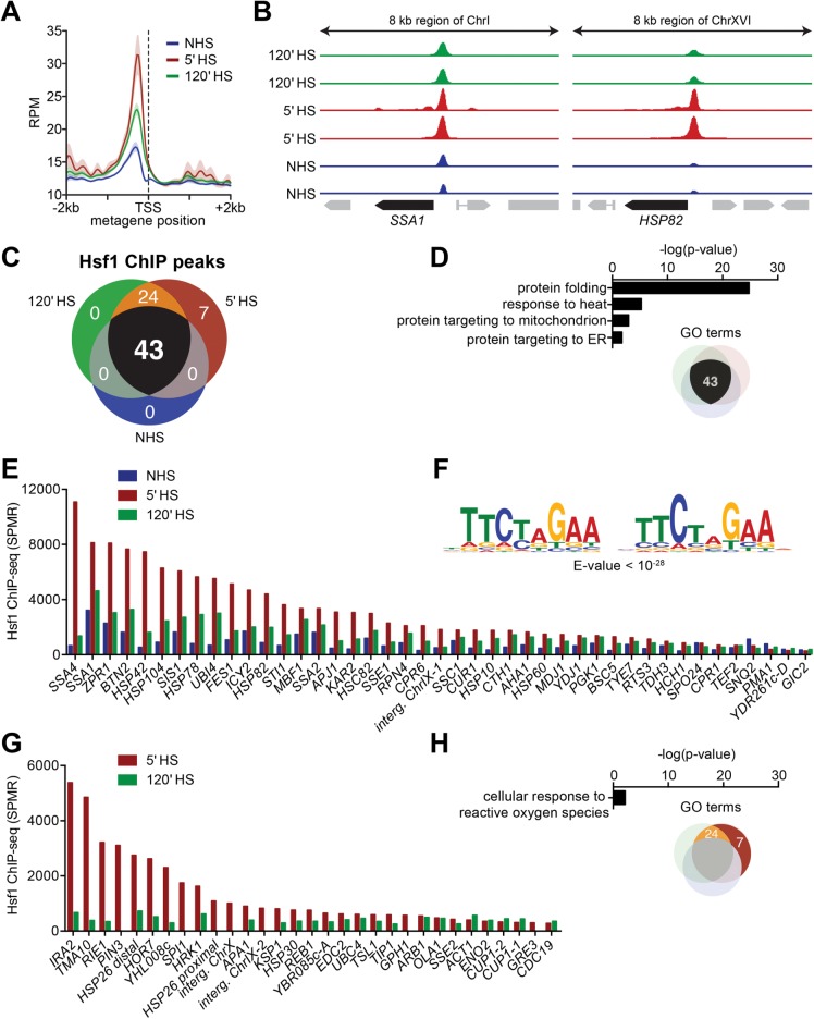
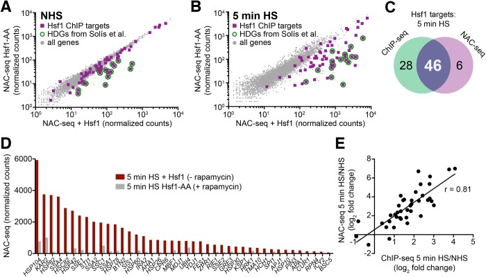
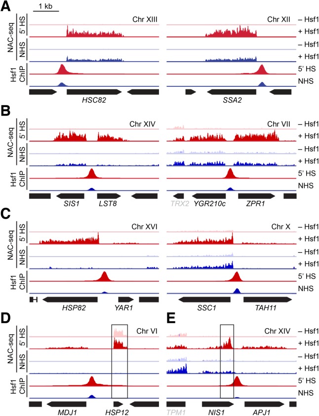
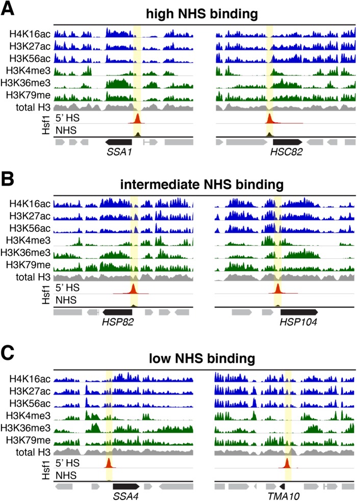
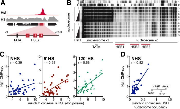
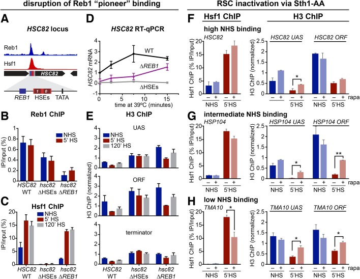
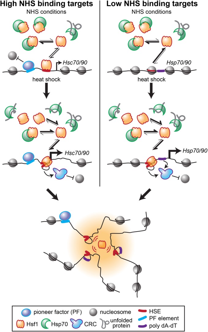
Heat shock factor 1 is the master transcriptional regulator of molecular chaperones and binds to the same cis-acting heat shock element (HSE) across the eukaryotic lineage. In budding yeast, Hsf1 drives the transcription of ∼20 genes essential to maintain proteostasis under basal conditions, yet its specific targets and extent of inducible binding during heat shock remain unclear. Here we combine Hsf1 chromatin immunoprecipitation sequencing (seq), nascent RNA-seq, and Hsf1 nuclear depletion to quantify Hsf1 binding and transcription across the yeast genome. We find that Hsf1 binds 74 loci during acute heat shock, and these are linked to 46 genes with strong Hsf1-dependent expression. Notably, Hsf1's induced DNA binding leads to a disproportionate (∼7.5-fold) increase in nascent transcription. Promoters with high basal Hsf1 occupancy have nucleosome-depleted regions due to the presence of "pioneer factors." These accessible sites are likely critical for Hsf1 occupancy as the activator is incapable of binding HSEs within a stably positioned, reconstituted nucleosome. In response to heat shock, however, Hsf1 accesses nucleosomal sites and promotes chromatin disassembly in concert with the Remodels Structure of Chromatin (RSC) complex. Our data suggest that the interplay between nucleosome positioning, HSE strength, and active Hsf1 levels allows cells to precisely tune expression of the proteostasis network.


Figures

FIGURE 1:
FIGURE 1:
Hsf1 ChIP-seq reveals differential basal and heat-shock-inducible binding across the Hsf1 regulon. (A) Metagene plot of Hsf1 ChIP-seq signal genomewide with respect to the TSS under NHS conditions and following 5-min and 120-min HS. Strain BY4741 was used for this and all other Hsf1 ChIP-seq assays in this work. RPM, reads per million mapped reads. (B) IGV browser images of the SSA1 and HSP82 loci showing Hsf1 ChIP-seq signal in two biological replicates under NHS conditions and following 5-min and 120-min HS. The y-axes are normalized to the maximum displayed signal in the 5-min time point. Preimmune serum ChIP samples (not shown) were used for peak calling (see Materials and Methods). (C) Venn diagram showing the number of Hsf1 ChIP peaks that surpassed the background cutoff in both biological replicates under each condition. (D) Gene ontology (GO) term enrichment values for genes immediately downstream of the 43 ChIP peaks. (E) Normalized Hsf1 ChIP signal at the 43 peaks identified under all three conditions; represent the mean of two biological replicates. SPMR, signal per million mapped reads. (F) Consensus motif identified under Hsf1 ChIP peaks detected under all conditions. (G) Normalized Hsf1 ChIP signal at the 31 peaks detected only under HS conditions; presented as in E. (H) GO term enrichment values for the 31 Hsf1 ChIP peaks detected only under HS conditions.
FIGURE 2:
FIGURE 2:
NAC-seq coupled with Hsf1-Anchor Away reveals genes dependent on Hsf1 for their basal and induced transcription. (A) NAC-seq counts transcriptome-wide under NHS conditions in the presence and absence of nuclear Hsf1 using the Hsf1 Anchor Away system (Hsf1-AA). Rapamycin (1 µM) was added for 45 min to deplete Hsf1 from the nucleus. Purple squares are genes with Hsf1 ChIP peaks; green circles show Hsf1 targets identified previously under NHS conditions (Solis et al., 2016). Genes with Hsf1 ChIP peaks above background (≥250 SPMR) and whose expression was significantly reduced by rapamycin treatment (p < 0.01; two-tailed t test) were designated as Hsf1-dependent genes (HDGs). Eighteen fell into this category: AHA1, BTN2, CUR1, CPR6, FES1, HCH1, HSC82, HSP104, HSP42, HSP78, HSP82, MBF1, MDJ1, SIS1, SSA1, SSA2, STI1, and YDJ1. (B) Analysis, representation, and designations as in A, except following 5 min HS. (C) Venn diagram comparing Hsf1 ChIP-seq gene targets and NAC-seq targets following a 5-min HS. ChIP-seq gene targets were derived from 74 total peaks, three of which were intergenic, two of which were linked to a single gene (HSP26), and four of which were linked to Hsf1-dependent, bidirectionally transcribed genes (see Figure 3, B and C). (D) NAC-seq counts for shared ChIP-seq/NAC-seq Hsf1 targets following a 5-min HS in the presence and absence of nuclear Hsf1. (Note: NAC-seq counts represent total nascent transcription and are not normalized for gene length.) Only one gene from the four Hsf1-dependent bidirectional genes is shown. (E) Scatter plot showing the correlation between HS-inducible Hsf1 DNA binding and HS-inducible transcription of shared ChIP-seq/NAC-seq Hsf1 targets. ChIP-seq signals represent the mean of two biological replicates.
FIGURE 3:
FIGURE 3:
Hsf1 stimulates both unidirectional and bidirectional transcription. (A) IGV browser images of 5-kb windows at Hsf1-dependent loci. Tracks show NAC-seq and Hsf1 ChIP-seq under NHS and 5-min HS conditions; NAC-seq was conducted in the presence and absence of nuclear Hsf1 using the Hsf1-AA system. ChIP-seq and NAC-seq tracks were normalized to the maximum displayed value for each locus in the 5-min heat-shock sample. NAC-seq tracks represent both sense and anti-sense transcription yet for simplicity are shown above the line for each gene. Hsf1 was anchored away with 1 µM rapamycin for 45 min. (B) As in A but for loci that show near-stoichiometric, Hsf1-dependent bidirectional transcription. (C) As in B but for loci that show substoichiometric, Hsf1-dependent transcription of the nonchaperone gene. (D) As in C but for the MDJ1/HSP12 locus on chromosome VI. Although both MDJ1 and HSP12 are induced by heat shock, only MDJ1 is Hsf1 dependent. (E) As in C but for the NIS1/APJ1 locus on chromosome XIV. Demonstrates heat-shock- and Hsf1-dependent bidirectional transcription in the sense direction for APJ1 and in the antisense direction for NIS1.
FIGURE 4:
FIGURE 4:
Hsf1’s constitutive occupancy correlates with preset accessible chromatin, while its heat-inducible occupancy correlates with preexisting histone acetylation and partial nucleosome occupancy. (A) ChIP-seq profiles of the indicated covalently modified histones, total H3 and Hsf1 over 10 kb windows of representative High NHS Binding loci (high Hsf1 occupancy under control conditions). Location of Hsf1 binding peak is highlighted in yellow. All tracks except Hsf1 were normalized to their own maximum displayed signal and are from publicly available data sets (NHS state; see Materials and Methods). Hsf1 tracks depict both NHS and 5-min HS states and are normalized to the 5-min HS sample (this study). (B) As in A, except depicted are Intermediate Hsf1 Binding loci. (C) As in A, except depicted are Low Hsf1 Binding loci.
FIGURE 5:
FIGURE 5:
Hsf1 DNA binding is impeded by nucleosomes, both in vitro and in vivo. (A) Hsf1 and H3 ChIP-seq signal at HSP82 under NHS conditions, normalized to their maximum displayed values. The region used for nucleosome reconstitution and DNase I footprinting is highlighted. (B) DNA corresponding to the HSP82 upstream region depicted in A (spanning –9 to –353 [ATG = +1]) and 32P-end labeled on the upper strand was either reacted directly with GST-Hsf1 (lanes 4–7) or following its reconstitution into a dinucleosome (lanes 9–12). Reconstitution was achieved using a 1:1 (wt/wt) HeLa histone: DNA ratio and salt dilution, followed by purification over a glycerol gradient (see Supplemental Figure S4). Both naked DNA and chromatin templates were challenged with increasing amounts of recombinant Hsf1 (lanes 3 and 8 are -Hsf1 controls) and then subjected to DNase I digestion. DNA was purified and electrophoresed on an 8% sequencing gel. (C) Scatter plots of Hsf1 ChIP-seq signal as a function of the strength of the HSE for the NHS, 5-min HS, and 120-min HS states. HSE strength was determined by MEME as a p value corresponding to how well the binding site beneath the summit of each ChIP peak matched the consensus HSE motif (Figure 1F). ChIP-seq signals represent the mean of two biological replicates. (D) As in C, but here each p value was divided by the H3 ChIP-seq signal below the summit of the Hsf1 peak under NHS conditions (H3 ChIP-seq data from Qiu et al. [2016]). Outliers (gray) consist of Hsf1-independent housekeeping genes.
FIGURE 6:
FIGURE 6:
The pioneer factor Reb1 enables Hsf1 binding to a high NHS binding target while RSC cooperates with Hsf1 to displace nucleosomes during heat shock. (A) Browser shot showing Reb1 ChEC-seq (Zentner et al., 2015) and Hsf1 ChIP-seq signal under NHS conditions at a 5-kb window around the HSC82 locus. Both tracks are normalized to their maximum displayed values. Expanded view of the HSC82 promoter shows the locations of the Reb1 binding site, HSEs, and TATA box (centered at –249, –193, and –138, respectively; ATG = +1). (B) Reb1-myc9 ChIP-qPCR analysis of the hsc82 promoter under NHS and 5-min HS conditions in isogenic HSC82, hsc82-∆HSEs, and hsc82-∆REB1 cells conducted as described under Materials and Methods. The hsc82-∆HSEs allele bears multiple point substitutions within HSE0 and HSE1 while hsc82-∆REB1 bears a 10-base-pair chromosomal substitution of the Reb1 binding site (Erkine et al., 1996). Shown are means + SD (N = 2 biological replicates; qPCR = 4). (C) Hsf1 ChIP analysis of the hsc82 UAS region, conducted and analyzed as in B. (D) Quantification of HSC82 mRNA expression level by RT-qPCR over a heat-shock time course. Plotted are means ± SD (N = 2; qPCR = 4). (E) Histone H3 ChIP analysis of the HSC82 promoter, midcoding region (open reading frame [ORF]), and 3′-UTR-terminator region. H3 ChIP signals were normalized to those detected at a nontranscribed locus, ARS504, which served as an internal recovery control. (F–H) Hsf1 and H3 ChIP at HSC82, HSP104, and TMA10 (as indicated) under NHS and 5-min HS conditions, in the presence and absence of the RSC catalytic subunit, Sth1 (rapa - and rapa +, respectively). Conditional nuclear depletion of Sth1 was achieved using an Sth1-AA strain that was pretreated with 1 µM rapamycin (rapa) for 2.5 h. Analysis and display as in B. *, p < 0.05; **, p < 0.01, two-tailed t test.
FIGURE 7:
FIGURE 7:
Model for differential basal and inducible Hsf1 binding across the genome. Target genes with high levels of Hsf1 binding under non-heat-shock conditions (High NHS binding targets) have nucleosome-free (or depleted) regions upstream of the TSS due to the presence of pioneer factors. Such NFRs are occupied by the small fraction of DNA binding-competent Hsf1 present, stimulating basal transcription of linked HSC70/90 genes (e.g., SSA1, SSA2, HSC82). These genes nonetheless show approximately twofold increases in Hsf1 binding on acute heat shock when the bulk of Hsf1 is liberated from repressive association with Hsp70. This inducible binding further restructures the loci and drives increased transcription. In contrast, Low NHS binding targets (e.g., SSA4, HSP26, TMA10) lack proximal pioneer factor binding, and Hsf1 binding sites are occluded by nucleosomes. The fraction of free Hsf1 available under NHS conditions is insufficient to invade the chromatin at these sites. On heat shock, the large increase in DNA-binding competent Hsf1 allows Hsf1 to cooperatively bind its cognate HSEs, and along with poly(dA-dT) tracts that help recruit CRCs, to restructure the loci and drive high-level transcription. Active Hsf1 then drives looping and coalescence of its target loci into transcriptionally active foci that may form phase-separated assemblies.

Similar articles

Cited by

References

    1. Abravaya K, Myers MP, Murphy SP, Morimoto RI. (1992). The human heat shock protein hsp70 interacts with HSF, the transcription factor that regulates heat shock gene expression. Genes Dev , 1153–1164. - PubMed
    1. Adams CC, Gross DS. (1991). The yeast heat shock response is induced by the conversion of cells to spheroplasts and by potent transcriptional inhibitors. J Bacteriol , 7429–7435. - PMC - PubMed
    1. Bai L, Ondracka A, Cross FR. (2011). Multiple sequence-specific factors generate the nucleosome-depleted region on CLN2 promoter. Mol Cell , 465–476. - PMC - PubMed
    1. Buchman AR, Lue NF, Kornberg RD. (1988). Connections between transcriptional activators, silencers, and telomeres as revealed by functional analysis of a yeast DNA-binding protein. Mol Cell Biol , 5086–5099. - PMC - PubMed
    1. Cairns BR, Lorch Y, Li Y, Zhang M, Lacomis L, Erdjument-Bromage H, Tempst P, Du J, Laurent B, Kornberg RD. (1996). RSC, an essential, abundant chromatin-remodeling complex. Cell. , 1249–1260. - PubMed

Publication types

MeSH terms