. 2018 Oct 16;25(3):598-610.e5.
doi: 10.1016/j.celrep.2018.09.072.

Long-Term In Vitro Expansion of Epithelial Stem Cells Enabled by Pharmacological Inhibition of PAK1-ROCK-Myosin II and TGF-β Signaling

Affiliations

Long-Term In Vitro Expansion of Epithelial Stem Cells Enabled by Pharmacological Inhibition of PAK1-ROCK-Myosin II and TGF-β Signaling

Chengkang Zhang et al. Cell Rep. .

Abstract

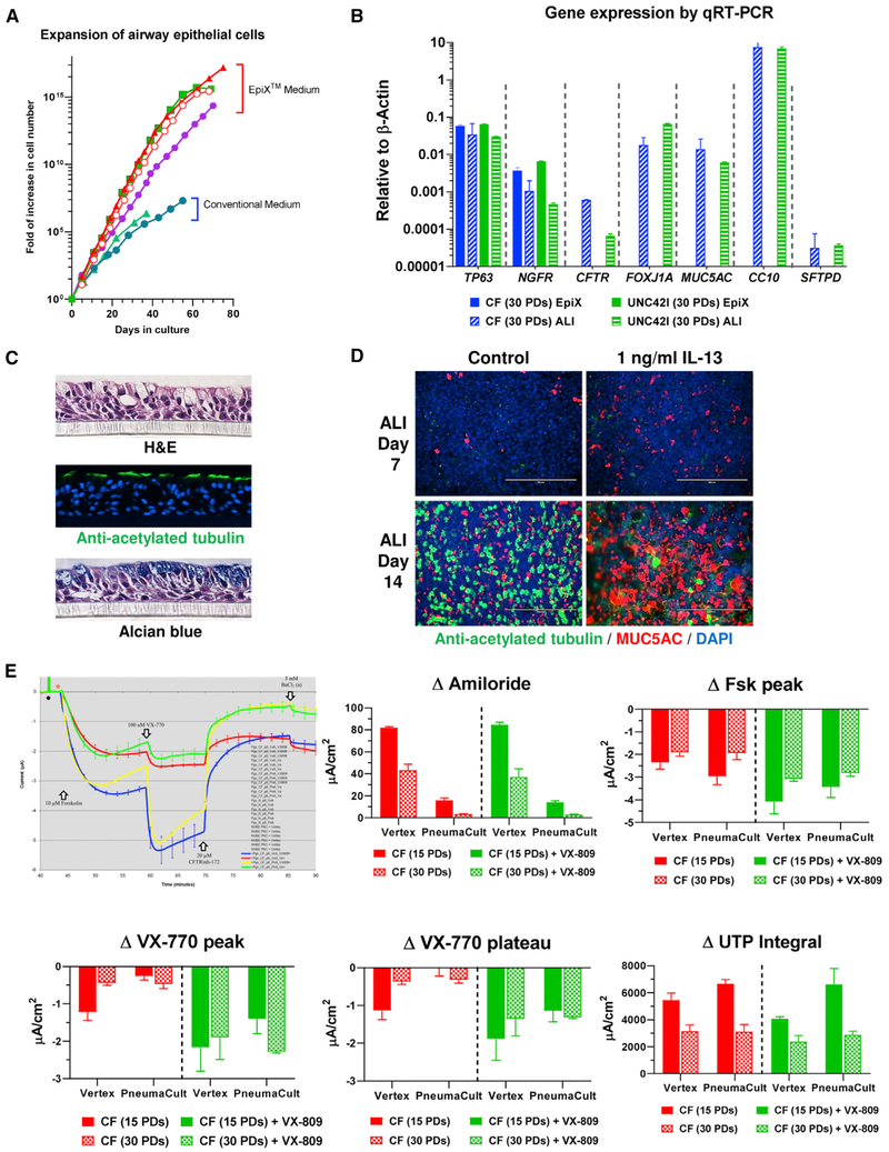
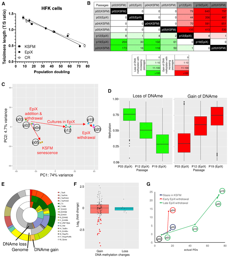
Despite substantial self-renewal capability in vivo, epithelial stem and progenitor cells located in various tissues expand for a few passages in vitro in feeder-free condition before they succumb to growth arrest. Here, we describe the EpiX method, which utilizes small molecules that inhibit PAK1-ROCK-Myosin II and TGF-β signaling to achieve over one trillion-fold expansion of human epithelial stem and progenitor cells from skin, airway, mammary, and prostate glands in the absence of feeder cells. Transcriptomic and epigenomic studies show that this condition helps epithelial cells to overcome stresses for continuous proliferation. EpiX-expanded basal epithelial cells differentiate into mature epithelial cells consistent with their tissue origins. Whole-genome sequencing reveals that the cells retain remarkable genome integrity after extensive in vitro expansion without acquiring tumorigenicity. EpiX technology provides a solution to exploit the potential of tissue-resident epithelial stem and progenitor cells for regenerative medicine.

Keywords: PAK1/ROCK/Myosin II; TGF-β; cell culture method; cell therapy; epithelial stem and progenitor cells; feeder-free; regenerative medicine.


Figures

Figure 1.
Figure 1.. TGF-β Signaling Inhibition, ROCK Inhibition, and Low Calcium Synergistically Support Long-Term Epithelial Cell Proliferation
(A) Small molecules inhibiting the TGF-β signaling or ROCK supported the proliferation of late-passage PrECs/nRFP cells in the absence of feeder cells in the F medium. Data are represented as mean ±SD, n = 4. (B) Synergy between A83-01 and Y-27632 in the F medium (four replicates per condition). (C and F) PrECs/nRFP cells proliferated for 10 PDs in the F medium plus Y-27632 and A83-01 (F+Y+A) but continued to proliferate in the CR condition (C). Many cells in F+Y+A exhibited differentiated morphology (F). (D) ROCK inhibitors synergistically promoted the proliferation of HBECs/nRFP cells in KSFM plus 1 μM A83-01. (E) Synergy between A83-01 and Y-27632 in KSFM (four replicates per condition). (G) Morphology of HFKs over successive passages in KSFM (P1 and P5) or the EpiX medium (P2, P11, and P20). (H) TP63 was ubiquitously expressed in late-passage HFKs (P16) cultured in the EpiX medium. (I-L) Expansion of HFKs (I), PrECs (J), HBECs (K), and mammary epithelial cells (L) in KSFM or EpiX.
Figure 2.
Figure 2.. Transcriptome Analysis of HFKs Expanded with the EpiX Medium
(A) Experimental scheme of HFK expansion in KSFM or the EpiX medium and time points for samples collection. HFKs underwent three different routes: (1)stasis in KSFM (P2→P5); (2) transient exposure and withdrawal from the EpiX medium (P2→P3→P4); and (3) expansion in the EpiX medium and withdrawal at late passage (P3→P12→P19→P20). Each sample was collected in duplicate. (B) PCA on HFK transcriptomes in various conditions. The red arrows represented the direction of culture condition changes. (C) MA plot of differentially expressed genes in two different media. The red dots represented differentially expressed genes. (D) Temporal expression changes of two top downregulated genes, VIM (solid line) and FN1 (dashed line). (E) Heatmap of 962 downregulated genes in HFKs expanded with the EpiX medium. These downregulated genes were de-repressed when the HFKs were withdrawn from EpiX medium. The red arrows indicated EpiX medium withdrawal. (F) Gene ontology (GO) and pathway enrichment analysis of upregulated and downregulated genes by metascape tool.
Figure 3.
Figure 3.. Genome Stability of Epithelial Cells Expanded with the EpiX Medium
(A) HFKs, HBECs, and PrECs expanded with the EpiX medium over 40 PDs retained diploid karyotypes. (B) EpiX-expanded HFKs did not form tumors after 12 weeks in female nude mice (1 × 10⁷ cells per mouse, n = 6 in each group). Data are represented as mean ±SD, n = 6. (C) Data analysis workflow for whole-genome sequencing. (D) Distributions of de novo SNVs identified by whole genome-sequencing in late-passage HFK and CF samples. Venn diagram showed the overlap between the CF and HFK samples. (E) Genes that were affected by missense de novo SNV (all heterozygous) after 40 PDs expansion in the EpiX medium. Two separate missense SNVs were found in the DDX11 gene in the CF and HFK samples.
Figure 4.
Figure 4.. Differentiation of HFKs Expanded with the EpiX Medium
(A) Addition of 1 mM CaCl2 to the EpiX medium induced the HFKs to differentiate in 24 hr. (B) Immunofluorescence staining of tight junctions (ZO-1 and Occludin) in HFKs cultured in EpiX plus 1 mM CaCl2 for 7 days. ZO-1, zonula occludens-1. (C and D) Confluent HFKs in EpiX (C) or EpiX plus 1 mM CaCl2 (D). Many domes with liquid accumulated underneath occurred in the culture with EpiX plus 1 mM CaCl2. (E) HFKs cultured in a T-75 flask in EpiX plus 1.5 mM CaCl2 for 7 days form an intact epithelium sheet, which was released from the flask after 30-min incubation in dispase at 37°C. (F) HFKs were differentiated at ALI for 14 days and formed a stratified epithelium. (G) EpiX-expanded HFKs were subcutaneously injected into immune-comprised mice. After 5 weeks, the cells formed cysts which resembled epidermis. (H) Most cells in the basal layer were stained positive for the proliferation marker Ki67. (I) The basal and supra-basal layers of the cystic epithelium stained positive for KRT14 (K14).
Figure 5.
Figure 5.. Differentiation of HBECs Expanded with the EpiX Medium
(A) HBECs from healthy and CF donors (n = 4) were expanded in the EpiX medium or conventional medium (BEGM). EpiX medium supported million-fold more expansion than BEGM (n = 2). (B) HBECs from a healthy (UNC42I) or a CF donor were expanded in the EpiX medium for 30 PDs and differentiated at ALI for 21 days. The expression of basal cell markers (TP63, NGFR), multiciliated cell markers (CFTR, FOXJ1A), goblet cell marker (MUC5AC), club cell marker (CC10), or type II cell marker (SFTPD) were checked by qRT-PCR. Gene expression levels were depicted as relative to that of β-actin, which was set at 1. Data are represented as mean ± SD, n = 3. (C) UNC42I cells (P8) were differentiated at ALI for 21 days. Paraffin sections were stained with H&E, or with an anti-acetylated tubulin antibody, or with alcian blue to show multiciliated cells and goblet cells. (D) UNC42I cells (P7) were differentiated at ALI and treated with 1 ng/mL IL-13. Multiciliated cells and goblet cells were stained with anti-acetylated tubulin (in green) or anti-MUC5AC (in red) antibodies respectively. IL-13 led to goblet cell hyperplasia and a decrease of multiciliated cells. (E) Early- (15 PDs) and late-passage (30 PDs) CF cells were differentiated at ALI for Ussing assays, using either the Vertex or the PneumaCult-ALI protocol. The activities of ENaC (ΔAmiloride, 30 μM amiloride), CFTR (ΔFsk peak, 10 μM forskolin; ΔVX-770 peak and ΔVX-770 plateau, 100 nM VX-770 and 3 μM VX-809) and CaCC (ΔUTP, 100 μM UTP) were measured. VX-809, a CFTR trafficking corrector; VX-770, a CFTR potentiator. The responses of mutant CFTR variants to the CFTR corrector (3 μM VX-809) and CFTR potentiator (100 nM VX-770) were similar between early and late passages. Data are represented as mean ± SD, n = 3.
Figure 6.
Figure 6.. Epigenetic Changes in HFKs Expanded with the EpiX Medium
(A) Telomere length (T/S ratio) gradually decreased in HFKs expanded in the EpiX medium or the CR method. Data are represented as mean ±SD, n = 3. (B) The number of differentially methylated regions (DMRs) identified between different culture conditions. The numbers in upper right cells were DMRs with DNA methylation gain, and the numbers in lower left cells were DMRs with DNA methylation loss. (C) PCA of DNA methylomes. DNA methylation levels of DMRs were used as input data. The first principal component (passage) explained most variances among the DNA methylomes. (D) DNA methylation levels in HFKs gradually changed over successive passages in the EpiX medium. The methylation levels of DMRs at P12 was intermediate between P3 and P19. Also see Figure S10. (E) The chromatin states of DMRs using 18 chromHMM expanded states in keratinocytes. Lots of DMRs were in regulatory regions such as promoters and enhancers. Also see Figure S10. (F) Expression changes of genes whose promoters underwent DNA methylation changes. Expression changes were calculated as log2 of fold change between P3 and P19. The red dots indicated the genes whose expression was downregulated as expected from the gain of promoter methylation. (G) Predicted cumulative population doublings (pcPDs) of HFKs based on the DNA methylation levels at six CpG sites correlated with the actual PDs at different passages.

Similar articles

Cited by

References

    1. Avior Y, Sagi I, and Benvenisty N (2016). Pluripotent stem cells in disease modelling and drug discovery. Nat. Rev. Mol. Cell Biol 17, 170–182. - PubMed
    1. Baar MP, Brandt RMC, Putavet DA, Klein JDD, Derks KWJ, Bourgeois BRM, Stryeck S, Rijksen Y, van Willigenburg H, Feijtel DA, et al. (2017). Targeted apoptosis of senescent cells restores tissue homeostasis in response to chemotoxicity and aging. Cell 169, 132–147.e16. - PMC - PubMed
    1. Bhowmick NA, Ghiassi M, Aakre M, Brown K, Singh V, and Moses HL (2003). TGF-β-induced RhoA and p160ROCK activation is involved in the inhibition of Cdc25A with resultant cell-cycle arrest. Proc. Natl. Acad. Sci. USA 100, 15548–15553. - PMC - PubMed
    1. Blanpain C, and Fuchs E (2014). Stem cell plasticity. Plasticity of epithelial stem cells in tissue regeneration. Science 344, 1242281. - PMC - PubMed
    1. Boj SF, Hwang C-I, Baker LA, Chio IIC, Engle DD, Corbo V, Jager M, Ponz-Sarvise M, Tiriac H, Spector MS, et al. (2015). Organoid models of human and mouse ductal pancreatic cancer. Cell 160, 324–338. - PMC - PubMed

Publication types

MeSH terms