. 2018 Oct 16;25(3):651-662.e5.
doi: 10.1016/j.celrep.2018.09.051.

Dopamine Neurons Mediate Learning and Forgetting through Bidirectional Modulation of a Memory Trace

Affiliations

Dopamine Neurons Mediate Learning and Forgetting through Bidirectional Modulation of a Memory Trace

Jacob A Berry et al. Cell Rep. .

Abstract

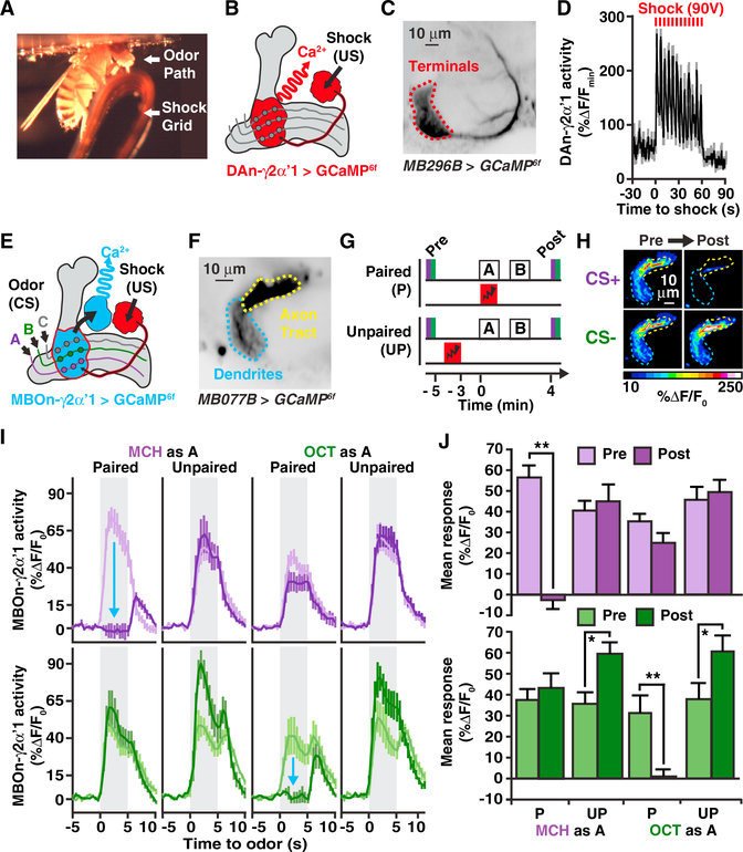
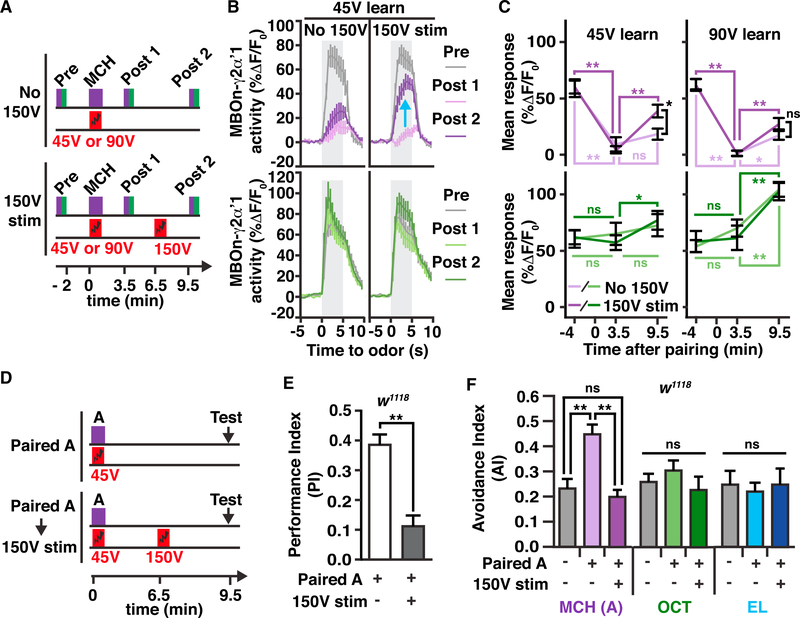
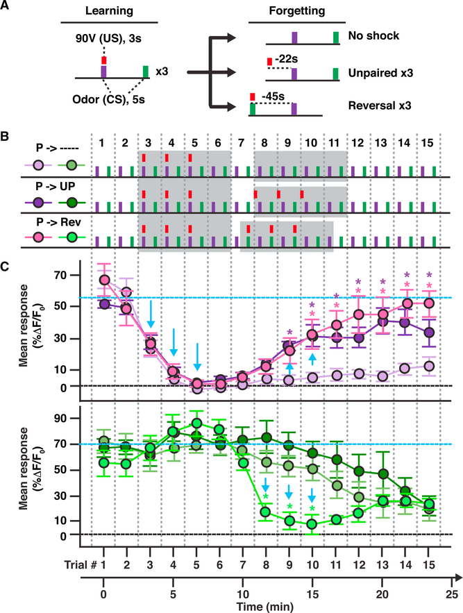
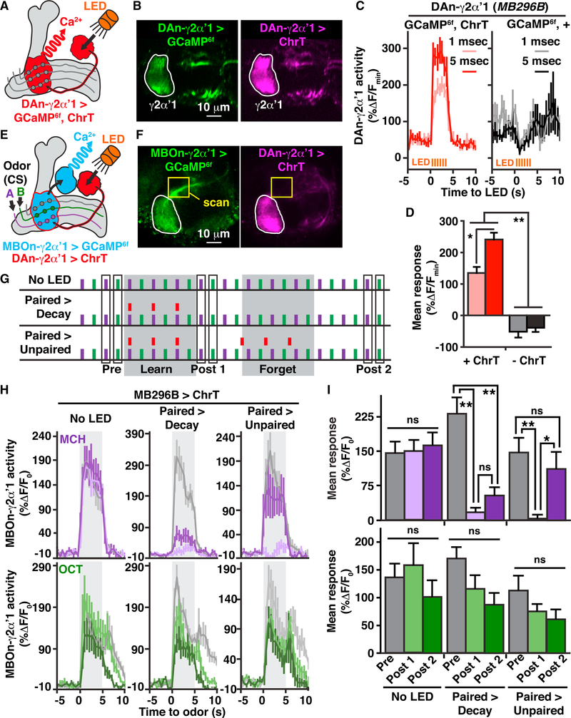
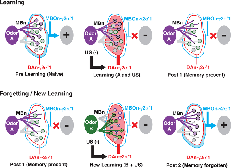
It remains unclear how memory engrams are altered by experience, such as new learning, to cause forgetting. Here, we report that short-term aversive memory in Drosophila is encoded by and retrieved from the mushroom body output neuron MBOn-γ2α'1. Pairing an odor with aversive electric shock creates a robust depression in the calcium response of MBOn-γ2α'1 and increases avoidance to the paired odor. Electric shock after learning, which activates the cognate dopamine neuron DAn-γ2α'1, restores the response properties of MBOn-γ2α'1 and causes behavioral forgetting. Conditioning with a second odor restores the responses of MBOn-γ2α'1 to a previously learned odor while depressing responses to the newly learned odor, showing that learning and forgetting can occur simultaneously. Moreover, optogenetic activation of DAn-γ2α'1 is sufficient for the bidirectional modulation of MBOn-γ2α'1 response properties. Thus, a single DAn can drive both learning and forgetting by bidirectionally modulating a cellular memory trace.

Keywords: dopamine neuron; forgetting; memory trace; memory updating.


Conflict of interest statement

DECLARATION OF INTERESTS

The authors declare no competing interests.

Figures

Figure 1.
Figure 1.. MBOn-γ2αʹ1 Receives Synaptic Input from MBns, and Its Output Is Required for Aversive Memory Retrieval
(A) Schematic diagram of the γ2αʹ1 compartment (also referred to as the junction) of the MB (gray shading) showing relevant neurons and pathways. The red objects represent DAn innervation, conveying information about electric shock to this neuropil compartment. The DAn axon terminals overlap the region of innervation by the dendrites of the MBOn-γ2αʹ1 neuron (red region outlined in blue). Circles represent MBn to MBOn synapses. Odors A, B, and C activate axon fibers from individual MBns (colored lines). (B) Simplified circuit diagram of (A), with odor inputbeing conveyed by MBns, and approach behavior biased by the output of MBOn-γ2αʹ1. (C) MBOn-γ2αʹ1 morphology visualized using MB077B-gal4 > myr:GFP (see inset of Figure S1A). D, dendrites; T, presynaptic terminals. (D) Reconstituted GFP across the MBn to MBOn-γ2αʹ1 synapses visualized using GRASP immunostaining (left) overlaid on the collection of MBn presynaptic terminals detected by syb::spGFP1–10 immunostaining (right). (E and F) MBOn-γ2αʹ1 output was blocked either during both acquisition, A, and retrieval, R, of short-term aversive memory (E), or specifically during the retrieval of short-term aversive memory (F). Time in protocols is with respect to the beginning of odor-shock association. Bar plots represent the mean with error bars equal to + SEM in this figure and others. Two-way ANOVA followed by Bonferroni post hoc tests. *p < 0.01, **p < 0.0001; n = 7–8. ns, not significant; UAS, upstream activation sequence. See also Figure S1.
Figure 2.
Figure 2.. Aversive Classical Conditioning Creates Robust Short-Term Plasticity in MBOn-γ2αʹ1
(A) Photograph of the in vivo functional imaging setup showing a fly standing on an electric shock grid prepared to receive odor and/or electric shock stimuli. The top of the head capsule is obscured but positioned just below the microscope objective. (B) Schematic of circuits and tools used to collect data in (C) and (D). (C) GCaMP6f specifically expressed in DAn-γ2αʹ1 axon terminals using MB296B-gal4 (red-dotted outline). Mean time series projection of GCaMP6f fluorescence from a 30-s time window before electric shock. (D) Time course of GCaMP6f responses in DAn-γ2αʹ1 terminals with electric shock exposure (90V, 12×) used in (G) showing the mean and SEM of n = 8 animals. (E) Schematic of circuits and tools used to collectdata shown in (F)–(J). Odors (CS) activate sparse MBns that stimulate MBOn-γ2αʹ1 (blue) via synaptic connections (circles). Electric shock (US) activates DAn-γ2αʹ1 (red) with DA release modulating MBn:MBOn-γ2αʹ1 functional connectivity. (F) Mean time series projection of baseline GCaMP6f driven by MB077B-gal4 in the MBOn-γ2αʹ1 dendrites and its axon tracts (tdTomato was also expressed but not used in the analysis). The area labeled “Axon Tract” resides outside the MB neuropil and probably contains axon terminals in addition to the axon tracts, explaining its breadth. (G) Paired and unpaired conditioning protocols used to collect data shown in panels (H)–(J). Odor A and B are defined as the first and second odors given during the training, respectively. GCaMP6f responses were monitored before conditioning (pre) to establish basal responses to odor stimulation. The CS+ was presented paired or unpaired with electric shock pulses. A CS odor was presented as a control. Changes in response properties were measured during odor stimulation after conditioning (post). Timeline (bottom) indicates time points relative to the start of CS/US association, including the start of pre and post responses and the end of shock in the unpaired protocol. (H) Pseudocolored peak responses of MBOn-γ2αʹ1 axons and dendrites to the CS+ (MCH) and CS(OCT) before and after a paired protocol. (I) Time course of the dendritic Ca2+ responses of MBOn-γ2αʹ1 during a 5-s exposure (gray-shaded regions) to either MCH (top) or OCT (bottom) before (light-colored line) and after (dark-colored line) paired or unpaired protocols. Traces show the average response (±SEM) across all flies tested. The blue arrows indicate the depressed response due to paired conditioning. (J) Mean dendritic response during odor exposure before (pre) and after (post) conditioning protocols from data shown in (I). Two-way repeated-measures ANOVA with Bonferroni post hoc tests. *p < 0.05, **p < 0.001; n = 8–10. See also Figure S2.
Figure 3.
Figure 3.. US Pathway Stimulation Restores MBOn-γ2αʹ1 Responses Previously Depressed by Learning and Causes Behavioral Forgetting
(A) Conditioning protocols used to collect data shown in (B) and (C) and Figures S3A–S3D. GCaMP6f responses in MBOn-γ2αʹ1 (MB077B-gal4 > GCaMP6f) to odor (pre, post 1, and post 2) were monitored before odor/shock pairing, after the pairing (45 V, 12× or 90 V, 12×), and after either strong 150 V, 12× electric shock (150 V stim) or no strong shock (no 150 V), respectively. Timeline (bottom) indicates time points relative to the start of CS/US association, including the start of pre, post 1, and post 2 responses, and the start of 150 V shock. (B) Time course of dendritic Ca2+ responses in MBOn-γ2αʹ1 for 45 V pairing experiments upon odor exposure (gray-shaded regions) to MCH or OCT before (pre, gray lines), after odor/45 V shock pairing (post 1, light-colored lines), and after either 150 V shock or no shock (post 2, dark-colored lines). The blue arrow indicates the recovered response due to the 150 V stimulation. (C) Mean dendritic response during the three odor exposures relative to time of learning for data from (A) (light-colored lines, no 150 V; dark-colored lines, 150 V stim). Two-way repeated-measures ANOVA with Bonferroni post hoc tests (comparing across 150 V stim or no 150 V conditions, black asterisk, *p < 0.05; or comparing across time points, colored asterisk matched to condition, *p < 0.05, **p < 0.001; ns, not significant; n = 10–11). (D) Behavioral conditioning protocols used to collect data in (E) and (F). Twelve electric shock pulses (45 V) were paired with an odor A (e.g., MCH) and then flies were tested for memory performance(E) or odor avoidance (F) after no intervening stimulus(paired A) or after electric shock stimulation(150 V,12×)(paired A→150 V stim).Timeline (bottom) indicates time points relative to the start of CS/US association, including the start of 150 V shock and the start of behavioral testing. (E) Performance index (PI) measuring preference of w1118 flies between the paired odor A (e.g., MCH) and a second odor (e.g., OCT) after the conditioning protocols illustrated in (D). A positive PI indicates aversion of the flies to odor A. One-way ANOVA with Bonferroni post hoc tests. **p < 0.0001; n = 8–12. (F) Avoidance index (AI) of w1118 flies measuring the preference between the paired odor A (MCH), or one of two novel odors, OCT and ethyl lactate (EL), and fresh air after the conditioning protocols illustrated in (D). One-way ANOVA with Bonferroni post hoc tests. *p = 0.0039, **p < 0.0001; n = 8–12. See also Figure S3.
Figure 4.
Figure 4.. New Learning Simultaneously Depresses MBOn-γ2αʹ1 Responses toa Paired Odor While Restoring Responses to a Previously Depressed Odor
(A) Simplified diagram of conditioning protocolsused to collect data for (C) and Figure S4, consisting of three short odor-shock pairings (learning) with a 45-s intertrial interval (ITI) between CS+ and CS followed by several forgetting protocols (forgetting). The red bar represents a single 3-s, 90-V shock. (B) Conditioning protocols used to collect data in (C). P, paired; UP, unpaired; Rev, reversal. The gray-shaded areas of the protocols highlight the learning trials (trials 3–6) and the forgetting trials (trials 7–11). (C) The mean dendritic Ca2+ responses of MBOn-γ2αʹ1 (MB077B-gal4 > GCaMP6f) during odor trials 1–15 for MCH (top) and OCT (bottom) depicted using the color scheme shown in (B). Blue arrows indicate depression or recovery of responses. Blue dashed lines show the initial response level. Two-way repeated-measures ANOVA with Bonferroni post hoc tests. *p < 0.05 between both unpaired and Rev groups relative to the control decay; n = 8. See also Figure S4.
Figure 5.
Figure 5.. Optogenetic Activation of DAn-γ2αʹ1 Fully Depresses or Restores MBOn-γ2αʹ1 Odor Responses When Paired or Unpaired with Odors, Respectively
(A) Schematic illustration of circuits and tools used to collect data in (B)–(D). (B) GCaMP6f (left) and Chrimson::tdTomato (ChrT, right) expression in DAn-γ2αʹ1 with the region of interest outlined and analyzed in (C) and (D). (C) Time course of the mean (±SEM) for axonal GCaMP6f responses in DAn-γ2αʹ1 during a 3-s train (2 Hz) of 1 or 5 ms LED pulses presented to flies with (ChrT) or without ChrT expression (n = 8). (D) Mean response of DAn-γ2αʹ1 during LED light exposure from data shown in (C). Two-way repeated-measures ANOVA with Bonferroni post hoc tests. *p < 0.01, **p < 0.0001; n = 8. (E) Schematic of circuits and tools and stimuli used to collect data shown in (F)–(I). (F) Example images of GCaMP6f (left) expression in MBOn-γ2αʹ1 using R25D02-lexA and Chrimson:tdTomato (right, ChrT) expression in DAn-γ2αʹ1 using MB296B-gal4. The yellow box indicates the two-photon scanning region of the MBOn-γ2αʹ1 axon to prevent unwanted activation of ChrT expressed in the DAn-γ2αʹ1 terminals (area outlined in white). (G) Stimulus protocols used for experiments shown in (H) and (I). MCH (5 s) and OCT (5 s) odor stimuli were presented with a 45-s ITI. The red bars represent 3-slight stimuli (LED). The MBOn-γ2αʹ1 axon was scanned before (pre) and after (post 1) a learning period (learn) during which odor and light stimuli were paired or light was not presented (no LED), and after (post 2) a forgetting period (forget) during which either three light pulses were presented unpaired with MCH (22 s prior) (paired > unpaired) or light was not presented (no LED and paired > decay). Timing of odor trials is the same as the timeline in Figure 4C. (H) Time course of axonal GCaMP6f responses during a 5-s odor exposure (gray-shaded region) to MCH or OCT at the pre (gray lines), post 1 (light-colored lines), and post 2 (dark-colored lines) time points shown in (G) for flies expressing ChrT in DAn-γ2αʹ1 and GCaMP6f in MBOn-γ2αʹ1. (I) Mean response during odor exposure of MBOn-γ2αʹ1 colored to match the data in (H). Two-way repeated-measures ANOVA with Bonferroni post hoc tests. *p < 0.01, **p < 0.001; n = 8–9; ns, not significant.
Figure 6.
Figure 6.. Model for DAn-Mediated Memory Updating in the γ2αʹ1Compartment
Learning: associative learning specifically depresses MBn/odor synaptic input to MBOn-γ2αʹ1, thus inhibiting CS+-odor-driven approach behavior. In the naive or prelearning state, MBn:MBOn connectivity is strong and similar between novel odors (magenta and green), indicated by the large circles, to bias odor-driven approach. However, when odor A (magenta) activation of MBn:MBOn synapses is paired with aversive stimulus induced DAn-γ2αʹ1 activation (US[-], red), (learning [A and US]), the co-activated synapses become depressed (- sign within circle), blocking approach behavior driven by odor A in this compartment (post 1 [memory present]). Forgetting/New Learning: when synaptic memory is present, aversive stimuli by themselves or when associated with a new odor can drive forgetting by restoring the depressed synapses. After initial learning to odor A (post 1 [memory present]), if DAn-γ2αʹ1 activation occurs in conjunction with a new odor (B, green), new learning occurs (new learning [B + US]), with DAn triggering depression of odor B synapses (- sign within circle) and the restoration of previously depressed synapses (+ sign within circle), thus disrupting the memory trace and causing forgetting (post 2). Hence, during one learning event (new learning), two relatively independent sets of synapses (those not overlapping between odors A and B) are either depressed or restored, so that learning and forgetting can occur in parallel. Note that this model represents events occurring with weak memory traces. Strong memory traces may also be removed via similar mechanisms over longer periods of time or by stronger “forgetting” stimuli.

References

    1. Aso Y, and Rubin GM (2016). Dopaminergic neurons write and update memories with cell-type-specific rules. eLife 5, e16135. - PMC - PubMed
    1. Aso Y, Hattori D, Yu Y, Johnston RM, Iyer NA, Ngo TT, Dionne H, Abbott LF, Axel R, Tanimoto H, and Rubin GM (2014a). The neuronal architecture of the mushroom body provides a logic for associative learning. eLife 3, e04577. - PMC - PubMed
    1. Aso Y, Sitaraman D, Ichinose T, Kaun KR, Vogt K, Belliart-Gue´ rin G, Plac¸ ais PY, Robie AA, Yamagata N, Schnaitmann C, et al. (2014b). Mushroom body output neurons encode valence and guide memory-based action selection in Drosophila. eLife 3, e04580. - PMC - PubMed
    1. Beck CD, Schroeder B, and Davis RL (2000). Learning performance of normal and mutant Drosophila after repeated conditioning trials with discrete stimuli. J. Neurosci 20, 2944–2953. - PMC - PubMed
    1. Berry JA, Cervantes-Sandoval I, Nicholas EP, and Davis RL (2012). Dopamine is required for learning and forgetting in Drosophila. Neuron 74, 530–542. - PMC - PubMed

Publication types

Substances