. 2018 Oct 16;19(10):3187.
doi: 10.3390/ijms19103187.

Ciprofloxacin Enhances TRAIL-Induced Apoptosis in Lung Cancer Cells by Upregulating the Expression and Protein Stability of Death Receptors through CHOP Expression

Affiliations

Ciprofloxacin Enhances TRAIL-Induced Apoptosis in Lung Cancer Cells by Upregulating the Expression and Protein Stability of Death Receptors through CHOP Expression

Eun Jin Lim et al. Int J Mol Sci. .

Abstract

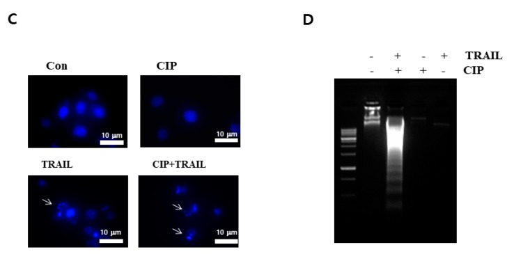
Ciprofloxacin (CIP) is a potent antimicrobial agent with multiple effects on host cells and tissues. Previous studies have highlighted their proapoptotic effect on human cancer cells. The current study showed that subtoxic doses of CIP effectively sensitized multiple cancer cells to tumor necrosis factor-related apoptosis-inducing ligand (TRAIL)-induced apoptosis. Although TRAIL alone mediated the partial proteolytic processing of procaspase-3 in lung cancer cells, co-treatment with CIP and TRAIL efficiently restored the complete activation of caspases. We found that treatment of lung cancer with CIP significantly upregulated the expression and protein stability of death receptor (DR) 5. These effects were mediated through the regulation of transcription factor CCAT enhancer-binding protein homologous protein (CHOP) since the silencing of these signaling molecules abrogated the effect of CIP. Taken together, these results indicated that the upregulation of death receptor expression and protein stability by CIP contributed to the restoration of TRAIL-sensitivity in lung cancer cells.

Keywords: CHOP; TRAIL; ciprofloxacin; death receptor.


Conflict of interest statement

The authors declare no conflicts of interest.

Figures

Figure 1
Figure 1
CIP enhanced TRAIL-induced A549 cell death. (A) Cells were pretreated with various concentrations of CIP for 20 h before exposure to TRAIL (10 and 50 ng/mL) for 4 h. Cell viability was analyzed by trypan blue exclusion assay. Data represent the mean ± SD of 3 samples. * p < 0.05 compared to the CIP + TRAIL-treated cells. (B) Cells were treated with TRAIL in the presence or absence of CIP for 24 h. After treatment, change in cell morphology was detected by light microscopy. Scale bar = 20 μm. (C) Microscopic examination was performed to detect apoptosis by nuclear staining with DAPI. The images shown are representatives of three independent experiments. Scale bar = 10 μm. (D) Cells were treated with TRAIL for 4 h in the presence or absence of CIP for 20 h. For analyzing DNA fragmentation, fragmented DNA was separated by using 1.5% agarose gel.
Figure 1
Figure 1
CIP enhanced TRAIL-induced A549 cell death. (A) Cells were pretreated with various concentrations of CIP for 20 h before exposure to TRAIL (10 and 50 ng/mL) for 4 h. Cell viability was analyzed by trypan blue exclusion assay. Data represent the mean ± SD of 3 samples. * p < 0.05 compared to the CIP + TRAIL-treated cells. (B) Cells were treated with TRAIL in the presence or absence of CIP for 24 h. After treatment, change in cell morphology was detected by light microscopy. Scale bar = 20 μm. (C) Microscopic examination was performed to detect apoptosis by nuclear staining with DAPI. The images shown are representatives of three independent experiments. Scale bar = 10 μm. (D) Cells were treated with TRAIL for 4 h in the presence or absence of CIP for 20 h. For analyzing DNA fragmentation, fragmented DNA was separated by using 1.5% agarose gel.
Figure 2
Figure 2
CIP treatment-induced caspase activation in A549 cells. (A) The protein expression of caspase-3, caspase-8, caspase-9, caspase-7, and PARP after treatment with different doses of CIP+TRAIL for 24 h. The total cells were collected and the lysates were subjected to western blotting with specific antibodies. Actin was used as a loading control. The proteolytic cleavages in PARP, cas-3, cas-8, cas-7, and cas-9 are indicated by arrows. (B) A549 cells were incubated with 50 µM z-VAD-fmk for 1 h before treatment with CIP + TRAIL. Equal amounts of cell lysates (40 µg) were electrophoresed and analyzed for PARP-1 by western blotting. The proteolytic cleavage of PARP is indicated by an arrow. (C) For analyzing DNA fragmentation, fragmented DNA was separated by using 1.5% agarose gel.
Figure 2
Figure 2
CIP treatment-induced caspase activation in A549 cells. (A) The protein expression of caspase-3, caspase-8, caspase-9, caspase-7, and PARP after treatment with different doses of CIP+TRAIL for 24 h. The total cells were collected and the lysates were subjected to western blotting with specific antibodies. Actin was used as a loading control. The proteolytic cleavages in PARP, cas-3, cas-8, cas-7, and cas-9 are indicated by arrows. (B) A549 cells were incubated with 50 µM z-VAD-fmk for 1 h before treatment with CIP + TRAIL. Equal amounts of cell lysates (40 µg) were electrophoresed and analyzed for PARP-1 by western blotting. The proteolytic cleavage of PARP is indicated by an arrow. (C) For analyzing DNA fragmentation, fragmented DNA was separated by using 1.5% agarose gel.
Figure 3
Figure 3
CIP-induced DR5 and DR4 expression. (A) A549 cells were treated with various concentrations of CIP (left) and with CIP 100 µg/mL for various time periods (right). Whole cell extracts were analyzed for DR4 and DR5 expression by western blotting. (B) CIP-induced DR4 and DR5 gene expression. A549 cells were treated with various concentrations of CIP (left) and with CIP 100 µg/mL for various time periods (right). Total RNA was extracted and examined for DR4 and DR5 expression by RT-PCR. Actin was used as an internal control to show equal RNA loading. (C) Various cancer cell lines were treated with CIP for 24 h and whole cell extracts were analyzed by western blotting. Equal amounts of protein (40 µg) were separated by SDS-PAGE and immunoblotted.
Figure 4
Figure 4
Effect of death receptors knockdown on CIP-induced sensitization to TRAIL. (A) A549 cells were transfected with DR5 siRNA, DR4 siRNA, and combined DR5 and DR4 siRNA. After 48 h, the cells were pretreated with CIP for 20 h and then treated with TRAIL for 4 h. Whole cell extracts were analyzed by western blotting using antibodies against PARP, DR4, and DR5. (B) Cell death was determined by Annexin V/PI staining. Scale bar = 20 μm. The bar represents the mean ± SD. * p < 0.01 indicates a significant difference between the untreated control and CIP + TRAIL-treated samples.
Figure 5
Figure 5
CIP exposure led to the regulation of CHOP proteins expression. (A) The effects of CIP on the expression levels of CHOP proteins. A549 cells were treated with various concentrations of CIP. Equal amounts (40 µg) of cell lysates were separated by SDS-PAGE. The images shown are representatives of two additional experiments which yielded similar results. (B) A549 cells were transfected with CHOP siRNA. After 48 h, the cells were pretreated with CIP for 24 h. Whole cell extracts were analyzed by western blotting using antibodies against CHOP, DR4, and DR5. (C) A549 cells were transfected with CHOP siRNA. After 48 h, cells were treated with CIP + TRAIL for 24 h. Cell death was determined by Annexin V/PI staining. The bar represents the mean ± SD. * p < 0.01 indicates a significant difference between the untreated group and CIP + TRAIL-treated samples.
Figure 6
Figure 6
Effect of CIP on DR4 and DR5 protein stability. (A) A549 cells were treated with or without CIP for 24 h in the presence or absence of CHX for the indicated time periods. DR5, DR4, and actin protein levels were determined by western blotting. Actin was used as a loading control. The band intensity of DR5 (B) and DR4 (C) proteins was measured using the public domain JAVA image-processing program Image J. Each value represents the mean ± SD of three independent experiments. * p < 0.05 compared to that of CHX.

Similar articles

Cited by

References

    1. Sheridan J.P., Marsters S.A., Pitti R.M., Gurney A., Skubatch M., Baldwin D., Ramakrishnan L., Gray C.L., Baker K., Wood W.I., et al. Control of TRAIL-induced apoptosis by a family of signaling and decoy receptors. Science. 1997;277:818–821. doi: 10.1126/science.277.5327.818. - DOI - PubMed
    1. Ashkenazi A., Pai R.C., Fong S., Leung S., Lawrence D.A., Marsters S.A., Blackie C., Chang L., McMurtrey A.E., Hebert A., et al. Safety and antitumor activity of recombinant soluble Apo2 ligand. J. Clin. Investig. 1999;104:155–162. doi: 10.1172/JCI6926. - DOI - PMC - PubMed
    1. Walczak H., Miller R.E., Ariail K., Gliniak B., Griffith T.S., Kubin M., Chin W., Jones J., Woodward A., Le T., et al. Tumoricidal activity of tumor necrosis factor-related apoptosis-inducing ligand in vivo. Nat. Med. 1999;5:157–163. doi: 10.1038/5517. - DOI - PubMed
    1. Nagata S. Apoptosis by death factor. Cell. 1997;88:355–365. doi: 10.1016/S0092-8674(00)81874-7. - DOI - PubMed
    1. Ashkenazi A., Dixit V.M. Death receptors: Signaling and modulation. Science. 1998;281:1305–1308. doi: 10.1126/science.281.5381.1305. - DOI - PubMed

MeSH terms