

High-resolution chromosome painting with repetitive and single-copy oligonucleotides in Arachis species identifies structural rearrangements and genome differentiation
- PMID: 30333010
- PMCID: PMC6192370
- DOI: 10.1186/s12870-018-1468-1
High-resolution chromosome painting with repetitive and single-copy oligonucleotides in Arachis species identifies structural rearrangements and genome differentiation
Abstract
Background: Arachis contains 80 species that carry many beneficial genes that can be utilized in the genetic improvement of peanut (Arachis hypogaea L. 2n = 4x = 40, genome AABB). Chromosome engineering is a powerful technique by which these genes can be transferred and utilized in cultivated peanut. However, their small chromosomes and insufficient cytological markers have made chromosome identification and studies relating to genome evolution quite difficult. The development of efficient cytological markers or probes is very necessary for both chromosome engineering and genome discrimination in cultivated peanut.
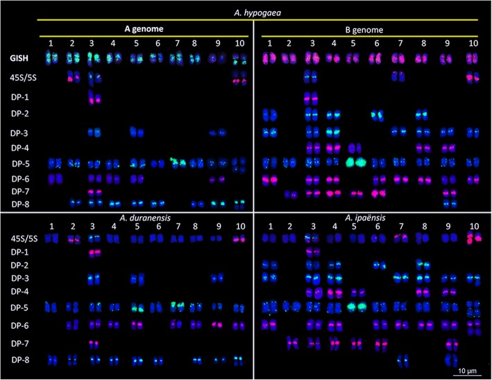
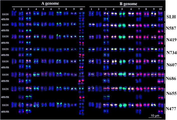
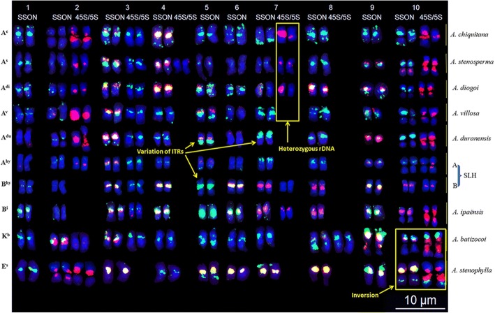
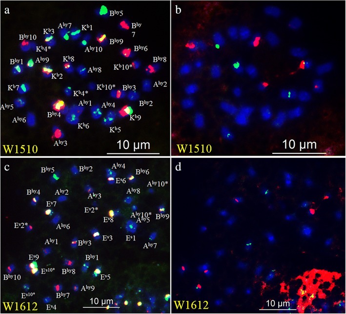
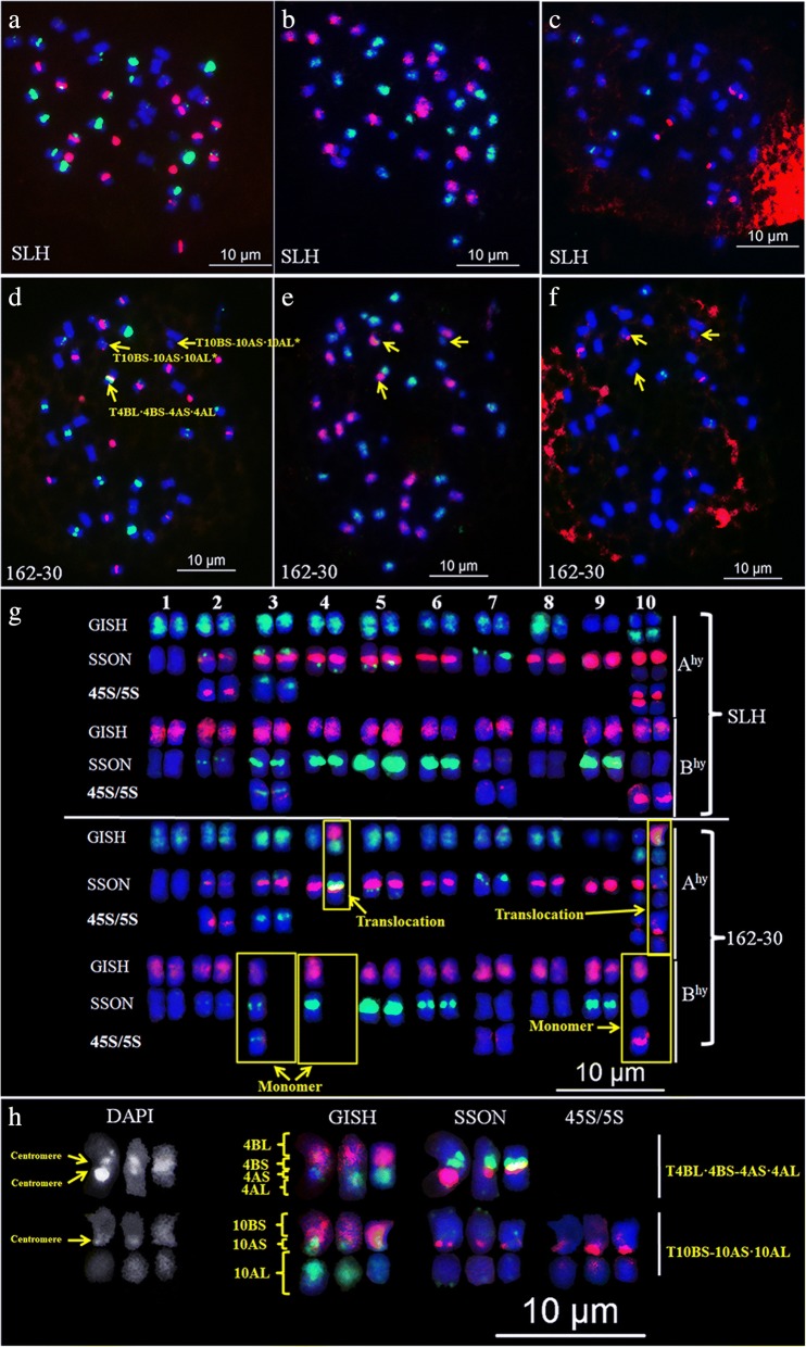
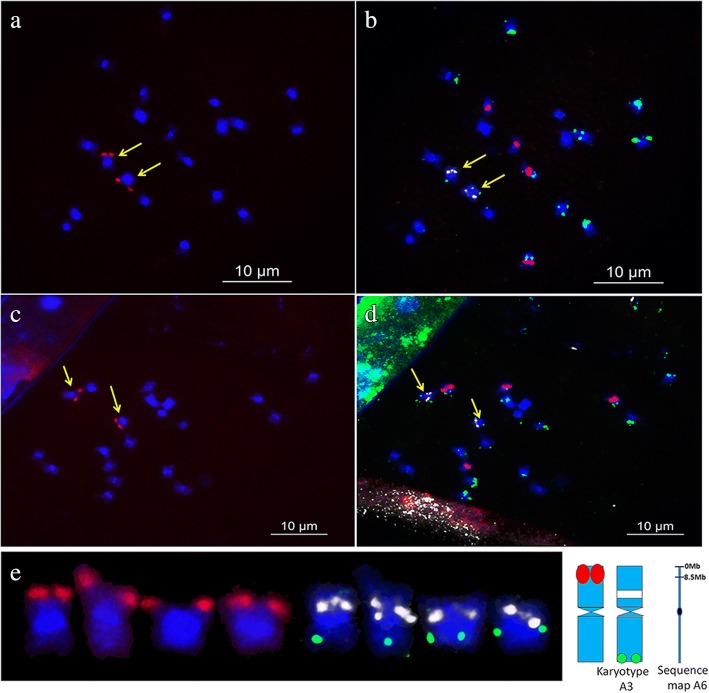
Results: A simple and efficient oligonucleotide multiplex probe to distinguish genomes, chromosomes, and chromosomal aberrations of peanut was developed based on eight single-stranded oligonucleotides (SSONs) derived from repetitive sequences. High-resolution karyotypes of 16 Arachis species, two interspecific F1 hybrids, and one radiation-induced M1 plant were then developed by fluorescence in situ hybridization (FISH) using oligonucleotide multiplex, 45S and 5S rDNAs, and genomic in situ hybridization (GISH) using total genomic DNA of A. duranensis (2n = 2x = 20, AA) and A. ipaënsis (2n = 2x = 20, BB) as probes. Genomes, chromosomes, and aberrations were clearly identifiable in the established karyotypes. All eight cultivars had similar karyotypes, whereas the eight wild species exhibited various chromosomal variations. In addition, a chromosome-specific SSON library was developed based on the single-copy sequence of chromosome 6A of A. duranensis. In combination with repetitive SSONs and rDNA FISH, the single-copy SSON library was applied to identify the corresponding A3 chromosome in the A. duranensis karyotype.
Conclusions: The development of repetitive and single-copy SSON probes for FISH and GISH provides useful tools for the differentiation of chromosomes and identification of structural chromosomal rearrangement. It facilitates the development of high-resolution karyotypes and detection of chromosomal variations in Arachis species. To our knowledge, the methodology presented in this study demonstrates for the first time the correlation between a sequenced chromosome region and a cytologically identified chromosome in peanut.
Keywords: Arachis species; Chromosome painting; Genomic evolution; High-resolution karyotype; Oligonucleotide multiplex.
Conflict of interest statement
Ethics approval and consent to participate
Not applicable.
Consent for publication
Not applicable.
Competing interests
The authors declare that they have no competing interests.
Publisher’s Note
Springer Nature remains neutral with regard to jurisdictional claims in published maps and institutional affiliations.
Figures
References
-
- Stalker HT. Utilizing wild species for peanut improvement. Crop Sci. 2017;57:1102–1120. doi: 10.2135/cropsci2016.09.0824. - DOI
-
- Food and Agriculture Organization of the United Nations. http://www.fao.org/faostat/en/#data/QC. Accessed 20 Jan 2018.
-
- Silvestri MC, Ortiz AM, Lavia GI. rDNA loci and heterochromatin positions support a distinct genome type for ‘x = 9 species’ of section Arachis (Arachis, Leguminosae) Plant Syst Evol. 2015;301:555–562. doi: 10.1007/s00606-014-1092-y. - DOI
-
- Raina SN, Mukai Y. Genomic in situ hybridization in Arachis (Fabaceae) identifies the diploid wild progenitors of cultivated (A. hypogaea) and related wild (A. monticola) peanut species. Plant Syst Evol. 1999;214:251–262. doi: 10.1007/BF00985743. - DOI
MeSH terms
Substances
Grants and funding
LinkOut - more resources
Full Text Sources
