. 2018 Oct 17;16(1):98.
doi: 10.1186/s12958-018-0416-0.

Rosiglitazone ameliorates palmitic acid-induced cytotoxicity in TM4 Sertoli cells

Affiliations

Rosiglitazone ameliorates palmitic acid-induced cytotoxicity in TM4 Sertoli cells

Xie Ge et al. Reprod Biol Endocrinol. .

Abstract

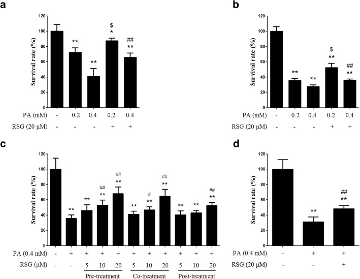
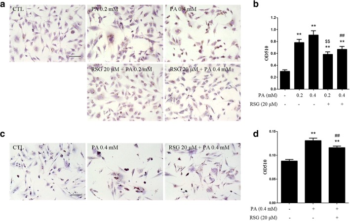
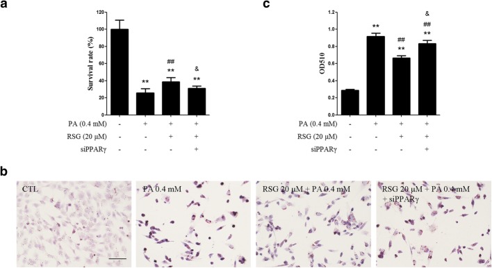
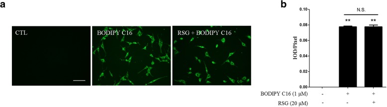
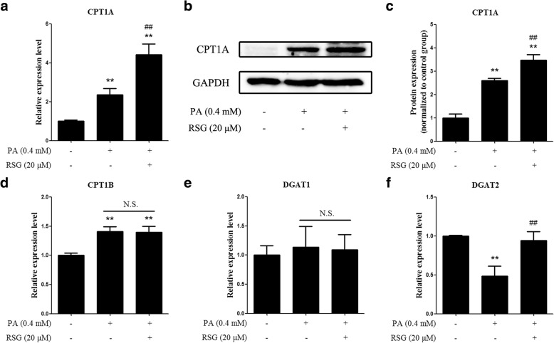
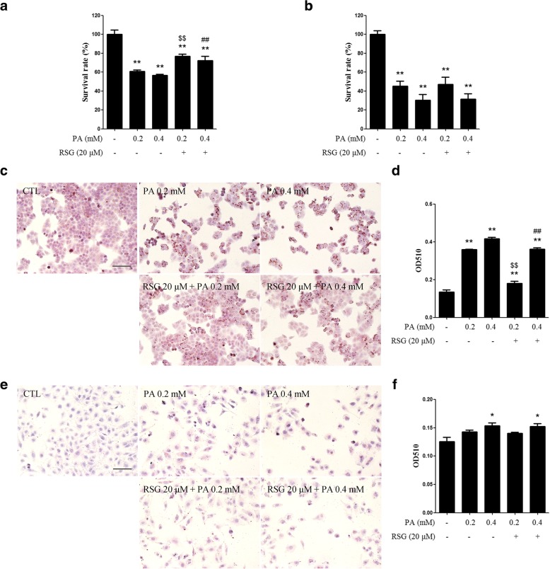
The Sertoli cell is the only somatic cell within the seminiferous tubules, and is vital for testis development and spermatogenesis. Rosiglitazone (RSG) is a member of the thiazolidinedione family and is a peroxisome proliferator-activated receptor-γ (PPARγ) agonist. It has been reported that RSG protects various types of cells from fatty acid-induced damage. However, whether RSG serves a protective role in Sertoli cells against palmitic acid (PA)-induced toxicity remains to be elucidated. Therefore, the aim of the present study was to investigate the effect of RSG on PA-induced cytotoxicity in Sertoli cells. MTT assay and Oil Red O staining revealed that RSG ameliorated the PA-induced decrease in TM4 cell viability, which was accompanied by an alleviation of PA-induced lipid accumulation in cells. In primary mouse Sertoli cells, RSG also showed similar protective effects against PA-induced lipotoxicity. Knockdown of PPARγ verified that RSG exerted its protective role in TM4 cells through a PPARγ-dependent pathway. To evaluate the mechanism underlying the protective role of RSG on PA-induced lipotoxicity, the present study analyzed the effects of RSG on PA uptake, and the expression of genes associated with both fatty acid oxidation and triglyceride synthesis. The results demonstrated that although RSG did not affect the endocytosis of PA, it significantly elevated the expression of carnitine palmitoyltransferase (CPT)-1A, a key enzyme involved in fatty acid oxidation, which indicated that the protective effect of RSG may have an important role in fatty acid oxidation. On the other hand, the expression of CPT1B was not affected by RSG. Moreover, the expression levels of diacylglycerol O-acyltransferase (DGAT)-1 and DGAT2, both of which encode enzymes catalyzing the synthesis of triglycerides, were not suppressed by RSG. The results indicated that RSG reduced PA-induced lipid accumulation by promoting fatty acid oxidation mediated by CPT1A. The effect of RSG in protecting cells from lipotoxicity was also found to be specific to Sertoli cells and hepatocytes, and not to other cell types that do not store excess lipid in large quantities, such as human umbilical vein endothelial cells. These findings provide insights into the cytoprotective effects of RSG on Sertoli cells and suggest that PPARγ activation may be a useful therapeutic method for the treatment of Sertoli cell dysfunction caused by dyslipidemia.

Keywords: Cytotoxicity; Palmitic acid; Rosiglitazone; Sertoli cells.


Conflict of interest statement

Ethics approval and consent to participate

Not applicable.

Consent for publication

Not applicable.

Competing interests

The authors declare that they have no competing interests.

Publisher’s Note

Springer Nature remains neutral with regard to jurisdictional claims in published maps and institutional affiliations.

Figures

Fig. 1
Fig. 1
RSG ameliorates the PA-induced decrease in Sertoli cell viability. (a and b) An MTT assay was performed with TM4 cells treated with PA for (a) 12 h or (b) 24 h, with or without RSG pre-treatment. c Dose- and time-dependent analysis of the effect of RSG on PA-induced cytotoxicity (MTT assay). d An MTT assay was conducted in primary mouse sertoli cells treated with PA for 24 h with or without RSG pre-treatment. Data are presented as the mean ± standard deviation of three independently prepared samples, each with three measurements. *P < 0.05 and **P < 0.01 vs. control group; $P < 0.05 vs. 0.2 mM-PA group; #P < 0.05 and ##P < 0.01 vs. 0.4 mM-PA group. RSG rosiglitazone, PA palmitic acid, MTT 3-(4,5-dimethyl-2-thiazolyl)-2,5-diphenyl-2-H-tetrazolium bromide
Fig. 2
Fig. 2
RSG alleviates PA-induced lipid accumulation in Sertoli cells. TM4 cells (a and b) and primary mouse Sertoli cells (c and d) were pre-treated with 20 μM RSG for 2 h, and then treated with 0.2 or 0.4 mM PA for 24 h. a and b ORO staining of TM4 cells (a) and quantification of neutral lipids (b). c and d ORO staining of primary mouse Sertoli cells (c) and quantification of neutral lipids (d). Data are presented as the mean ± standard deviation of three independently prepared samples, each with three measurements. Scale bar, 100 μm.**P < 0.01 vs. control group; $$P < 0.01 vs. 0.2-mM PA group; ##P < 0.01 vs. 0.4 mM-PA group. RSG rosiglitazone, PA palmitic acid, ORO oil red O
Fig. 3
Fig. 3
Knockdown of PPARγ alleviated the protective effects of RSG on PA-induced lipotoxicity in Sertoli cells. TM4 cells were transfected with NC-siRNA or siPPARγ. 6 h after transfection, cells were pretreated with (or without) RSG for 2 h, and then treated with PA for 24 h. a MTT assay of TM4 cells. b and c ORO staining of primary mouse Sertoli cells (b) and quantification of neutral lipids (c). Data are presented as the mean ± standard deviation of three independently prepared samples, each with three measurements. Scale bar, 100 μm. **P < 0.01 vs. control group; ##P < 0.01 vs. 0.4 mM-PA group; &P < 0.05 vs. 0.4 mM-PA + 20 μM-RSG group. RSG rosiglitazone, PA palmitic acid, ORO oil red O
Fig. 4
Fig. 4
RSG did not affect PA endocytosis. TM4 cells were pretreated with or without 20 μM RSG for 24 h, and then treated with 1 μM BODIPY FL C16 for 30 min. a Fluorescent images were captured with a fluorescence microscope (scale bar, 100 μm), and (b) the mean fluorescence intensities were quantified. Data are presented as the mean ± standard deviation of three independent experiments. **P < 0.01 vs. control group. N.S., not significant; RSG, rosiglitazone; PA, palmitic acid; BODIPY FL C16, 4,4-difluoro-5,7-dimethyl-4-bora-3a,4a-diaza-s-indacene-3-hexadecanoic acid
Fig. 5
Fig. 5
Effects of RSG on lipid metabolic genes. mRNA expression levels of (a) CPT1A, (d) CPT1B, (e) DGAT1 and (f) DGAT2 were assessed using the reverse transcription-quantitative polymerase chain reaction method. b Protein expression levels of CPT1A were validated using western blot analysis, and (c) quantified by densitometry. Data are presented as the mean ± standard deviation of three independent experiments. **P < 0.01 vs. control group; ##P < 0.01 vs. 0.4 mM-PA group. N.S., not significant; RSG, rosiglitazone; PA, palmitic acid; CPT1A/1B, carnitine palmitoyltransferase 1A/1B; DGAT1/2, diacylglycerol O-acyltransferase 1/2
Fig. 6
Fig. 6
Effects of RSG on PA-induced cytotoxicity in HepG2 cells and HUVECs. a and b MTT assay of (a) HepG2 cells and (b) HUVECs treated with PA for 24 h with or without RSG pre-treatment. c-f ORO staining of (c and d) HepG2 cells and (e and f) HUVECs. Cells were pre-treated with 20 μM RSG for 2 h, and then treated with 0.2 or 0.4 mM PA for 24 h. c and e Scale bar, 100 μm. d and f Quantifications of neutral lipids in cells stained with ORO were also presented. Data are presented as the mean ± standard deviation of three independently prepared samples, each with three measurements. *P < 0.05 and **P < 0.01 vs. control group; $$P < 0.01 vs. 0.2 mM-PA group; ##P < 0.01 vs. 0.4 mM-PA group. RSG, rosiglitazone; PA, palmitic acid; ORO, oil red O; HUVECs, human umbilical vein endothelial cells

References

    1. Nery SF, Vieira MA, Dela Cruz C, Lobach VN, Del Puerto HL, Torres PB, Rocha AL, Reis AB, Reis FM. Seminal plasma concentrations of anti-Mullerian hormone and inhibin B predict motile sperm recovery from cryopreserved semen in asthenozoospermic men: a prospective cohort study. Andrology. 2014;2(6):918–923. doi: 10.1111/andr.278. - DOI - PubMed
    1. Garcia TX, Farmaha JK, Kow S, Hofmann MC. RBPJ in mouse Sertoli cells is required for proper regulation of the testis stem cell niche. Development. 2014;141(23):4468–4478. doi: 10.1242/dev.113969. - DOI - PMC - PubMed
    1. McCabe MJ, Tarulli GA, Laven-Law G, Matthiesson KL, Meachem SJ, McLachlan RI, Dinger ME, Stanton PG. Gonadotropin suppression in men leads to a reduction in claudin-11 at the Sertoli cell tight junction. Hum Reprod. 2016;31(4):875–886. doi: 10.1093/humrep/dew009. - DOI - PubMed
    1. Wang X, Zhao F, Lv ZM, Shi WQ, Zhang LY, Yan M. Triptolide disrupts the actin-based Sertoli-germ cells adherens junctions by inhibiting rho GTPases expression. Toxicol Appl Pharmacol. 2016;310:32–40. doi: 10.1016/j.taap.2016.08.017. - DOI - PubMed
    1. Keber R, Rozman D, Horvat S. Sterols in spermatogenesis and sperm maturation. J Lipid Res. 2013;54(1):20–33. doi: 10.1194/jlr.R032326. - DOI - PMC - PubMed

LinkOut - more resources