. 2018 Oct 17;11(1):60.
doi: 10.1186/s13072-018-0224-y.

Oxidative stress in sperm affects the epigenetic reprogramming in early embryonic development

Affiliations

Oxidative stress in sperm affects the epigenetic reprogramming in early embryonic development

Sarah Wyck et al. Epigenetics Chromatin. .

Abstract

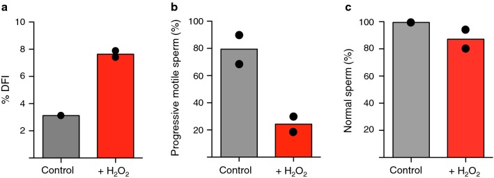
Background: Reactive oxygen species (ROS)-induced oxidative stress is well known to play a major role in male infertility. Sperm are sensitive to ROS damaging effects because as male germ cells form mature sperm they progressively lose the ability to repair DNA damage. However, how oxidative DNA lesions in sperm affect early embryonic development remains elusive.

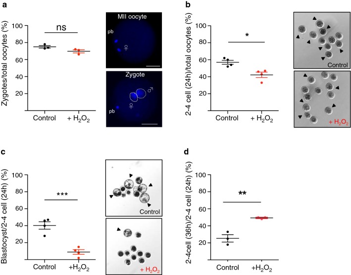
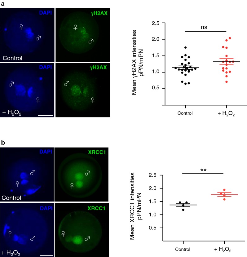
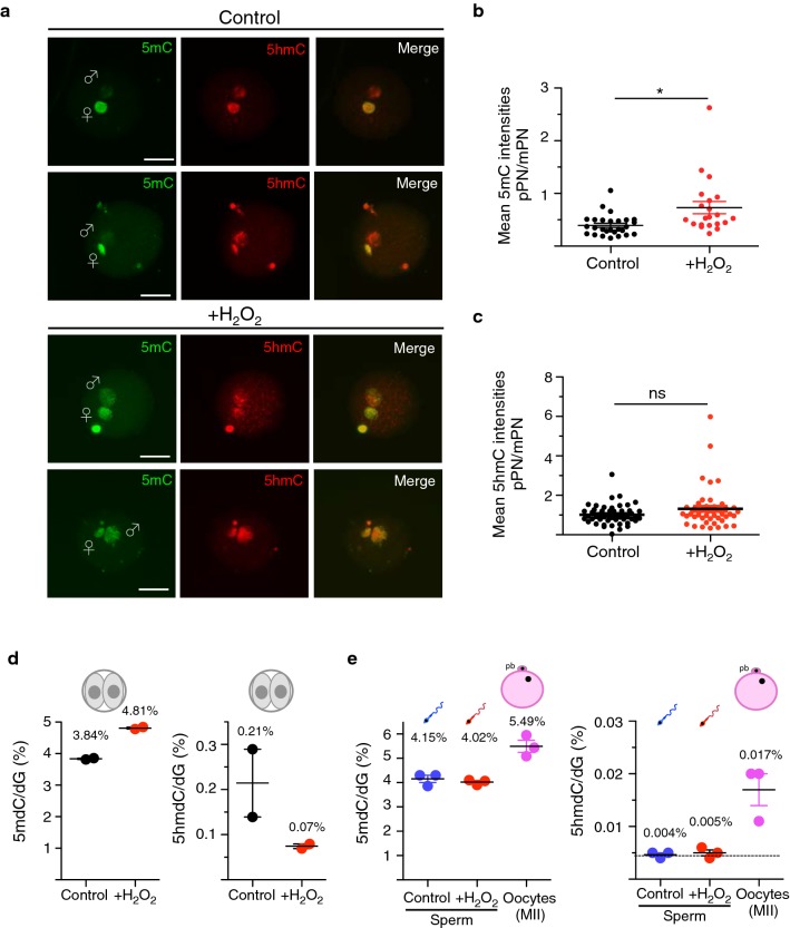
Results: Using cattle as model, we show that fertilization using sperm exposed to oxidative stress caused a major developmental arrest at the time of embryonic genome activation. The levels of DNA damage response did not directly correlate with the degree of developmental defects. The early cellular response for DNA damage, γH2AX, is already present at high levels in zygotes that progress normally in development and did not significantly increase at the paternal genome containing oxidative DNA lesions. Moreover, XRCC1, a factor implicated in the last step of base excision repair (BER) pathway, was recruited to the damaged paternal genome, indicating that the maternal BER machinery can repair these DNA lesions induced in sperm. Remarkably, the paternal genome with oxidative DNA lesions showed an impairment of zygotic active DNA demethylation, a process that previous studies linked to BER. Quantitative immunofluorescence analysis and ultrasensitive LC-MS-based measurements revealed that oxidative DNA lesions in sperm impair active DNA demethylation at paternal pronuclei, without affecting 5-hydroxymethylcytosine (5hmC), a 5-methylcytosine modification that has been implicated in paternal active DNA demethylation in mouse zygotes. Thus, other 5hmC-independent processes are implicated in active DNA demethylation in bovine embryos. The recruitment of XRCC1 to damaged paternal pronuclei indicates that oxidative DNA lesions drive BER to repair DNA at the expense of DNA demethylation. Finally, this study highlighted striking differences in DNA methylation dynamics between bovine and mouse zygotes that will facilitate the understanding of the dynamics of DNA methylation in early development.

Conclusions: The data demonstrate that oxidative stress in sperm has an impact not only on DNA integrity but also on the dynamics of epigenetic reprogramming, which may harm the paternal genetic and epigenetic contribution to the developing embryo and affect embryo development and embryo quality.

Keywords: BER; DNA methylation; Epigenetic reprogramming; Oxidative stress.


Figures

Fig. 1
Fig. 1
Oxidative stress reduces motility and increases DNA damage in sperm. a Sperm chromatin structure measurements (%DFI) of sperm treated without (control) and with H2O2 (+H2O2) were performed using the sperm chromatin structure assay (SCSA™). Mean and values of two independent experiments are shown. Progressive motility (b) and morphology (c) of sperm treated without and with H2O2 were analysed by computer-assisted sperm analysis (CASA). Mean and values of two independent experiments are shown
Fig. 2
Fig. 2
The developmental capacity of embryos is drastically reduced upon oxidative stress in sperm. a Fertilization rates of sperm treated without (control) or with H2O2 (+H2O2) were evaluated 20 h after IVF by staining the pronuclei with DAPI. Images were acquired with an inverted Leica CTR6000 microscope (software: Leica Microsystems LAS-AF6000; Leica Microsystems, Bensheim, Germany). The fertilization capacity (%) was quantified by calculating the number of obtained zygotes versus the total number of oocytes used in each IVF experiment. Each data point represents the mean of one of four independent experiments. Total amount of oocytes: 225 (control) and 209 (+H2O2). b Cleavage rate (%) was quantified by calculating the number of cleaved embryos (two-cell stage and further) obtained 24 h after IVF versus the total number of oocytes used in each IVF experiment. Each data point represents the mean of one of the four independent experiments. Total amount of oocytes: 100 (control), 123 (+H2O2). Arrows indicate the cleaved embryo stages. c Blastocyst rate (%) was quantified by calculating the number of blastocysts obtained 8 days after IVF versus the number of embryos that reached the cleavage stage 24 h after IVF. Each data point represents the mean of one of the four independent experiments. Total amount of oocytes: 148 (control) and 165 (+H2O2). Arrows indicate the blastocyst stage embryos. d Arrested embryos were evaluated 36 h after IVF. Quantifications were assessed by counting the number of embryos that were arrested at two- to four-cell stages 36 h after IVF versus the number of embryos that already reached two- to four-cell stage 24 h after IVF. Each data point represents the mean of one of three independent experiments. Total amount of oocytes: 127 (control) and 141 (+H2O2). Statistical analyses were performed using Student’s t-test (two-tailed). Error bars indicate s.d. ns: non-significant and refers to P = 0.091; *P < 0.05; **P < 0.01; ***P < 0.0001. Maternal pronucleus: mPN/♀/continuous line; paternal pronucleus: pPN/♂/dashed line. Scale bars, 50 μm
Fig. 3
Fig. 3
The BER machinery is sequestered to paternal pronuclei of zygotes derived from sperm exposed to oxidative stress. a γH2AX (green) levels were assessed by immunofluorescence of zygotes 20 h after IVF using γH2AX antibodies. Representative images of PN3/4 zygotes are shown. Quantification of γH2AX is represented as a ratio of the mean signal intensity between the two pronuclei signal (mean γH2AX intensities pPN/mPN). (n = 24 zygotes control and n = 17 H2O2-treated zygotes; experiment replicated three times independently.) Each data point represents the mean signal of the ratio (pPN/mPN) within an independent zygote. b Representative immunofluorescence images of PN3/4 zygotes 20 h after IVF stained with XRCC1 antibody (green) are shown. Quantifications of XRCC1 are shown as ratio of the mean signal intensity between the two pronuclei signal (pPN/mPN). Each data point represents the value of four independent experiments, using 120 zygotes for the control group and 85 zygotes from +H2O2 groups. Statistical analysis was performed using Student’s t-test (two-tailed). Error bars indicate s.d.; **P < 0.01; ns: non-significant and refers to P = 0.075. Maternal pronucleus: mPN/♀; paternal pronucleus: pPN/♂. Scale bars, 50 μm
Fig. 4
Fig. 4
Oxidative stress in sperm impairs active DNA demethylation on the paternal pronucleus. a Representative immunofluorescence images showing the levels of 5mC (green) and 5hmC (red) in PN3/4 zygotes using 5mC- and 5hmC-specific antibodies. Zygotes were analysed 20 h after IVF. Quantifications of 5mC (b) and 5hmC (c) signals are shown as ratio of the signal of the mean intensity of the paternal pronucleus (pPN) over the mean intensity signal of the maternal pronucleus (mPN) after background subtraction. Each data point represents an independent zygote (5mC control n = 27 zygotes and 5mC +H2O2 = 21 zygotes, three independent experiments; 5hmC control n  = 52 and 5hmC +H2O2 n = 48, three independent experiments). Statistical analysis was performed using Student’s t-test (two-tailed). Error bars indicate s.d., ns: non-significant **P < 0.01. Maternal pronucleus: mPN/♀; paternal pronucleus: pPN/♂. Scale bars, 50 μm. d Quantification of 5mC/dG by LC–MS of two-cell embryos obtained upon IVF with control sperm and sperm treated with H2O2. Data are from two independent experiments. Each data point represents the mean of two technical replicates of a pool of 50 embryos. e Quantification of 5mC and 5hmC by LC–MS of bovine sperm untreated or treated with H2O2 and MII oocytes. Data are from three independent experiments. Each data point represents the mean of two technical replicates
Fig. 5
Fig. 5
Replacement of cytosines in pre-replicative zygotes. a Schema describes the strategy to measure replication and DNA demethylation via the incorporation of EdU and EdC in zygotes and its readout. Representative images showing zygotes with EdU (green) and EdC (green) incorporation 12 h after IVF. Numbers refer to zygotes showing EdU or EdC incorporation relative to the total number of analysed zygotes, respectively. b Schema describes the strategy to measure replication and DNA demethylation via the incorporation of BrdU and EdC in zygotes and its readout. Representative images showing zygotes stained for BrdU (red) and EdC (green) incorporation 12 h after IVF. Numbers refer to zygotes positive for EdC signal and negative for BrdU signal relative to the total number of analysed zygotes. Zygotes 24 h pIVF were stained and used as post-replicative control zygotes. Scale bars, 50 μm
Fig. 6
Fig. 6
Oxidative lesions in sperm impair active DNA demethylation at the paternal genome in zygotes. The model shows the link of BER to DNA damage and active DNA demethylation. On the left, it is shown how a putative DNA glycosylase recognizes 5mC or its modified forms, giving rise to abasic sites that via the subsequent enzymes of the BER pathway (i.e. APE1 and XRCC1) allows the incorporation of unmodified cytosines. On the right, it is shown how oxidative lesions in sperm can impair DNA demethylation. Two models are here shown. In the first case, the presence of abasic sites that are established by OGG1 prior fertilization in sperm sequester XRCC1 at the expense of DNA demethylation activities, where recognition and excision of 5mC or its derivative is initiated only post-fertilization. The second model suggests that DNA lesions inhibit the activity of the DNA glycosylase(s) responsible for the DNA demethylation. In both cases, the final product is a DNA that is repaired but still contains methylated cytosines

References

    1. Adiga SK, Toyoshima M, Shiraishi K, Shimura T, Takeda J, Taga M, Nagai H, Kumar P, Niwa O. p21 provides stage specific DNA damage control to preimplantation embryos. Oncogene. 2007;26:6141–6149. doi: 10.1038/sj.onc.1210444. - DOI - PubMed
    1. Agarwal A, Allamaneni SS, Nallella KP, George AT, Mascha E. Correlation of reactive oxygen species levels with the fertilization rate after in vitro fertilization: a qualified meta-analysis. Fertil Steril. 2005;84:228–231. doi: 10.1016/j.fertnstert.2004.12.057. - DOI - PubMed
    1. Agarwal A, Durairajanayagam D, Halabi J, Peng J, Vazquez-Levin M. Proteomics, oxidative stress and male infertility. Reprod Biomed Online. 2014;29:32–58. doi: 10.1016/j.rbmo.2014.02.013. - DOI - PubMed
    1. Agarwal A, Saleh RA. Role of oxidants in male infertility: rationale, significance, and treatment. Urol Clin North Am. 2002;29:817–827. doi: 10.1016/S0094-0143(02)00081-2. - DOI - PubMed
    1. Aitken RJ. Reactive oxygen species as mediators of sperm capacitation and pathological damage. Mol Reprod Dev. 2017;84:1039–1052. doi: 10.1002/mrd.22871. - DOI - PubMed

Publication types

Substances