. 2019 Jan 4;93(2):e01574-18.
doi: 10.1128/JVI.01574-18. Print 2019 Jan 15.

Acute Infection and Subsequent Subclinical Reactivation of Herpes Simplex Virus 2 after Vaginal Inoculation of Rhesus Macaques

Affiliations

Acute Infection and Subsequent Subclinical Reactivation of Herpes Simplex Virus 2 after Vaginal Inoculation of Rhesus Macaques

Ming Lo et al. J Virol. .

Abstract

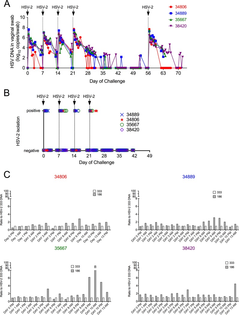
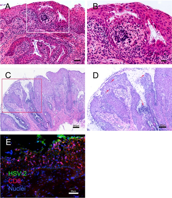
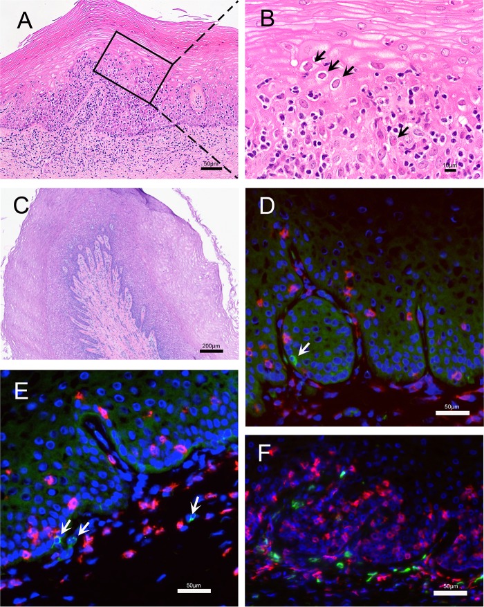
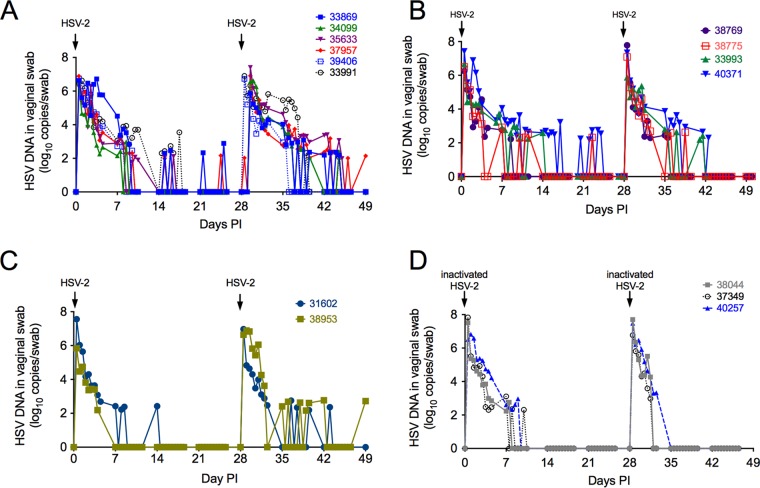
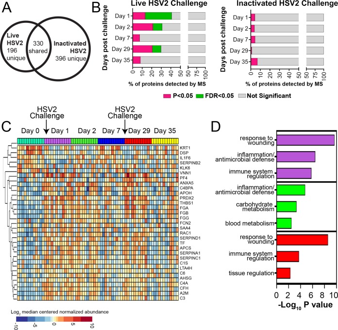
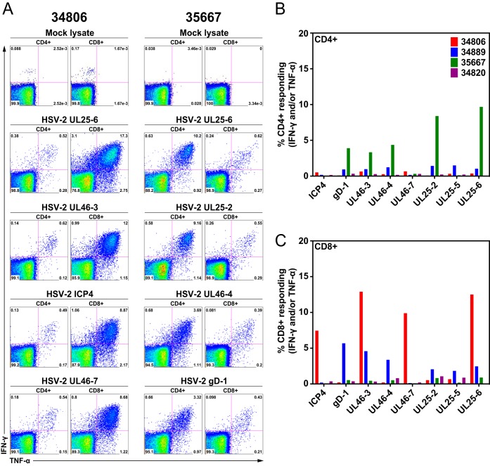
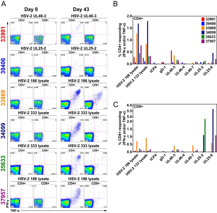
Herpes simplex virus 2 (HSV-2) is a common sexually transmitted infection with a highly variable clinical course. Many infections quickly become subclinical, with episodes of spontaneous virus reactivation. To study host-HSV-2 interactions, an animal model of subclinical HSV-2 infection is needed. In an effort to develop a relevant model, rhesus macaques (RM) were inoculated intravaginally with two or three HSV-2 strains (186, 333, and/or G) at a total dose of 1 × 10⁷ PFU of HSV-2 per animal. Infectious HSV-2 and HSV-2 DNA were consistently shed in vaginal swabs for the first 7 to 14 days after each inoculation. Proteins associated with wound healing, innate immunity, and inflammation were significantly increased in cervical secretions immediately after HSV-2 inoculation. There was histologic evidence of acute herpesvirus pathology, including acantholysis in the squamous epithelium and ballooning degeneration of and intranuclear inclusion bodies in epithelial cells, with HSV antigen in mucosal epithelial cells and keratinocytes. Further, an intense inflammatory infiltrate was found in the cervix and vulva. Evidence of latent infection and reactivation was demonstrated by the detection of spontaneous HSV-2 shedding post-acute inoculation (102 to 103 DNA copies/swab) in 80% of RM. Further, HSV-2 DNA was detected in ganglia in most necropsied animals. HSV-2-specifc T-cell responses were detected in all animals, although antibodies to HSV-2 were detected in only 30% of the animals. Thus, HSV-2 infection of RM recapitulates many of the key features of subclinical HSV-2 infection in women but seems to be more limited, as virus shedding was undetectable more than 40 days after the last virus inoculation.IMPORTANCE Herpes simplex virus 2 (HSV-2) infects nearly 500 million persons globally, with an estimated 21 million incident cases each year, making it one of the most common sexually transmitted infections (STIs). HSV-2 is associated with increased human immunodeficiency virus type 1 (HIV-1) acquisition, and this risk does not decline with the use of antiherpes drugs. As initial acquisition of both HIV and HSV-2 infections is subclinical, study of the initial molecular interactions of the two agents requires an animal model. We found that HSV-2 can infect RM after vaginal inoculation, establish latency in the nervous system, and spontaneously reactivate; these features mimic some of the key features of HSV-2 infection in women. RM may provide an animal model to develop strategies to prevent HSV-2 acquisition and reactivation.

Keywords: T-cell responses; cervix; female reproductive tract; herpes simplex virus; proteomics; subclinical infection; vaginal swabs.


Figures

FIG 1
FIG 1
HSV-2 shedding in cervicovaginal secretions of rhesus macaques after HSV-2 inoculation (group 1). Each of the four female rhesus macaques was intravaginally challenged with total 1 × 107 PFU of HSV-2 in 1 ml of inoculum containing a 1:1 mixture of HSV-2 186 and HSV-2 333 once a week for 4 weeks. After a 5-week interval, the animals were reinoculated with the same HSV-2 mixture and necropsied 1 to 2 weeks later. Twice-daily vaginal swabs were collected (except weekends) after the first HSV-2 inoculation. Animal numbers are indicated and color-coded to matched graphed data. (A) Results of PCR to quantitate HSV-2 gB DNA levels in vaginal swabs. (B) Results of attempt to isolate infectious HSV-2 from vaginal swabs by tissue culture. (C) Ratio of HSV-2 strains in the cervicovaginal secretions. PCR was used to differentiate the strains of HSV-2 detected in secretions. HSV-2 strain 333 levels were used as a baseline (white bars, ratio of 333 to 333 = 1), the ratio of HSV-2 186 levels to HSV-2 333 levels is shown by gray bars.
FIG 2
FIG 2
Histopathology and localization of HSV-2 antigen and CD8+ T cells in cervical biopsy tissue obtained 2 and 7 days after vaginal HSV-2 inoculation. Panels A and B show formalin-fixed paraffin-embedded sections of a cervical biopsy specimen collected at 2 days p.i. (A) H&E stain of ectocervix. The overlying epithelium is intact but is compromised by microvesicles and clefts filled with fibrin and cellular debris. Inflammatory cell infiltrates surround small blood vessels in the lamina propria and extend into the stratified epithelium of the ectocervix. Lymphatic vessels in the lamina propria are distended with inflammatory cells and edema fluid. (B) Higher magnification of the inset from panel A. The inflammatory cell infiltrate is focused on the epithelium and is comprised mainly of mononuclear cells, with fewer neutrophils. There is widespread acantholysis in the stratified squamous epithelium, with ballooning degeneration of epithelial cells. The microvesicles within the epithelium contain fibrin, cell debris, acantholytic epithelial cells, and inflammatory cells. Many of the epithelial cells at the edge of the microvesicles have large ground-glass nuclei with marginated chromatin. Panels C, D, and E show frozen sections of a cervical biopsy specimen collected at 7 days p.i. (C) H&E stain of the transformation zone at the border of the endocervix and ectocervix. Much of the overlying epithelium is lost or degenerating, replaced by fibrin, inflammatory cells, and cellular debris. The remaining epithelium contains acellular areas filled with fibrin, cellular debris, and edema. Inflammatory cell infiltrates surround small blood vessels in the lamina propria and extend into the columnar epithelium of the endocervix and the stratified squamous epithelium of the ectocervix. Lymphatic vessels in the lamina propria are distended with inflammatory cells and fluid. (D) Higher magnification of the inset from panel C. The inflammatory cell infiltrate is focused on the epithelium and is comprised mainly of mononuclear cells, with fewer neutrophils. There is widespread acantholysis in the stratified squamous epithelium, with ballooning degeneration of epithelial cells. The arrows indicate microvesicles within the epithelium that contain fibrin, cell debris, and inflammatory cells. (E) Immunofluorescent staining for HSV-2 antigen (green) and CD8+ T cells (red). Note that the HSV-2 antigen is expressed in the middle layer of the epithelium by acantholytic or detached epithelial cells at the edge of intraepithelial vesicles.
FIG 3
FIG 3
Histopathology and localization of HSV-2 antigen and T cells in vaginal mucosa obtained at necropsy. (A and B) Images are from a region of vaginal mucosa with obvious histopathology. (A) Within the squamous epithelium and lamina propria there were multiple focal areas with mononuclear inflammatory cell infiltrates. In the lamina propria, the inflammatory cells surrounded small blood and lymphatic vessels that were distended by edema. In the stratified squamous epithelium, the inflammatory cell infiltrates were focused in areas with acantholytic and degenerating epithelial cells. (B) Higher magnification of the inset from panel A. There is widespread acantholysis in the stratified squamous epithelium with ballooning degeneration of epithelial cells. The arrows indicate some of the numerous eosinophilic (red) intranuclear inclusion bodies and marginated chromatin within epithelial cells that are characteristic of herpesvirus infection. (C to F) Images are from a region of the vaginal mucosa with mild histopathology. (C) Normal epithelium with numerous layers characteristic of the follicular phase of the menstrual cycle. There is a mild mononuclear cell infiltrate with edema centered on the basal epithelial layer, superficial lamina propria, and around small vessels. Lymphatic vessels are distended with fluid. (D and E) Immunofluorescent staining for HSV-2 antigen (green) and CD8+ T cells (red). HSV-2 antigen is expressed in a few basal epithelial cells and mononuclear cells in the lamina propria (arrows). (F) Immunofluorescent staining for CD4+ (green) and CD8+ (red) T cells.
FIG 4
FIG 4
Spontaneous reactivation of HSV-2 in RM is detected after infectious viral challenge but not after heat-inactivated HSV-2 challenge. (A) Four of the six female monkeys in group 2 were intravaginally inoculated with a mixture of HSV-2 strains 186 and 333 (total of 1 × 107 PFU in the 1-ml inoculum) (solid lines and solid symbols); the remaining 2 animals were inoculated with a mixture of HSV-2 strains 186, 333, and G (dashed lines and open symbols). Inoculations were separated by 28 days. (B) Group 3. These 4 animals were treated with medroxyprogesterone acetate and then inoculated 2 times with HSV-2 (days 0 and 28). (C) Group 4. These 2 animals were treated with a depleting antibody that targets CD8α+ lymphocytes (T cells and NK cells) and then inoculated 2 times with HSV-2 (days 0 and 28). (D) Group 5. These three female monkeys were intravaginally inoculated with a mixture of heat-killed HSV-2 strains 186, 333, and G (1 × 107 PFU of HSV-2/ml prior to inactivation). Note that 11 of 12 RM inoculated with live HSV-2, but none of the 3 animals inoculated with heat-killed HSV-2, shed HSV-2 DNA in genital secretions at least once between days 14 to 28 and 35 to 49 postinoculation.
FIG 5
FIG 5
Mucosal wound healing and inflammatory responses were observed in CVS after live HSV-2 inoculation. (A) Using mass spectrometry, a total of 526 and 726 proteins were detected in CVS samples from live HSV-2-challenged RM (groups 2, 3, and 4; n = 12) and inactivated HSV-2-challenged macaques (group 5; n = 3), respectively. (B) Significantly differentially expressed proteins were identified on days 1 and 2 after live HSV-2 challenge relative to baseline (day 0). The proportion of proteins that were significantly different at an unadjusted P value of <0.05 (pink) and adjusted P value of <0.05 (FDR = 5% [green]) are displayed. Significant protein changes were also observed 1 day after a second live HSV-2 challenge (day 29). No significant protein alterations were observed after challenge with inactivated HSV-2 (P > 0.05). (C) Differentially expressed proteins in CVS of 12 RM (groups 2, 3, and 4) after live HSV-2 challenge displayed as a heat map. (D) The top 3 functions associated with proteins that were significantly altered in groups 2, 3, and 4 on days 1 (purple bars), 2 (green bars), and 29 (red bars) by live HSV-2 challenge were related to wound healing, innate immunity, and inflammation.
FIG 6
FIG 6
HSV-2-specific CD4+ and CD8+ T-cell responses in splenocytes of four HSV-2-infected rhesus macaques (group 1). Splenocytes of four monkeys were stimulated with various HSV-2 peptide pools. HSV-2-specific, TNF-α- and IFN-γ-secreting CD4+ and CD8+ T cells were detected by flow cytometric intracellular cytokine staining. (A) Representative data from 2 animals (35667 and 34806) stimulated with various HSV-2 peptide pools or a mock cellular lysate as indicated above each dot plot. For each animal, CD4+ T-cell responses are on the left and CD8+ T-cell responses are on the right. (B) Frequency of CD4+ T cells producing TNF-α and IFN-γ in response to stimulation with the peptide pools indicated on the x axis. (C) Frequency of CD8+ T cells producing TNF-α and IFN-γ in response to stimulation with the peptide pools indicated on the x axis.
FIG 7
FIG 7
HSV-2-specific CD4+ and CD8+ T-cell responses in PBMC from six HSV-2-infected rhesus macaques (group 2). PBMC collected prior to HSV-2 inoculation and 43 days after the first HSV-2 inoculation were stimulated with a lysate of HSV-2-infected cells or various HSV-2 peptide pools. HSV-2-specific, TNF-α- and IFN-γ-secreting CD4+ and CD8+ T cells were detected by flow cytometric intracellular cytokine staining. (A) Representative data from 6 animals stimulated with various HSV-2 peptide pools or HSV-2 lysates as indicated above each dot plot. For each time point, CD4+ T-cell responses are on the left and CD8+ T-cell responses are on the right. (B) Frequency of CD4+ T cells in day 63 PBMC producing TNF-α and IFN-γ in response to stimulation with the HSV-2 lysates and HSV-2 peptide pools indicated on the x axis. (C) Frequency of CD8+ T cells in day 63 PBMC producing TNF-α and IFN-γ in response to stimulation with the HSV-2 lysates and HSV-2 peptide pools indicated on the x axis.

References

    1. Looker KJ, Magaret AS, Turner KM, Vickerman P, Gottlieb SL, Newman LM. 2015. Global estimates of prevalent and incident herpes simplex virus type 2 infections in 2012. PLoS One 10:e114989. doi: 10.1371/journal.pone.0114989. - DOI - PMC - PubMed
    1. Gottlieb MS, Schanker HM, Fan PT. 1981. Pneumocystis pneumonia—Los Angeles. MMWR Morb Mortal Wkly Rep 30:250–252. - PubMed
    1. Gottlieb MS, Schroff R, Schanker HM, Weisman JD, Fan PT, Wolf RA, Saxon A. 1981. Pneumocystis carinii pneumonia and mucosal candidiasis in previously healthy homosexual men. N Engl J Med 305:1425–1431. doi: 10.1056/NEJM198112103052401. - DOI - PubMed
    1. Stamm WE, Handsfield HH, Rompalo AM, Ashley RL, Roberts PL, Corey L. 1988. The association between genital ulcer disease and acquisition of HIV infection in homosexual men. JAMA 260:1429–1433. doi: 10.1001/jama.1988.03410100119036. - DOI - PubMed
    1. Freeman EE, Weiss HA, Glynn JR, Cross PL, Whitworth JA, Hayes RJ. 2006. Herpes simplex virus 2 infection increases HIV acquisition in men and women: systematic review and meta-analysis of longitudinal studies. AIDS 20:73–83. doi: 10.1097/01.aids.0000198081.09337.a7. - DOI - PubMed

Publication types