. 2018 Oct 18;3(20):e121900.
doi: 10.1172/jci.insight.121900.

Stress response protein GJA1-20k promotes mitochondrial biogenesis, metabolic quiescence, and cardioprotection against ischemia/reperfusion injury

Affiliations

Stress response protein GJA1-20k promotes mitochondrial biogenesis, metabolic quiescence, and cardioprotection against ischemia/reperfusion injury

Wassim A Basheer et al. JCI Insight. .

Abstract

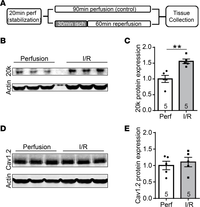
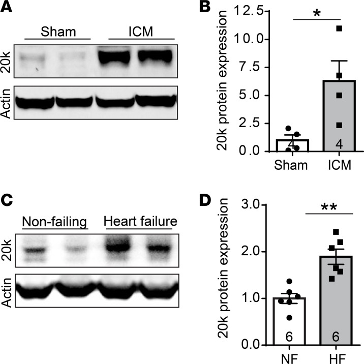
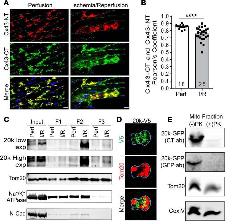
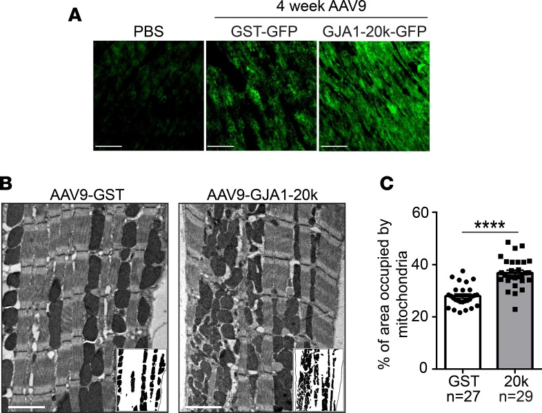
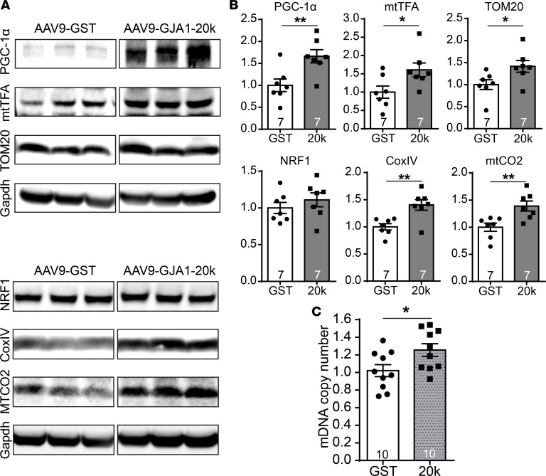
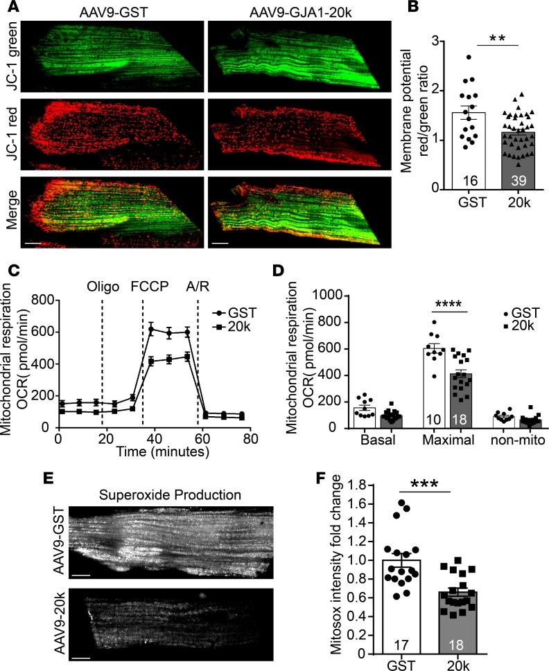
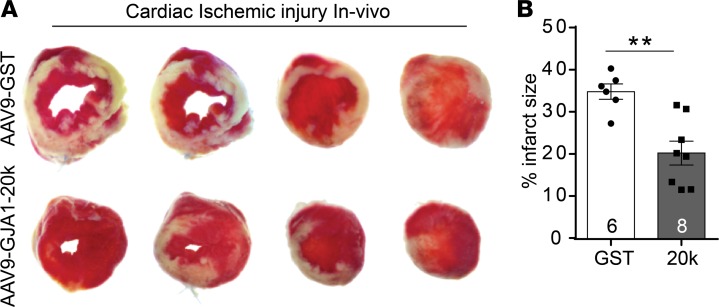
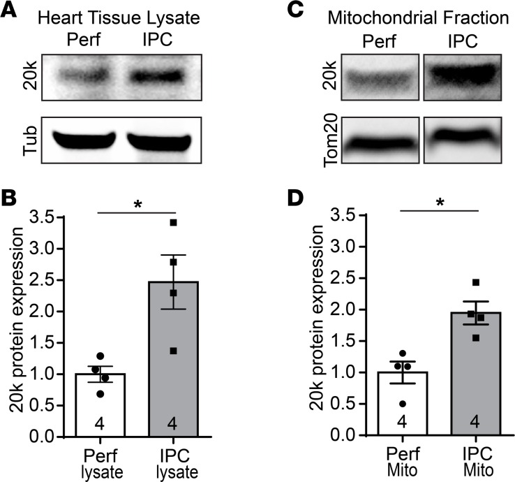
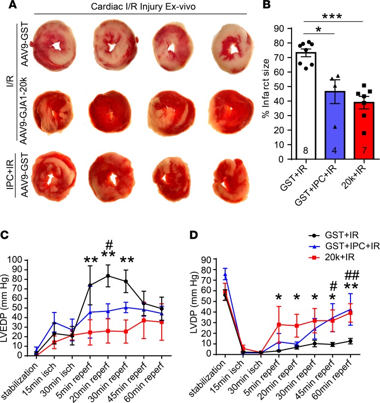
Connexin 43 (Cx43), a product of the GJA1 gene, is a gap junction protein facilitating intercellular communication between cardiomyocytes. Cx43 protects the heart from ischemic injury by mechanisms that are not well understood. GJA1 mRNA can undergo alternative translation, generating smaller isoforms in the heart, with GJA1-20k being the most abundant. Here, we report that ischemic and ischemia/reperfusion (I/R) injuries upregulate endogenous GJA1-20k protein in the heart, which targets to cardiac mitochondria and associates with the outer mitochondrial membrane. Exploring the functional consequence of increased GJA1-20k, we found that AAV9-mediated gene transfer of GJA1-20k in mouse hearts increases mitochondrial biogenesis while reducing mitochondrial membrane potential, respiration, and ROS production. By doing so, GJA1-20k promotes a protective mitochondrial phenotype, as seen with ischemic preconditioning (IPC), which also increases endogenous GJA1-20k in heart lysates and mitochondrial fractions. As a result, AAV9-GJA1-20k pretreatment reduces myocardial infarct size in mouse hearts subjected to in vivo ischemic injury or ex vivo I/R injury, similar to an IPC-induced cardioprotective effect. In conclusion, GJA1-20k is an endogenous stress response protein that induces mitochondrial biogenesis and metabolic hibernation, preconditioning the heart against I/R insults. Introduction of exogenous GJA1-20k is a putative therapeutic strategy for patients undergoing anticipated ischemic injury.

Keywords: Heart failure; Ion channels; Metabolism; Muscle Biology; Translation.


Conflict of interest statement

Conflict of interest: The authors have declared that no conflict of interest exists.

Figures

Figure 1
Figure 1. GJA1-20k is upregulated with acute cardiac I/R injury.
(A) Schematic of the protocol used for I/R injury in Langendorff-perfused mouse hearts. After a 20-minute stabilization period, mouse hearts were subjected to continuous perfusion for 90 minutes as a control, or subjected to 30 minutes of ischemia followed by 60 minutes of reperfusion for I/R injury. (B) Western blot of heart tissue lysates after either I/R injury or control perfusion. The blots are probed with an antibody for Cx43 C-terminus and an anti-actin antibody. (C) Protein expression for GJA1-20k is normalized to actin and shown as fold change after I/R injury in the quantified graphs. (D) Western blot showing Cav1.2 protein levels in the control perfused hearts and after I/R injury. (E) Cav1.2 expression is normalized to actin and shown as fold change after I/R injury in the quantified graph. The number of hearts examined in C and E is shown on the graphs, and data are presented as mean fold change ± SEM. **P < 0.01, Mann-Whitney U test.
Figure 2
Figure 2. GJA1-20k is upregulated after subacute and chronic heart failure.
(A) Western blot of protein lysates isolated from mouse hearts with subacute ischemic cardiomyopathy (ICM) induced by 3-week occlusion of the LAD coronary artery. The blots are probed with an antibody for Cx43 C-terminus and an actin antibody. (B) GJA1-20k expression is normalized to actin, and the data are shown as mean fold change ± SEM (n = 4). *P < 0.05, Mann-Whitney U test. (C) Western blot of protein lysates isolated from either nonfailing human hearts or hearts with end-stage ischemic cardiomyopathy. The blots are probed with a Cx43 C-terminus antibody and an actin antibody. (D) GJA1-20k expression is normalized to actin, and the data are shown as mean fold change ± SEM (n = 6). **P < 0.01, Mann-Whitney U test.
Figure 3
Figure 3. GJA1-20k targets mitochondria after I/R injury and associates with the OMM.
(A) Confocal images of mouse heart cryosections following I/R injury. Tissue is costained with a Cx43 N-terminus antibody (Cx43-NT, red) and a Cx43 C-terminus antibody (Cx43-CT, green), and cell membranes are labeled with WGA (white dotted line). Scale bar: 10 μm. Pearson’s coefficient for the colocalization signal (yellow, merged images) is quantified in B. Data are mean ± SEM (number of cells quantified is shown on the graph). ****P < 0.0001, unpaired t test. (C) Biochemical fractionation of isolated hearts after I/R, showing GJA1-20k protein distribution in the supernatant prior to ultracentrifugation (F1) versus mitochondrial fractions (F2, F3). Low- and high-exposure blots of GJA1-20k are shown as well as mitochondrial marker (Tom20) and plasma membrane markers (Na+/K+ATPase and N-cadherin). (D) 3D STORM images of a single cardiac mitochondrion (outlined with a blue line) expressing V5-tagged GJA1-20k (green). Tom20 is shown in red. Scale bar: 0.5 μm. (E) Western blot of mitochondrial fractions isolated from HEK cells transfected with GFP-tagged GJA1-20k and subjected to a proteinase K protection assay (1 μg/ml PK on ice for 30 minutes). PK treatment completely abolished GJA1-20k at the outer mitochondrial membrane (OMM), as assessed using GFP and Cx43-CT antibodies. Tom20 is used as an OMM marker and CoxIV is used as an inner mitochondrial membrane marker.
Figure 4
Figure 4. GJA1-20k increases mitochondrial content.
(A) GST-GFP control or GJA1-20k-GFP are introduced into the heart by the recombinant AAV9 gene delivery system at 3 × 10¹⁰ vector genomes per mouse. Immunofluorescence confocal imaging of heart tissue confirms the expression of the GFP-tagged proteins at 4 weeks after AAV9 expression as compared with a PBS control heart with no AAV9. Scale bar: 50 μm. (B) Electron microscope images of left ventricular tissue isolated from AAV9-GJA1-20k– or AAV9-GST–expressing mouse hearts. Insets show mitochondria, with cardiomyocyte area outlined using a black line. Scale bar: 2 μm. (C) Quantification of the percentage area of the cardiomyocyte occupied by mitochondria in each group (number of images assessed is shown on the graph). Data in C are mean ± SEM. **P < 0.01, ****P < 0.0001, unpaired t test.
Figure 5
Figure 5. GJA1-20k promotes mitochondrial biogenesis.
(A) Western blots showing the expression level of critical transcription factors involved in mitochondrial biogenesis (PGC1-1α, mtTFA, and NRF1) and mitochondrial proteins (TOM20, CoxIV, and mtCO2) in heart lysates from AAV9-GJA1-20k or AAV9-GST mice. Quantification of protein expression is shown in B as fold change after normalization to Gapdh (n = 7 hearts for each treatment). (C) Real-Time PCR data showing mitochondrial DNA copy number in heart tissue isolated from AAV9-GJA1-20k and AAV9-GST mice (n = 10). Data in B and C are mean ± SEM. *P < 0.05, **P < 0.01, unpaired t test.
Figure 6
Figure 6. GJA1-20k does not affect the amount of active Drp1 fission protein or Mitofusin1 fusion protein.
(A) Western blot showing amount of active Drp1 fission protein, Mitofusin1 fusion protein, and Tom20 in the F3 mitochondrial fraction isolated from AAV9-GST or AAV9-GJA1-20k hearts. Quantification of protein band intensity is shown in B as fold change (n = 5 for GST and n = 6 for 20k hearts). Data in B are mean ± SEM. **P < 0.01, Kruskal-Wallis test.
Figure 7
Figure 7. GJA1-20k promotes mitochondrial quiescence.
(A) Cardiomyocytes isolated from AAV9-GJA1-20k– or AAV9-GST–expressing hearts were loaded with JC-1 MitoTracker dye, which selectively enters mitochondria and reversibly changes color from JC-1 red to JC-1 green as the membrane potential decreases. (B) Ratiometric measurement (red/green) using JC-1 in the two groups (number of cells quantified is shown on the graph). Data are mean ± SEM. **P < 0.01, unpaired t test. (C) Seahorse XF Cell Mito Stress assay is used for characterization of oxygen consumption rate (OCR) in cardiomyocytes isolated from AAV9-GJA1-20k or AAV9-GST hearts. OCR under basal conditions and in response to the indicated mitochondrial inhibitors is shown overtime. (D) Quantification of basal respiration, maximal respiratory, and nonmitochondrial respiration between the two groups. Data are mean ± SEM (number of wells assessed per group is shown on the graph). ****P < 0.0001, 2-way ANOVA. (E) Cardiomyocytes isolated from AAV9-GJA1-20k– or AAV9-GST–expressing hearts were loaded with the fluorogenic dye MitoSOX Red to examine mitochondrial superoxide production by assessing fluorescence intensity, which is quantified in F as fold change (n = 17 cells for GST and n = 18 cells for 20k hearts). Data in F are mean ± SEM. ***P < 0.001, unpaired t test. Scale bar: 10 μm.
Figure 8
Figure 8. GJA1-20k protects the heart from ischemic injury in vivo.
(A) Representative images of transverse slices of TTC-stained hearts from AAV9-GJA1-20k– and AAV9-GST–expressing mice at 72 hours after in vivo myocardial infarction induced by LAD coronary artery occlusion. TTC stains viable heart muscle deep red while areas of infarction are pale. (B) Infarct size is quantified as a percentage of total slice area, corrected by slice weight (GST, n = 6 hearts; 20k, n = 8 hearts). Data are shown as mean ± SEM. **P < 0.01, Mann-Whitney U test.
Figure 9
Figure 9. GJA1-20k increases with ischemic preconditioning.
Western blots of total tissue lysates (A) or F3 mitochondrial fractions (C) isolated from Langendorff-perfused mouse hearts, which were subjected to either ischemic preconditioning (IPC) or continuous perfusion (Perf) as a control. The blots are probed with a Cx43 C-terminus antibody to detect endogenous GJA1-20k, with a tubulin antibody, or with a Tom20 antibody. (B) Protein expression for GJA1-20k in A is normalized to tubulin and shown as fold change. n = 4 hearts per group. Data are shown as mean ± SEM. *P < 0.05, Mann-Whitney U test. (D) Protein expression for GJA1-20k in C is normalized to Tom20 and shown as fold change. n = 4 hearts per group. Data are shown as mean ± SEM. *P < 0.05, Mann-Whitney U test.
Figure 10
Figure 10. GJA1-20k mimics the IPC protective phenotype seen in heart ex vivo I/R injury.
(A) Representative transverse slices of TTC-stained cardiac sections from Langendorff-perfused hearts isolated from AAV9-GST– or AAV9-GJA1-20k–expressing mice and subjected to ex vivo I/R injury alone (30-minute ischemia and 60-minute reperfusion) or to IPC stimulus prior to the prolonged I/R injury. (B) Infarct size is quantified as a percentage of total slice area, corrected by slice weight (number of hearts quantified per group in shown on the graph). Data are shown as mean ± SEM. *P < 0.05, ***P < 0.001, Kruskal-Wallis test. (C) Left ventricular end diastolic pressure (LVEDP) and (D) left ventricular developed pressure (LVDP) measurements during I/R in Langendorff-perfused hearts isolated from AAV9-GST– or AAV9-GJA1-20k–expressing mice. The hearts were subjected to ex vivo I/R injury alone or to 4 cycles of IPC followed by I/R. GST+IR compared with 20k+IR is indicated by either * or **, and GST+IR compared with GST+IPC+IR is indicated by either # or ##. *P < 0.05, #P < 0.05, **P < 0.01, ##P < 0.01, 2-way ANOVA. Data are mean ± SEM (GST+I/R, n = 6 hearts; GST+IPC+IR, n = 4 hearts; 20k+IR, n = 5 hearts).

Similar articles

Cited by

References

    1. Wong ND. Epidemiological studies of CHD and the evolution of preventive cardiology. Nat Rev Cardiol. 2014;11(5):276–289. doi: 10.1038/nrcardio.2014.26. - DOI - PubMed
    1. Bolli R, Patel BS, Jeroudi MO, Lai EK, McCay PB. Demonstration of free radical generation in “stunned” myocardium of intact dogs with the use of the spin trap alpha-phenyl N-tert-butyl nitrone. J Clin Invest. 1988;82(2):476–485. doi: 10.1172/JCI113621. - DOI - PMC - PubMed
    1. Walters AM, Porter GA, Brookes PS. Mitochondria as a drug target in ischemic heart disease and cardiomyopathy. Circ Res. 2012;111(9):1222–1236. doi: 10.1161/CIRCRESAHA.112.265660. - DOI - PMC - PubMed
    1. Griffiths EJ, et al. Mitochondrial calcium transporting pathways during hypoxia and reoxygenation in single rat cardiomyocytes. Cardiovasc Res. 1998;39(2):423–433. doi: 10.1016/S0008-6363(98)00104-7. - DOI - PubMed
    1. Halestrap AP, Clarke SJ, Javadov SA. Mitochondrial permeability transition pore opening during myocardial reperfusion--a target for cardioprotection. Cardiovasc Res. 2004;61(3):372–385. doi: 10.1016/S0008-6363(03)00533-9. - DOI - PubMed

Publication types

MeSH terms