. 2018 Oct 18;3(20):e122857.
doi: 10.1172/jci.insight.122857.

Combining STING-based neoantigen-targeted vaccine with checkpoint modulators enhances antitumor immunity in murine pancreatic cancer

Affiliations

Combining STING-based neoantigen-targeted vaccine with checkpoint modulators enhances antitumor immunity in murine pancreatic cancer

Heather L Kinkead et al. JCI Insight. .

Abstract

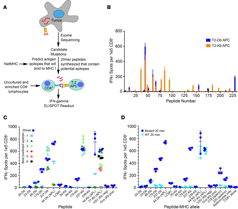
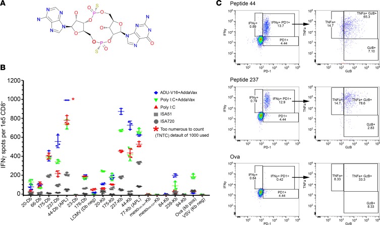
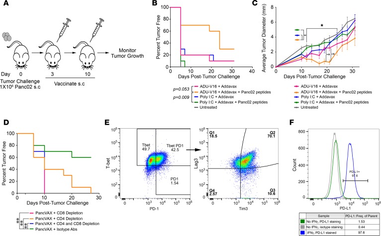
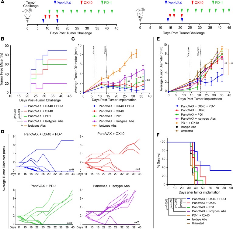
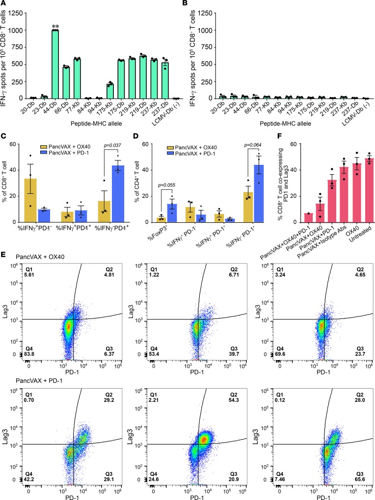
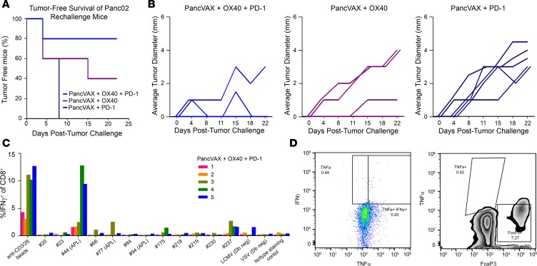
Tumor neoantigens arising from somatic mutations in the cancer genome are less likely to be subject to central immune tolerance and are therefore attractive targets for vaccine immunotherapy. We utilized whole-exome sequencing, RNA sequencing (RNASeq), and an in silico immunogenicity prediction algorithm, NetMHC, to generate a neoantigen-targeted vaccine, PancVAX, which was administered together with the STING adjuvant ADU-V16 to mice bearing pancreatic adenocarcinoma (Panc02) cells. PancVAX activated a neoepitope-specific T cell repertoire within the tumor and caused transient tumor regression. When given in combination with two checkpoint modulators, namely anti-PD-1 and agonist OX40 antibodies, PancVAX resulted in enhanced and more durable tumor regression and a survival benefit. The addition of OX40 to vaccine reduced the coexpression of T cell exhaustion markers, Lag3 and PD-1, and resulted in rejection of tumors upon contralateral rechallenge, suggesting the induction of T cell memory. Together, these data provide the framework for testing personalized neoantigen-based combinatorial vaccine strategies in patients with pancreatic and other nonimmunogenic cancers.

Keywords: Cancer immunotherapy; Oncology.


Conflict of interest statement

Conflict of interest: Under a licensing agreement between Aduro Biotech Inc. and Johns Hopkins University and EMJ, Johns Hopkins University is entitled to milestone payments and royalties on sales of certain cancer vaccine products.

Figures

Figure 1
Figure 1. Neoantigen prediction pipeline and estimation of immunogenicity.
(A) Tumor neoepitope identification pipeline from whole-exome sequencing (WES) to in vivo verification. (B) C57BL6J mice were immunized with 20-mer peptides corresponding to mutant Panc02 peptides identified in Supplemental Table 4. Isolated CD8+ T cells were stimulated with T2-Db or Kb APCs pulsed with cognate peptides on an IFN-γ capture plate and resulting spots were counted (ELISPOT assay, see Methods). (C) Mice were immunized with pooled 20-mer peptides, and isolated CD8+ cells were tested for reactivity to minimal epitopes that were predicted from NetMHC algorithm (Supplemental Table 3). Altered peptide ligands (APLs) are indicated in the graph. The peptide number is followed by the MHC-restricted allele (Db or Kb) on the x axis. Symbols represent a single mouse together with mean ± SEM (n = 3 mice per group). Negative controls were VSV and LCMV peptides (see Methods). (D) Mice were immunized with pooled 20-mer peptides corresponding to the mutant neoepitopes and APLs, and isolated CD8+ T cells were analyzed for cross-reactivity to the wild-type 20-mer.
Figure 2
Figure 2. Optimizing adjuvant choice in combination with peptide vaccine.
(A) Chemical structure of the STING-based agonist adjuvant ADU-V16 (Aduro Biotech). (B) Naive C57BL6J mice were immunized with pooled 20-mer peptides corresponding to the mutant neoepitopes and altered peptide ligands (APLs), together with adjuvants, namely ADU-V16, R287, poly I:C, ISA51, ISA720, or AddaVax. Isolated CD8+ T cells were analyzed for IFN-γ production (ELISPOT, see Methods). Wells that were saturated were marked as too numerous to count (TNTC), with a default value of 1,000 spots. APLs are indicated in the graph after the peptide identifier, followed by the MHC-restricted allele (Db or Kb). Symbols represent a single mouse; mean ± SEM (n = 3 mice per group). (C) Naive C57BL6J mice were immunized with Panc02 peptides (50 μg of each if the twelve 20-mer peptides), ADU-V16, and AddaVax. Overnight stimulation with 2 Panc02 peptides, 44 and 237, presented by T2 APCs resulted in a robust, polyfunctional CD8+ T cell response (flow cytometry, representative data from single mouse). Live, CD3+CD8+ T cells were gated and graphed for IFN-γ and PD-1 expression. The granzyme B and TNF-α graph shows cells gated on both IFN-γ and PD-1, as denoted by the arrows. Ovalbumin stimulation was used a negative control.
Figure 3
Figure 3. Tumor-specific peptides in combination with ADU-V16 and AddaVax result in tumor regression and a survival benefit.
(A) Schematic representation of the vaccination schedule. Mice were challenged with 106 Panc02 tumor cells in the right hind leg on day 0, followed by vaccination, at days 3 and 10, with Panc02 peptides together with AddaVax (50 μg) and either Poly I:C (10 μg) or ADU-V16 (5 μg). Mice were euthanized when the tumors reached 10 × 10 mm or when tumors began to impair mobility or ulcerate. (B) Kaplan-Meier curves showing the percentage of mice that were tumor free. Statistics by log-rank Mantel-Cox test. (C) Tumor growth was measured every 3 to 4 days with calipers until tumors reached 10 × 10 mm. *P < 0.001, Student’s t test, corrected for multiple comparisons. n = 10 mice in each treated group, 5 mice in untreated group for both B and C. (D) Mice were similarly vaccinated as in A and were either depleted of CD4+ or CD8+ T cells or both with the respective antibodies (see Methods). Kaplan-Meier curves showing the percentage of mice that were tumor free. n = 10 mice per group at outset. Statistics by log-rank Mantel-Cox test. (E) Flow cytometry for cell surface exhaustion markers of tumor-infiltrating lymphocytes from mice treated with PancVAX, ADU-V16, and AddaVax. Live, CD3+CD8+Tbet+PD1+ T cells were gated and graphed for Tim3 and Lag3 expression (representative data shown). (F) Panc02 cells were cultured in the presence or absence of 10 ng/ml mouse IFN-γ for 72 hours. Cells were stained with anti-mouse PD-L1 or control isotype antibody and analyzed by flow cytometry (shown as representative traces).
Figure 4
Figure 4. Checkpoint modulation with PancVAX results in tumor clearance.
(A) Treatment timeline for Panc02-bearing C57BL6 mice. On the left, 1 week following Panc02 implantation, mice received PancVAX (50 μg each of the 12 20-mer peptides), ADU-V16, AddaVax, agonist OX40 antibody (150 μg), and anti–PD-1 antibody (100 μg), as shown (results in BD). On the right, PancVAX, ADU-V16, AddaVax, low-dose agonist OX40 antibody (50 μg), and anti–PD-1 antibody (100 μg) were given 2 weeks after transplantation, as shown (results in E and F). (B) Kaplan-Meier curves displaying the percentage of tumor-free mice following various treatments (as shown). Statistics by log-rank Mantel-Cox test. (C) Average tumor diameter (mm) was measured in the same group of mice (as in B) by calipers every 3 to 4 days, starting day 11, until tumors reached 10 × 10 mm. Statistical comparisons were made with PancVAX plus isotype Abs. *P < 0.05 for triple therapy from day 25 onward, **P < 0.002 at day 35, triple therapy versus PancVax + Isotype Abs; other groups, namely, PancVAX plus OX40, PancVAX plus PD-1, were nonsignificant, except as noted. Statistics by Student’s t test, corrected for multiple comparisons. (D) Average tumor diameter for each mouse for the treatments, as noted. The number indicates the number of mice that were tumor free at day 43. (E) Average tumor diameter (mm) was measured by calipers every 3 to 4 days until tumors reached 10 × 10 mm; Statistics by Student’s t test adjusted for multiple comparisons. *P < 0.05, comparing PancVAX, OX40, and PD-1 versus OX40 and PD-1 or PancVAX and isotype Abs from day 24 onward (except day 28, P = 0.082 for the latter comparison). In all cases 10 mice per group were used at the outset. Of note, mice in whom tumor diameter could not be reliably measured due to poor demarcation of the tumor edge were excluded. (F) Kaplan-Meier curves displaying the percentage of tumor-free mice following various treatments. Statistics by log-rank Mantel-Cox test.
Figure 5
Figure 5. Tumor-infiltrating T cells in mice treated with PancVAX and checkpoint modulators.
ELISPOT assays showing IFN-γ–producing CD8+ T cells from tumors isolated from mice receiving triple therapy (PancVAX + anti–PD-1 + OX40). ** indicates too many spots to count (A) or isotype control is shown in (B). Isolated CD8+ T cells were incubated with T2-Db or T2-Kb antigen-presenting cells (APCs) that were pulsed with individual PancVAX peptides (as shown). Tumors from 10 mice per group were pooled; technical replicates are shown. (C) Flow cytometry showing the percentage of CD8+ T cells expressing IFN-γ, IFN-γ with PD-1, or PD-1 alone. (D) Flow cytometry showing the percentage of CD4+ T cells expressing FoxP3, IFN-γ, IFN-γ and PD-1, or PD-1 alone. Each bar represents cells isolated from a single tumor for C and D. Statistics by unpaired Student’s t test. (E) Tumor-infiltrating T cells were harvested and stained for the surface expression of the exhaustion markers Lag3 and PD-1 (flow cytometry) (representative traces from single tumor). (F) Flow cytometry showing the percentage of CD8+ T cells coexpressing PD-1 and Lag3 following treatment of mice with PancVAX, ADU-V16, AddaVax, low-dose OX40 (50 μg), and/or anti–PD-1 (100 μg). Relevant isotype antibodies were used as controls. For E and F, cells were gated by size for T cells and then gated for live CD8+ T cells. Statistics by Student’s t test, corrected for Bonferroni. P < 0.05; n = 3 mice per group (except the triple treatment as the other 2 tumors were cleared); individual mice and mean ± SEM are shown.
Figure 6
Figure 6. Tumor rechallenge with Panc02 results in sustained protection.
(A) 106 Panc02 cells were implanted in the contralateral hind legs of mice that had cleared tumor following initial treatments with PancVAX, ADU-V16, AddaVax, OX40, and/or anti–PD-1 (as in Figure 4, B–D) but without additional treatment (n = 5 per group). (B) Tumor diameter of individual mice from the groups in A as a function of time. (C) Splenocytes were harvested from each mouse in the triple treatment group (PancVAX, OX40, and anti–PD-1), plated overnight at 2 × 10⁶ cells in 1 ml CTL medium with each PancVAX peptide (2 μg/ml) or anti-CD3/28 magnetic beads in the presence of protein transport inhibitors. The following day, cells were washed and stained for intracellular cytokine staining to determine peptide specificities. Each number represents a single mouse; mouse 1 did not eradicate the rechallenge tumor. (D) Tumor-infiltrating lymphocytes were harvested from mouse 1, which did not respond to tumor rechallenge following triple therapy and showed the absence of IFN-γ and TNF-α production (left) as well as a marked number of immunosuppressive FoxP3+CD4+ Tregs (right).

Similar articles

Cited by

References

    1. Le DT, et al. PD-1 Blockade in tumors with mismatch-repair deficiency. N Engl J Med. 2015;372(26):2509–2520. doi: 10.1056/NEJMoa1500596. - DOI - PMC - PubMed
    1. Schumacher TN, Schreiber RD. Neoantigens in cancer immunotherapy. Science. 2015;348(6230):69–74. doi: 10.1126/science.aaa4971. - DOI - PubMed
    1. Yarchoan M, Johnson BA, Lutz ER, Laheru DA, Jaffee EM. Targeting neoantigens to augment antitumour immunity. Nat Rev Cancer. 2017;17(4):209–222. doi: 10.1038/nrc.2016.154. - DOI - PMC - PubMed
    1. Carreno BM, et al. Cancer immunotherapy. A dendritic cell vaccine increases the breadth and diversity of melanoma neoantigen-specific T cells. Science. 2015;348(6236):803–808. doi: 10.1126/science.aaa3828. - DOI - PMC - PubMed
    1. Ott PA, et al. An immunogenic personal neoantigen vaccine for patients with melanoma. Nature. 2017;547(7662):217–221. doi: 10.1038/nature22991. - DOI - PMC - PubMed

Publication types

MeSH terms