Rectal Lymphoid Follicle Aphthous Lesions Frequently Progress to Ulcerative Colitis with Proximal Extension
- PMID: 30333412
- PMCID: PMC6443555
- DOI: 10.2169/internalmedicine.1635-18
Rectal Lymphoid Follicle Aphthous Lesions Frequently Progress to Ulcerative Colitis with Proximal Extension
Abstract
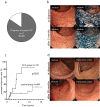
Objective Rectal lymphoid follicular aphthous (LFA) lesions are related to ulcerative colitis (UC) and can be initial lesions of UC. We investigated the clinical course and prognosis of rectal LFA lesions. Methods This is a retrospective analysis of the clinical records at a single center. Patients Thirteen consecutive cases with LFA lesions treated at Hiroshima University Hospital between 1998 and 2015 were evaluated. Another 49 consecutive cases with ulcerative proctitis treated in the same period were enrolled as the control group. The clinical course and prognosis of both groups were evaluated. Results The group with LFA lesions included 9 women and 4 men with a median age of 39.9 years (range, 21-70 years). A total of 11 cases progressed to typical UC at 5-51 months. Proximal extension of these typical UC lesions was observed in 7 (53.8%) cases, which was significantly higher than in the control group (10 cases, 20.8%). Three cases (5-year accumulation incidence rate, 27.3%) progressed to steroid-intractable UC, a significantly higher incidence than that of the control group (3 cases; 5-year accumulation incidence rate, 6.9%). Conclusion Rectal LFA lesions frequently progress to typical UC with proximal extension, some of which become intractable to corticosteroid treatment.
Keywords: aphthous lesions; proctitis; proximal extension; ulcerative colitis.
Conflict of interest statement
Figures


References
-
- Danese S, Fiocchi C. Ulcerative colitis. N Engl J Med 365: 1713-1725, 2011. - PubMed
-
- Okawa K, Aoki T, Sano K, Harihara S, Kitano A, Kuroki T. Ulcerative colitis with skip lesion at the mouth of the appendix. Am J Gatroenterol 93: 2405-2410, 1998. - PubMed
-
- Matsumoto T, Kubokura N, Yada S, et al. . Aphthous lesions of the colon as a precursor of ulcerative colitis. I to Cho (Stomach and Intestine) 44: 1514-1521, 2009(in Japanese, Abstract in English).
-
- Yokoyama J, Watanabe K, Ajioka Y, et al. . Aphthous lesions of the colorectum-clinical significance of the initial lesion of ulcerative colitis. I to Cho (Stomach and Intestine) 44: 1523-1533, 2009(in Japanese, Abstract in English).

