

3D-cultured human placenta-derived mesenchymal stem cell spheroids enhance ovary function by inducing folliculogenesis
- PMID: 30333505
- PMCID: PMC6193033
- DOI: 10.1038/s41598-018-33575-9
3D-cultured human placenta-derived mesenchymal stem cell spheroids enhance ovary function by inducing folliculogenesis
Abstract
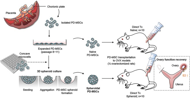
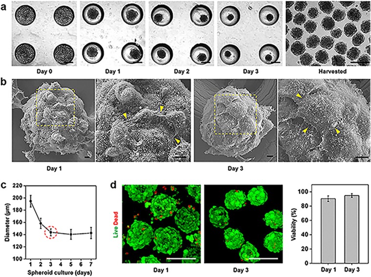
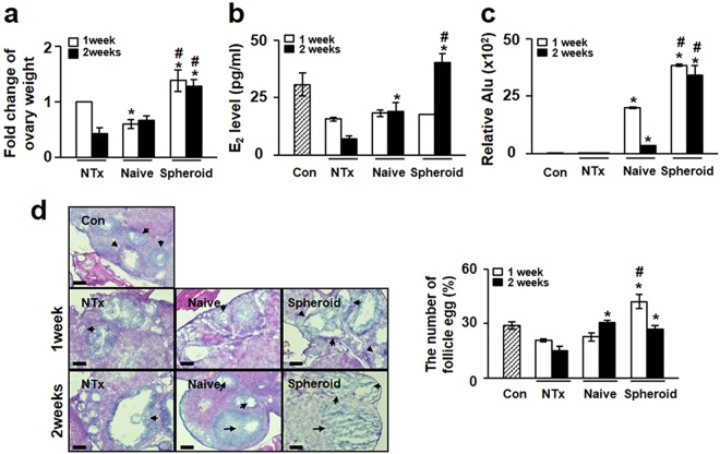
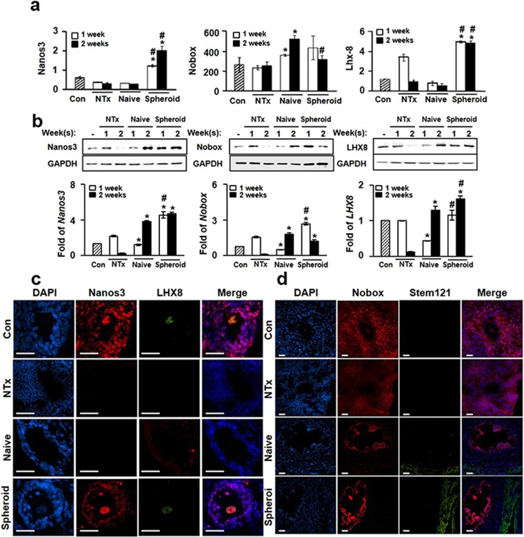
Placenta-derived mesenchymal stem cells (PD-MSCs) have numerous advantages over other adult MSCs that make them an attractive cell source for regenerative medicine. Here, we demonstrate the therapeutic effect of PD-MSCs in ovariectomized (Ovx) rats and compare their efficacy when generated via a conventional monolayer culture system (2D, naïve) and a spheroid culture system (3D, spheroid). PD-MSC transplantation significantly increased the estradiol level in Ovx rats compared with the non-transplantation (NTx) group. In particular, the estradiol level in the Spheroid group was significantly higher than that in the Naïve group at 2 weeks. Spheroid PD-MSCs exhibited a significantly higher efficiency of engraftment onto ovarian tissues at 2 weeks. The mRNA and protein expression levels of Nanos3, Nobox, and Lhx8 were also significantly increased in the Spheroid group compared with those in the NTx group at 1 and 2 weeks. These results suggest that PD-MSC transplantation can restore ovarian function in Ovx rats by increasing estrogen production and enhancing folliculogenesis-related gene expression levels and further indicate that spheroid-cultured PD-MSCs have enhanced therapeutic potential via increased engraftment efficiency. These findings improve our understanding of stem-cell-based therapies for reproductive systems and may suggest new avenues for developing efficient therapies using 3D cultivation systems.
Conflict of interest statement
The authors declare no competing interests.
Figures
Similar articles
-
Vascular remodeling by placenta-derived mesenchymal stem cells restores ovarian function in ovariectomized rat model via the VEGF pathway.Lab Invest. 2021 Mar;101(3):304-317. doi: 10.1038/s41374-020-00513-1. Epub 2020 Dec 10. Lab Invest. 2021. PMID: 33303971 Free PMC article.
-
Microenvironmental changes induced by placenta-derived mesenchymal stem cells restore ovarian function in ovariectomized rats via activation of the PI3K-FOXO3 pathway.Stem Cell Res Ther. 2020 Nov 16;11(1):486. doi: 10.1186/s13287-020-02002-0. Stem Cell Res Ther. 2020. PMID: 33198818 Free PMC article.
-
Increased phosphatase regenerating liver-1 trigger vascular remodeling in injured ovary via platelet-derived growth factor signaling pathway.Stem Cell Res Ther. 2022 Mar 7;13(1):95. doi: 10.1186/s13287-022-02772-9. Stem Cell Res Ther. 2022. PMID: 35255961 Free PMC article.
-
Mesenchymal stromal/stem cells spheroid culture effect on the therapeutic efficacy of these cells and their exosomes: A new strategy to overcome cell therapy limitations.Biomed Pharmacother. 2022 Aug;152:113211. doi: 10.1016/j.biopha.2022.113211. Epub 2022 Jun 10. Biomed Pharmacother. 2022. PMID: 35696942 Review.
-
Dual-Double Stem Cell Ovarian Therapy: A Comprehensive Approach in Regenerative Medicine.Int J Mol Sci. 2024 Dec 25;26(1):69. doi: 10.3390/ijms26010069. Int J Mol Sci. 2024. PMID: 39795929 Free PMC article. Review.
Cited by
-
Placenta-Derived Mesenchymal Stem Cells Restore the Ovary Function in an Ovariectomized Rat Model via an Antioxidant Effect.Antioxidants (Basel). 2020 Jul 6;9(7):591. doi: 10.3390/antiox9070591. Antioxidants (Basel). 2020. PMID: 32640638 Free PMC article.
-
Recent trends in stem cell therapy for premature ovarian insufficiency and its therapeutic potential: a review.J Ovarian Res. 2020 Jun 23;13(1):74. doi: 10.1186/s13048-020-00671-2. J Ovarian Res. 2020. PMID: 32576209 Free PMC article. Review.
-
Engineering mesenchymal stem cells for premature ovarian failure: overcoming challenges and innovating therapeutic strategies.Theranostics. 2024 Oct 7;14(17):6487-6515. doi: 10.7150/thno.102641. eCollection 2024. Theranostics. 2024. PMID: 39479455 Free PMC article. Review.
-
Metabolic Rewiring by Human Placenta-Derived Mesenchymal Stem Cell Therapy Promotes Rejuvenation in Aged Female Rats.Int J Mol Sci. 2022 Jan 5;23(1):566. doi: 10.3390/ijms23010566. Int J Mol Sci. 2022. PMID: 35008991 Free PMC article.
-
Stem cell therapy: old challenges and new solutions.Mol Biol Rep. 2020 Apr;47(4):3117-3131. doi: 10.1007/s11033-020-05353-2. Epub 2020 Mar 3. Mol Biol Rep. 2020. PMID: 32128709 Review.
References
Publication types
MeSH terms
LinkOut - more resources
Full Text Sources
