Review
. 2018 Dec;15(12):731-747.
doi: 10.1038/s41571-018-0113-0.

NTRK fusion-positive cancers and TRK inhibitor therapy

Affiliations
Review

NTRK fusion-positive cancers and TRK inhibitor therapy

Emiliano Cocco et al. Nat Rev Clin Oncol. 2018 Dec.

Abstract

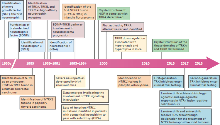
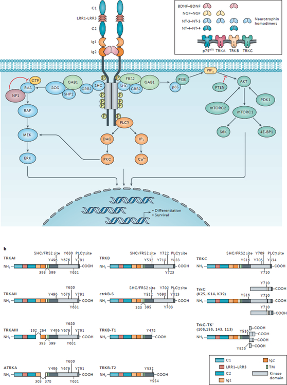
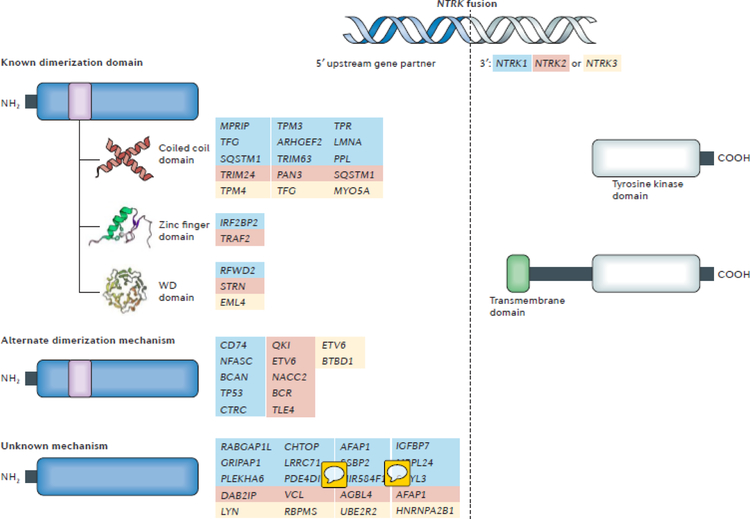
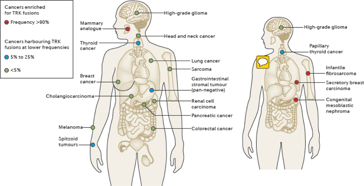
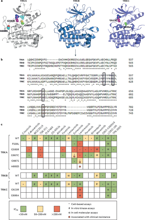
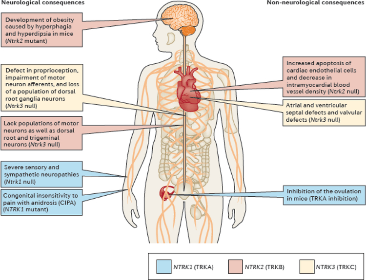
NTRK gene fusions involving either NTRK1, NTRK2 or NTRK3 (encoding the neurotrophin receptors TRKA, TRKB and TRKC, respectively) are oncogenic drivers of various adult and paediatric tumour types. These fusions can be detected in the clinic using a variety of methods, including tumour DNA and RNA sequencing and plasma cell-free DNA profiling. The treatment of patients with NTRK fusion-positive cancers with a first-generation TRK inhibitor, such as larotrectinib or entrectinib, is associated with high response rates (>75%), regardless of tumour histology. First-generation TRK inhibitors are well tolerated by most patients, with toxicity profiles characterized by occasional off-tumour, on-target adverse events (attributable to TRK inhibition in non-malignant tissues). Despite durable disease control in many patients, advanced-stage NTRK fusion-positive cancers eventually become refractory to TRK inhibition; resistance can be mediated by the acquisition of NTRK kinase domain mutations. Fortunately, certain resistance mutations can be overcome by second-generation TRK inhibitors, including LOXO-195 and TPX-0005 that are being explored in clinical trials. In this Review, we discuss the biology of NTRK fusions, strategies to target these drivers in the treatment-naive and acquired-resistance disease settings, and the unique safety profile of TRK inhibitors.


Conflict of interest statement

Competing interests

A.D. has received honoraria (as an advisory board member) from Bayer, Ignyta, Loxo Oncology, Pfizer, Roche/Genentech, and TP Therapeutics, and research funding from Loxo Oncology. M.S. has received research funding from Daiichi Sankyo and Puma Biotechnology.

Figures

Fig. 1 |
Fig. 1 |. Timeline of key advances relating to the biology and therapeutic targeting of TRK signalling.
Milestone discoveries that are relevant to normal TRK pathway biology (boxes above the timeline arrow) and NTRK fusions in cancer (boxes below the timeline arrow) are depicted. Key events relating to the following fields of study are colour coded as follows: neurotrophin identification (light blue), TRK function (red), TRK loss or NTRK-mutant phenotypes (yellow), TRK protein structure (green), TRK overexpression and splicing (grey), identification of NTRK fusions in clinical samples (orange), and clinical trials of TRK inhibitors (dark blue).
Fig. 2 |
Fig. 2 |. TRK biology and signalling in the nervous system.
a | The inset image indicates the ligand specificity of the TRK proteins for the neurotrophins, brain-derived neurotrophic factor (BDNF), nerve-growth factor (NGF), neutrotrophin 3 (NT-3), and/or neurotrophin 4 (NT-4), which each bind to their cognate receptors as a homodimer. TRKA is the high affinity receptor for NGF, whereas TRKB has high affinity for both BDNF and NT-4. NT-3 can bind to all TRK receptors but has highest affinity for TRKC and is the sole ligand of this receptor. Additionally, the TNF receptor superfamily member p75NTR can bind to all neurotrophins with low affinity, resulting in enhanced TRK signalling and/or the activation of distinct signalling pathways. The main image depicts the structure of the TRK–neurotrophin complex and the signalling pathways activated by TRK upon neurotrophin stimulation. The cysteine clusters C1 and C2, leucine-rich regions (LRR) 1–3, the Ig1 and Ig2 immunoglobulin-like motifs, and the kinase domain (KD) are indicated. The binding of neurotrophins to the extracellular region of TRK proteins, predominantly at the Ig2 domain, results in ligand-dependent receptor homodimerization followed by transactivation of the intracellular tyrosine kinase domains and the recruitment of various cytoplasmic adaptors. The phosphorylation events that mediate activation of the kinase domain or binding of SHC-transforming protein (SHC), fibroblast growth factor receptor substrate 2 (FRS2), and phosphoinositide phospholipase Cɣ (PLCɣ) to TRK proteins are depicted. The recruited adaptor proteins activate downstream signalling pathways, including the MAPK, PI3K, and PKC pathways. Each of these signalling pathways also activates the transcription of genes involved in the differentiation and survival of neurons. b | The schematic domain structures of known splice variants of TRKA, TRKB, and TRKC are shown. The docking residues for SHC–FRS2, and PLCɣ, and the three phosphorylated tyrosines within the activation loop of the kinase domain are also displayed. The ctrkB-S, TrkC, and TrkC-TK schematics were generated using the amino acid sequences of chicken TrkB and porcine TrkC, respectively — the organisms in which these variants were initially discovered. Of note, an alternative ATG transcription-initiation site in the NTRK2 gene has been reported; transcripts starting from this alternative site give rise to multiple additional TRKB-derived transcripts (not shown). 4E-BP1, eukaryotic translation initiation factor 4E-binding protein 1; DAG, diacylgycerol; GAB1, GRB2-associated-binding protein 1; GRB2, growth factor receptor-bound protein 2; IP3, inositol triphosphate; mTORC, mechanistic target of rapamycin complex; NF1, neurofibromin; p85, PI3K 85 kDa regulatory subunit α; PIP3, phosphatidylinositol 3,4,5-trisphosphate; SHP2, SH-PTP2 (also known as tyrosine-protein phosphatase non-receptor type 11); SOS, son of sevenless homologue.
Fig. 3 |
Fig. 3 |. Activating mechanisms of NTRK fusions.
The structure of a representative NTRK fusion gene with sequences of the NTRK gene (grey) and the upstream partner gene (blue) is shown. Most NTRK fusion partners are hypothesized to activate the downstream TRK kinase domain and thus aberrant TRK signalling via dimerization. A list of the known upstream partners, stratified according to the type of oligomerization domain that they contain (coiled coil, zinc finger, or WD domains), is provided. Of note, not all fusion partners harbour typical dimerization domains and the upstream genes for which an alternate mechanism of dimerization is potentially responsible for fusion activation, or the mechanism by which fusion activation is unknown are also listed. Partners of NTRK1, NTRK2, and NTRK3 are indicated by blue, red, and yellow text, respectively. The TRK kinase domain is always included in the oncogenic fusion protein. By contrast, the transmembrane domain of the TRK protein is only present in select fusions, suggesting that this domain is not required for activation of the TRK kinase; however, incorporation of the transmembrane domain might have an effect on cellular localization of the fusion protein (for example, to the plasma membrane).
Fig. 4 |
Fig. 4 |. Distribution and frequency of NTRK fusions in adult and paediatric tumours.
NTRK fusions are identified across multiple paediatric and adult cancer histologies. The frequency of these fusions varies from <1% in cancer types including lung, colorectal, pancreatic, breast cancers, melanoma, and other solid or haematological cancers (green circles), up to 25% in tumours including thyroid, spitzoid, and gastrointestinal stromal tumours (blue circles), to >90% in rare tumours types, specifically secretory breast carcinoma, mammary analogue secretory carcinoma (MASC), congenital infantile fibrosarcoma, and cellular or mixed congenital mesoblastic nephroma (red circles) for which the NTRK fusions are considered practically pathognomonic.
Fig. 5 |
Fig. 5 |. Mechanisms of acquired resistance to TRK inhibitors and profiles of TRK inhibitor activity.
a | Structural modelling of the tyrosine kinase domains of TRKA, TRKB, and TRKC showing amino acid substitutions resulting from somatic mutations in NTRK1, NTRK2, and NTRK3 fusions, respectively, that have been associated with acquired resistance to first-generation TRK inhibitors in patients: the TRKA G595R and TRKC G623R solvent-front substitutions; the TRKA F589L gatekeeper mutation; G667C and G696A substitutions within the xDFG-motif of the kinase activation loops of TRKA and TRKC, respectively; and the TRKA A608D mutation. No substitutions involving TRKB have yet been identified in patient samples. Paralogous mutations in TRKA and TRKC are shown in the same colour. b | Homology alignment of the kinase domains of TRKA, TRKB, and TRKC indicates that these resistant mutations affect highly conserved amino acid residues across the TRK proteins (black boxes). The arrow indicates the beginning of the kinase domains. c | A heat map of the half maximal inhibitory concentration (IC50) values of selected multi-kinase or TRK-specific inhibitors is shown. These drugs have different levels of activity against wild-type (WT) or mutant TRK proteins (for example, those with solvent-front, gatekeeper, or xDFG substitutions). The symbols in each box indicate which type of assay has been used to determine the IC50. Red circles represent reports of clinical resistance that have been observed for each mutant. Of note, some mutants that have been described to be sensitive to specific inhibitors in preclinical analyses were instead found to confer resistance to the same drugs in patients. xDFG, X-aspartate-phenylalanine-glycine.
Fig. 6 |
Fig. 6 |. Consequences of loss, decreased activity, or inhibition of TRK.
Genetic or pharmacological disruption of TRK signalling can cause a variety of neurological and non-neurological changes. These phenotypic effects deriving from impairments in TRK protein function were described in preclinical animal models or in patients with genetic conditions resulting in a decrease or loss of TRK activity. Consequences of loss of Ntrk1/NTRK1 (TRKA) include sensory and sympathetic neuropathies, insensitivity to pain, anhidrosis, and impairments in ovulation (blue boxes). Ntrk2/NTRK2 (TRKB) deficiency is associated with hyperphagia, the loss of specific populations of neurons, cardiac defects, and memory loss (red boxes). Similarly, loss of Ntrk3/NTRK3 (TRKC) results in defects in proprioception, a lack of certain populations of neurons, and cardiac dysfunction (yellow boxes). Consistent with these findings, paresthesias, weight gain, cognitive disturbance, and dizziness have been reported in patients treated with TRK inhibitors,.

Similar articles

Cited by

References

    1. Robert C et al. Improved Overall Survival in Melanoma with Combined Dabrafenib and Trametinib. The New England journal of medicine 372, 30–39, doi: 10.1056/NEJMoa1412690 (2015). - DOI - PubMed
    1. Geyer CE et al. Lapatinib plus capecitabine for HER2-positive advanced breast cancer. The New England journal of medicine 355, 2733–2743, doi:10.1056/NEJMoa064320 (2006). - DOI - PubMed
    1. Planchard D et al. Dabrafenib plus trametinib in patients with previously treated BRAF(V600E)-mutant metastatic non-small cell lung cancer: an open-label, multicentre phase 2 trial. The Lancet. Oncology 17, 984–993, doi:10.1016/S1470-2045(16)30146-2 (2016). - DOI - PMC - PubMed
    1. Shaw AT et al. Crizotinib in ROS1-rearranged non-small-cell lung cancer. The New England journal of medicine 371, 1963–1971, doi:10.1056/NEJMoa1406766 (2014). - DOI - PMC - PubMed
    1. Slamon DJ et al. Use of chemotherapy plus a monoclonal antibody against HER2 for metastatic breast cancer that overexpresses HER2. The New England journal of medicine 344, 783–792, doi:10.1056/NEJM200103153441101 (2001). - DOI - PubMed

Publication types

MeSH terms