Review
. 2018 Nov;19(11):1169-1178.
doi: 10.1038/s41590-018-0233-9. Epub 2018 Oct 17.

Antibody-mediated protection against Ebola virus

Affiliations
Review

Antibody-mediated protection against Ebola virus

Erica Ollmann Saphire et al. Nat Immunol. 2018 Nov.

Abstract

Recent Ebola virus disease epidemics have highlighted the need for effective vaccines and therapeutics to prevent future outbreaks. Antibodies are clearly critical for control of this deadly disease; however, the specific mechanisms of action of protective antibodies have yet to be defined. In this Perspective we discuss the antibody features that correlate with in vivo protection during infection with Ebola virus, based on the results of a systematic and comprehensive study of antibodies directed against this virus. Although neutralization activity mediated by the Fab domains of the antibody is strongly correlated with protection, recruitment of immune effector functions by the Fc domain has also emerged as a complementary, and sometimes alternative, route to protection. For a subset of antibodies, Fc-mediated clearance and killing of infected cells seems to be the main driver of protection after exposure and mirrors observations in vaccination studies. Continued analysis of antibodies that achieve protection partially or wholly through Fc-mediated functions, the precise functions required, the intersection with specificity and the importance of these functions in different animal models is needed to identify and begin to capitalize on Fc-mediated protection in vaccines and therapeutics alike.


Conflict of interest statement

COMPETING FINANCIAL INTERESTS

The authors declare no competing interests.

Figures

Figure 1.
Figure 1.
Evaluation of early antibody treatments evaluated for their protection of NHPs. In Figures 1–2, all mAbs that bind the GP core remaining after cathepsin cleavage (termed GPcl) are colored orange or yellow. All head- and glycan cap-binding mAbs are colored blue or purple. (a) Crystal structure of Ebola virus GP (grey) in complex with KZ52 (orange). KZ52 was produced in CHO cells for evaluation in rhesus macaques at 50 mg/kg one day prior and four days after viral challenge. (b) Superimposed negative stain EM structures of antibodies contained in the ZMAb cocktail (2G4, orange; 4G7 yellow; 1H3 blue) in complex with Ebola virus GP (grey). ZMAb antibodies were produced in murine hybridoma culture for evaluation in cynomolgus macaques at 25 mg/kg on days 1, 4 and 7 (100% survival) or 2, 5, and 8 after challenge (50% survival). (c) Model of the MB-003 cocktail made from the negative stain EM structure of the 13C6-GP complex and the crystal structure of 13F6 (purple molecular surface) bound to with its mucin-like domain linear epitope (white ball-and-stick). MB-003 antibodies were produced in CHO or Nicotiniana cells and evaluated in rhesus macaques at 50 mg/kg (CHO cell-produced, 50% survival) or 16.7 mg/kg (Nicotiniana-produced, 100% survival) at 1 hour and 4 and 7 days after challenge. Delivery of Nicotiniana-produced MB-003 at later time points (5, 7, and 10 days after challenge) resulted in 43% survival. No high-resolution structural information yet exists for the mucin-like domain or MB-003 component 6D8. These have been modeled with grey circles and a purple Fab fragment, respectively. The relative positions of the two mucin-binding antibodies 13F6 and 6D8 are as yet unknown.
Figure 2.
Figure 2.
Antibody treatments approved for evaluation in outbreaks of Ebola virus in 2018. (a) Superimposed CryoEM structures of the ZMapp antibodies 2G4 (green), 4G7 (yellow) and 13C6 (blue). Only the variable domains (Fv) could be built into cryoEM maps. ZMapp antibodies were produced in Nicotiniana for evaluation in rhesus macaques at 50 mg/kg on days 5, 8 and 11 after challenge. ZMapp was also evaluated in humans in 2014. (c) Crystal structure of mAb 114 (blue) in complex with Ebola virus GP, and its dosage information in rhesus macaques at 50 mg/kg on days . (D) REGN-EB3 containing antibodies 3470, 3471, and 3479 visualized by negative stain EM and dosage information in rhesus macaques. REGN-3B3 was evaluated in three-dose and single-dose regimens.
Figure 3.
Figure 3.
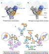
Structure, epitopes of Ebola virus GP, and antibody functions. (a) Ebola virus GP is cleaved in the endosome to remove the glycan cap and mucin like domains. The remaining GP core, outlined in black, is termed GPcl and is competent for receptor binding. Antibodies against GPcl are much more likely to be neutralizing. The base region of GP that encompasses the epitopes of KZ52, 2G4 and 4G7, is indicated by the orange circle. The glycan cap, including the 13C6 and 3471 epitopes, is deleted by enzymatic cleavage of GP and is not retained on GPcl. Light blue circles represent the mucin-like domain of GP (light blue), which is disordered in higher resolution structures. The viral membrane is indicated by a grey dotted line. (b) Antibodies against epitopes in the upper tier of GP, Tier 1, exhibit stronger effector functions on average than antibodies against the middle and lower regions of GP, as measured by immune ‘polyfunctionality’. This greater sum of Fc-mediated immune functions in Tier 1 is driven by measures of phagocytosis. Epitopes of antibodies with strong NK activities occur across the GP. (c) Mechanical neutralization and effector function protection mediated by antibodies. At left, IgG antibodies are shown anchoring to viral GP (blue) on Ebola virus (yellow). By anchoring to the GP, neutralizing antibodies prevent viral entry into potential target cells (purple). At right, IgG antibodies are illustrated as anchoring to viral GPs on Ebola virus (yellow) or an infected cell (purple). Through GP anchoring, the antibody Fc couples to neutrophils, monocytes, macrophages, NK cells and other immune mediators to mediate destruction and clearance of the virus and infected cells. Neutralization and effector-mediated clearance are independent functions and may occur on the same or different antibodies.
Figure 4.
Figure 4.
Outliers in the VIC study. At left, nine antibodies protect ≥60% of mice, yet offer little to no neutralization (neutralization score of 0 (no neutralization measured at all in any assay) to 1 (weak neutralization measured in one assay, no neutralization measured in the other three) or 2 (strong neutralization in only one assay). Seven of these nine recognize Tier 1 or unknown epitopes. At right are eight antibodies that protect ≤30% of mice, despite relatively strong neutralization scores of 5–7. These antibodies typically neutralize at least moderately, and often potently, in all four measures, and recognize Tier 2 and 3 epitopes. In parentheses are the average polyfunctionality, neutralization score and protection value for the protective/poorly neutralizing group at left and the neutralizing/poorly protective group on the right. Each antibody in the panel is labelled atop a vertical column of boxes representing polyfunctionality, immune effector functions scored, protection and overall neutralization. For clarity, each measure is indicated by a colored box rather than an absolute value. Blue, yellow and red boxes indicate strong, moderate and weak scores, respectively. Polyfunctionality is a sum of yes/no answers for each of the seven effector functions measured including markers of both phagocytosis and natural killer cell function. For polyfunctionality, one point was given for strong or moderate activity on each of the seven readouts, with a maximum polyfunctionality score of 7. Antibody neutralization scores were calculated with 2, 1 or 0 points given for strong, moderate and weak/no activity on the four neutralization readouts, for a maximum score of 8. Epitopes of each antibody are indicated at the bottom with the color code at right.
Figure 5.
Figure 5.
Specific glycan profiles are linked to distinct functional activity. (a) Hierarchical clustering of VIC mAbs using Fc glycan content revealed 17 clusters of antibodies within the panel. The dashed line across the dendogram indicates the cut-off used to define the clusters. Each cluster is represented by a different color, and cluster number is listed below each cluster. (b) Unsupervised principal component analysis of Fc glycan content of VIC mAbs. The mAbs are color-coded according to the clusters identified in (A) in the plot at left, and the loading plot showing the glycan features driving separation of the mAbs is illustrated at right. (c) Heatmap of the average total galactose (G0, G1, or G2), fucose, bisecting GlcNAc, or sialic acid content of the mAbs within each cluster. Dark blue represents the row minimum and red represents the row maximum. (d) The functional activity of mAbs within each cluster was averaged for each effector function measured, and categorized into high, medium, or low/negligible based on assay cutoffs. Each wedge is color coded by effector function, and the size of the wedge indicates the magnitude of response.

References

    1. Carter PJ & Lazar GA Next generation antibody drugs: pursuit of the ‘high-hanging fruit’. Nat. Rev. Drug Discov 17, 197–223 (2018). - PubMed
    1. Salazar G, Zhang N, Fu T-M & An Z Antibody therapies for the prevention and treatment of viral infections. NPJ Vaccines 2, 19 (2017). - PMC - PubMed
    1. Lu LL, Suscovich TJ, Fortune SM & Alter G Beyond binding: antibody effector functions in infectious diseases. Nat. Rev. Immunol 18, 46–61 (2018). - PMC - PubMed
    1. Casadevall A Antibody-based therapies for emerging infectious diseases. Emerg. Infect. Dis 2, 200–208 (1996). - PMC - PubMed
    1. Corti D et al. Prophylactic and postexposure efficacy of a potent human monoclonal antibody against MERS coronavirus. Proc. Natl. Acad. Sci. U. S. A 112, 10473–10478 (2015). - PMC - PubMed

Publication types