. 2018 Oct 2:9:2289.
doi: 10.3389/fimmu.2018.02289. eCollection 2018.

Metabolite-Sensing G Protein Coupled Receptor TGR5 Protects Host From Viral Infection Through Amplifying Type I Interferon Responses

Affiliations

Metabolite-Sensing G Protein Coupled Receptor TGR5 Protects Host From Viral Infection Through Amplifying Type I Interferon Responses

Qingqing Xiong et al. Front Immunol. .

Abstract

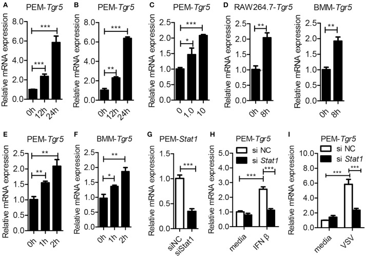
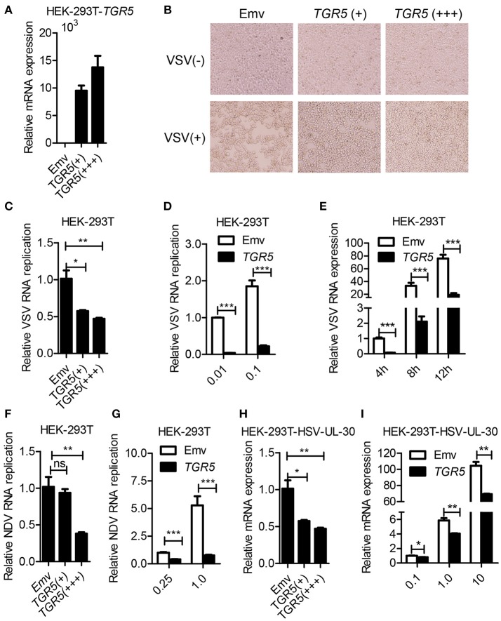
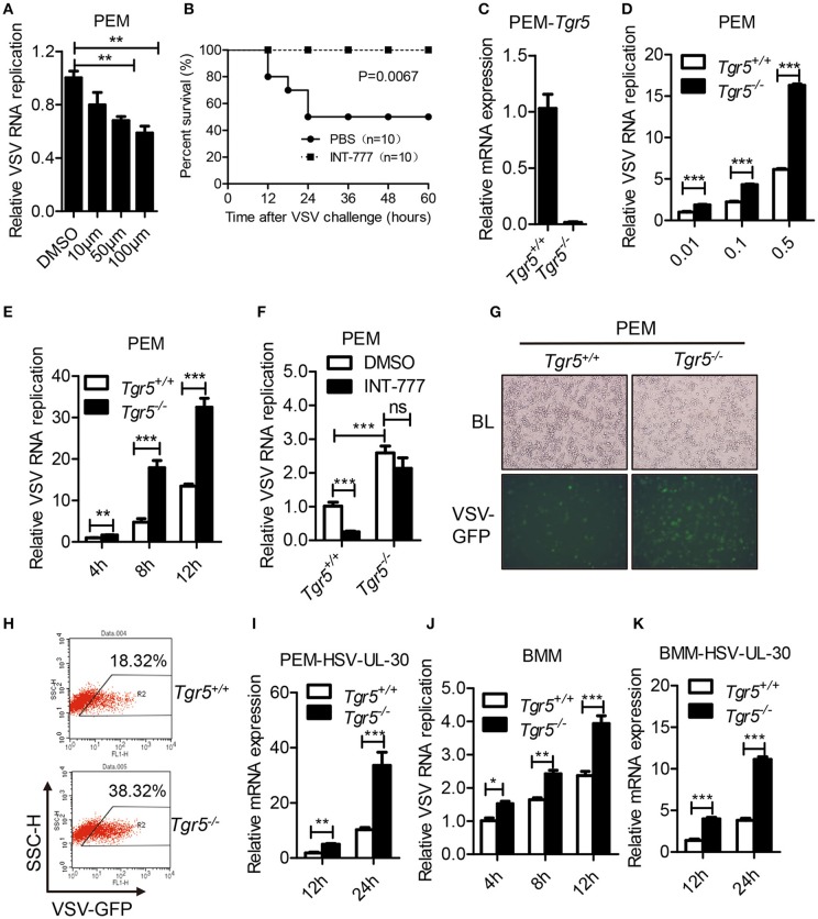
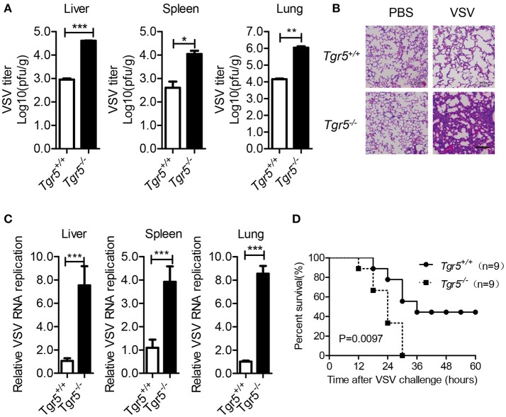
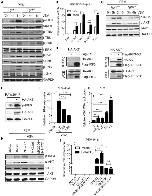
The metabolite-sensing G protein-coupled receptors (GPCRs) bind to various metabolites and transmit signals that are important for proper immune and metabolic functions. However, the roles of metabolite-sensing GPCRs in viral infection are not well characterized. Here, we identified metabolite-sensing GPCR TGR5 as an interferon (IFN)-stimulated gene (ISG) which had increased expression following viral infection or IFN-β stimulation in a STAT1-dependent manner. Most importantly, overexpression of TGR5 or treatment with the modified bile acid INT-777 broadly protected host cells from vesicular stomatitis virus (VSV), newcastle disease virus (NDV) and herpes simplex virus type 1 (HSV-1) infection. Furthermore, VSV and HSV-1 replication was increased significantly in Tgr5-deficient macrophages and the VSV distribution in liver, spleen and lungs was increased in Tgr5-deficient mice during VSV infection. Accordingly, Tgr5-deficient mice were much more susceptible to VSV infection than wild-type mice. Mechanistically, TGR5 facilitates type I interferon (IFN-I) production through the AKT/IRF3-signaling pathway, which is crucial in promoting antiviral innate immunity. Taken together, our data reveal a positive feedback loop regulating IRF3 signaling and suggest a potential therapeutic role for metabolite-sensing GPCRs in controlling viral diseases.

Keywords: IFN-I; ISG; TGR5; metabolite-sensing GPCRs; viral infection.


Figures

Figure 1
Figure 1
Tgr5 is an interferon-stimulated gene (ISG). (A,B) Quantitative PCR (Q-PCR) analysis of Tgr5 expression in PEMs infected with RNA virus VSV [1 MOI (multiplicity of infection), A] and DNA virus HSV-1 (1 MOI, B) for the indicated times. (C) Q-PCR analysis of Tgr5 expression in PEMs infected with the indicated VSV MOI for 8 h. (D) Q-PCR analysis of Tgr5 expression in RAW264.7 cells and BMMs infected with VSV (1 MOI) for 8 h. (E,F) Q-PCR analysis of Tgr5 expression in PEMs (E) and BMMs (F) stimulated with IFN-β (100 ng/ml) for the indicated hours. (G) Q-PCR analysis of Stat1 expression in PEMs transfected with Stat1 siRNA for 48 h. NC, negative control. (H,I) Q-PCR analysis of Tgr5 expression in PEMs transfected with Stat1 siRNA for 48 h and then stimulated with IFN-β (100 ng/ml) for 2 h (H) or infected with VSV (1 MOI) for 8 h (I). Glyceraldehyde-3-phosphate dehydrogenase (GAPDH) was used as an internal control for Q-PCR. The data are shown as the mean ± SD. *P < 0.05; **P < 0.01; ***P < 0.001. All experiments were performed three times with similar results.
Figure 2
Figure 2
Overexpression of TGR5 inhibits virus replication. (A) Q-PCR analysis of TGR5 expression in HEK-293T cells transfected for 28 h with different amounts (0.6 and 1.8 μg) of TGR5 expression plasmid. Emv, empty vector. (B) HEK-293T cells transfected with TGR5 plasmid as described in (A) were infected with VSV (0.1 MOI) for 8 h and then observed under a microscope. Original magnification 10 × . (C) Q-PCR analysis of VSV RNA replicates in (B). (D,E) Q-PCR analysis of VSV RNA replicates in TGR5-overexpressing (1.8 μg) HEK-293T cells infected by VSV with the indicated MOI (D) and times (E). (F) Q-PCR analysis of Newcastle disease virus (NDV) RNA replicates in HEK-293T cells transfected with TGR5 plasmids as described in (A) and infected with NDV (0.25 MOI) for 12 h. (G) Q-PCR analysis of NDV RNA replicates in TGR5-overexpressing (1.8 μg) HEK-293T cells infected by NDV with the indicated MOI for 18 h. (H) Q-PCR analysis of herpes simplex virus (HSV-1) UL-30 expression in HEK-293T cells transfected with TGR5 plasmids as described in (A) and infected with HSV-1 (0.5 MOI) for 18 h. (I) Q-PCR analysis of HSV-1 UL-30 expression in TGR5-overexpressing (1.8 μg) HEK-293T cells infected by HSV-1 with the indicated MOI for 18 h. GAPDH was used as an internal control for Q-PCR. The data are shown as the mean ± SD. ns, not significant; *P < 0.05; **P < 0.01; ***P < 0.001. All experiments were performed three times with similar results.
Figure 3
Figure 3
TGR5 deficiency promotes viral infection in vitro. (A) Q-PCR analysis of VSV RNA replicates in PEMs pretreated with INT-777 for 1 h at the indicated dose (μM) and then infected with VSV (0.1 MOI) for 8 h. DMSO, dimethyl sulfoxide. (B) Survival of 8-week-old control or INT-777 (30 mg/kg) pretreated mice given intraperitoneal injections of VSV [1 × 10⁸ plaque-forming units (pfu)/g] (n = 10 per group). (C) Q-PCR analysis of Tgr5 expression in Tgr5+/+- and Tgr5−/−-PEMs. (D,E) Q-PCR analysis of VSV RNA levels from Tgr5+/+- and Tgr5−/−-PEMs infected with the indicated VSV MOI for 8 h (D) and with 1 MOI for the indicated times (E). (F) Q-PCR analysis of VSV RNA levels from Tgr5+/+- and Tgr5−/−-PEMs pretreated with INT-777 (500 μM) for 1 h and then infected with VSV (1 MOI) for 8 h. (G,H) PEMs from Tgr5+/+- and Tgr5−/−-mice were infected VSV-GFP (0.01 MOI) for 12 h, and VSV-GFP was examined by light microscopy (G) or fluorescence-activated cell sorting (FACS) (H). Original magnification 10 × ; BL, bright light; SSC-H, side scatter-height. (I) Q-PCR analysis of HSV-1 UL-30 expression in Tgr5+/+- and Tgr5−/−-PEMs infected with the HSV-1 (1 MOI) for the indicated times. (J,K) Q-PCR analysis of VSV RNA levels (J) and HSV-1 UL-30 expression (K) in Tgr5+/+- and Tgr5−/−-BMMs infected with VSV (1 MOI) or HSV-1 (1 MOI) for the indicated times. GAPDH was used as an internal control for Q-PCR. The data are shown as the mean ± SD. ns, not significant; *P < 0.05; **P < 0.01; ***P < 0.001. All experiments were performed three times with similar results.
Figure 4
Figure 4
TGR5 deficiency promotes viral infection in vivo. (A) Determination of VSV loads in organs by standard plaque assays from Tgr5+/+- and Tgr5−/−-mice intraperitoneally infected with VSV (1 × 10⁸ pfu per mouse) for 24 h. (B) Haematoxylin & eosin staining of lung sections from mice in (A). Scale bar, 200 μm. PBS, phosphate-buffered saline. (C) Determination of VSV loads in organs by Q-PCR from Tgr5+/+- and Tgr5−/−-mice intravenously infected with VSV (1 × 10⁵ pfu per mouse) for 24 h. (D) Survival of 8-week-old Tgr5+/+- and Tgr5−/−-mice given intraperitoneal injections of VSV (1 × 10⁸ pfu/g) (n = 9 per group). GAPDH was used as an internal control for Q-PCR. The data are shown as the mean ± SD. *P < 0.05; **P < 0.01; ***P < 0.001. All experiments were performed three times with similar results.
Figure 5
Figure 5
TGR5 positively regulates IFN-I production. (A,B) Q-PCR analysis of Ifn-β (A) and Ifn-a4 (B) expression in Tgr5+/+- and Tgr5−/−-PEMs infected with VSV (1 MOI) for the indicated times. (C) Q-PCR analysis of Ifn-β expression in Tgr5+/+- and Tgr5−/−-PEMs infected with the indicated VSV MOI for 8 h. (D) Q-PCR analysis of Ifn-β expression in Tgr5+/+- and Tgr5−/−-BMMs infected with VSV (1 MOI) for the indicated times. (E) Q-PCR analysis of IFN-β expression in TGR5-overexpressing (1.8 μg) HEK-293T cells infected with VSV (1 MOI) for 8 h. (F,G) Q-PCR analysis of Ifn-β expression in Tgr5+/+- and Tgr5−/−-PEMs transfected with Poly (I:C) (1.0 μg/ml) (F) or stimulated with Poly (I:C) (10 μg/ml) (G) for the indicated times. (H) Q-PCR analysis of Ifn-β expression in Tgr5+/+- and Tgr5−/−-BMMs stimulated with Poly (I:C) (10 μg/ml) for the indicated times. (I) Q-PCR analysis of Ifn-β expression in PEMs pretreated with INT-777 (500 μM) for 1 h and then stimulated with Poly (I:C) (10 μg/ml) for 2 h. (J) Q-PCR analysis of Ifn-β expression in PEMs pretreated with INT-777 (500 μM) for 1 h and then infected with VSV (1 MOI) or HSV-1 (1 MOI) for 8 h. (K) ELISA of IFN-β in sera from Tgr5+/+- and Tgr5−/−-mice intraperitoneally injected with VSV (1 × 10⁸ pfu per mouse) for 24 h. nd, not detected. (L) Q-PCR analysis of Ifn-β expression in organs from Tgr5+/+- and Tgr5−/−-mice in (K). GAPDH was used as an internal control for Q-PCR. The data are shown as the mean ± SD. ns, not significant; *P < 0.05; **P < 0.01; ***P < 0.001. All experiments were performed three times with similar results.
Figure 6
Figure 6
TGR5 amplifies IFN-I signaling via AKT-mediated IRF3 activation. (A) Immunoblot analysis of phosphorylated (p-) or total (t-) proteins in lysates of Tgr5+/+- and Tgr5−/−-PEMs infected with VSV (1 MOI) for the indicated times. (B) TGR5 was co-transfected with RIG-I (N) (RIG-N), MAVS, STING, TBK1, or empty vectors, together with an IFN-β luciferase reporter, into HEK-293T cells for 28 h. IFN-β luciferase activity was detected and normalized to Renilla luciferase activity. (C) Immunoblot analysis of phosphorylated (p-) AKT or total (t-) AKT in lysates of Tgr5+/+- and Tgr5−/−-PEMs infected with VSV (1 MOI) for the indicated times. (D) HEK-293T cells were transfected with plasmids encoding HA-AKT and Flag-IRF3 or Flag-IRF3-5D for 28 h. The cell lysate supernatants were immunoprecipitated using M2 beads, and then immunoblotted with antibodies to HA or Flag tags. WCE, whole-cell extracts. (E) Immunoblot analysis of phosphorylated (p-) IRF3 and HA-AKT in lysates of RAW 264.7 cells transfected with plasmids encoding HA-AKT for 28 h. (F) Q-PCR analysis of Ifn-β expression in PEMs pretreated with MK 2206 (an inhibitor of AKT) at the indicated dose (μM) for 1 h and then infected with VSV (1 MOI) for 8 h. (G) Q-PCR analysis of VSV RNA replicates in PEMs pretreated with MK 2206 at the indicated dose (μM) for 1 h and then infected with VSV (1 MOI) for 8 h. (H) Immunoblot analysis of phosphorylated (p-) IRF3 and (p-) AKT in lysates of PEMs pretreated with MK 2206 (3 μM) or INT-777 (500 μM) for 1 h, and then infected with VSV (1 MOI) for 8 h. (I) Q-PCR analysis of Ifn-β expression in PEMs pretreated with MK 2206 (3 μM) or INT-777 (500 μM) for 1 h, and then stimulated with poly (I:C) (10 μg/ml) for 2 h. GAPDH was used as an internal control for Q-PCR. The data are shown as the mean ± SD. ns, not significant; **P < 0.01; ***P < 0.001. All experiments were performed three times with similar results.

Similar articles

Cited by

References

    1. Steed AL, Christophi GP, Kaiko GE, Sun L, Goodwin VM, Jain U, et al. . The microbial metabolite desaminotyrosine protects from influenza through type I interferon. Science (2017) 357:498–502. 10.1126/science.aam5336 - DOI - PMC - PubMed
    1. Yu Y, Clippinger AJ, Pierciey FJ, Jr, Alwine JC. Viruses and metabolism: alterations of glucose and glutamine metabolism mediated by human cytomegalovirus. Adv Virus Res. (2011) 80:49–67. 10.1016/B978-0-12-385987-7.00003-8 - DOI - PubMed
    1. Chalmers DT, Behan DP. The use of constitutively active GPCRs in drug discovery and functional genomics. Nat Rev Drug Discov. (2002) 1:599–608. 10.1038/nrd872 - DOI - PubMed
    1. George SR, O'Dowd BF, Lee SP. G-protein-coupled receptor oligomerization and its potential for drug discovery. Nat Rev Drug Discov. (2002) 1:808–20. 10.1038/nrd913 - DOI - PubMed
    1. Alvarez-Curto E, Milligan G. Metabolism meets immunity: the role of free fatty acid receptors in the immune system. Biochem Pharmacol. (2016) 114:3–13. 10.1016/j.bcp.2016.03.017 - DOI - PubMed

Publication types

MeSH terms