Review
. 2018 Oct 2:6:128.
doi: 10.3389/fcell.2018.00128. eCollection 2018.

Crosstalk Between Mammalian Autophagy and the Ubiquitin-Proteasome System

Affiliations
Review

Crosstalk Between Mammalian Autophagy and the Ubiquitin-Proteasome System

Nur Mehpare Kocaturk et al. Front Cell Dev Biol. .

Abstract

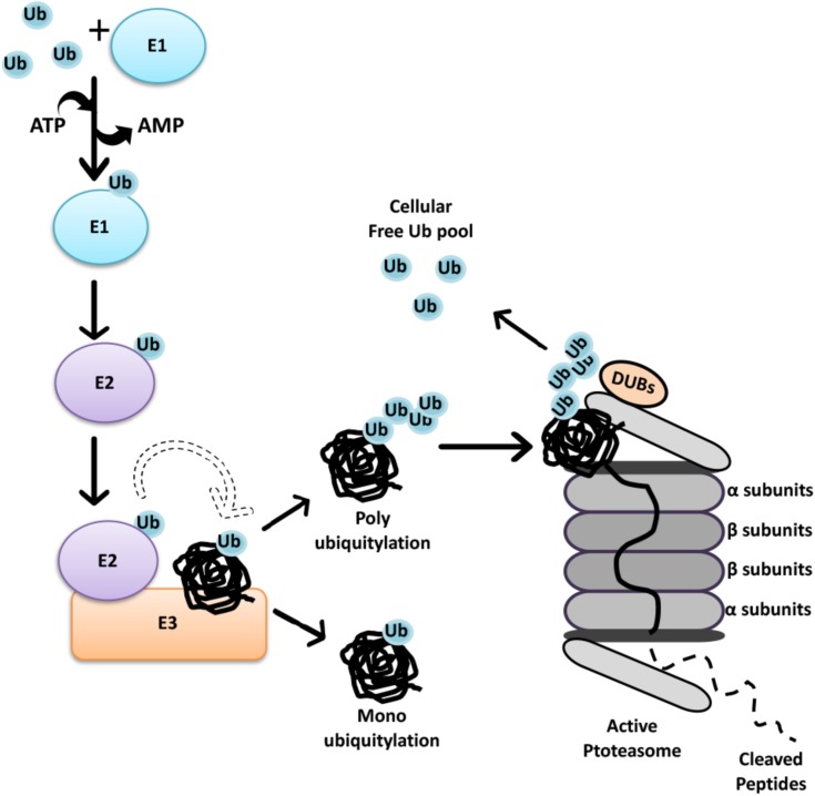
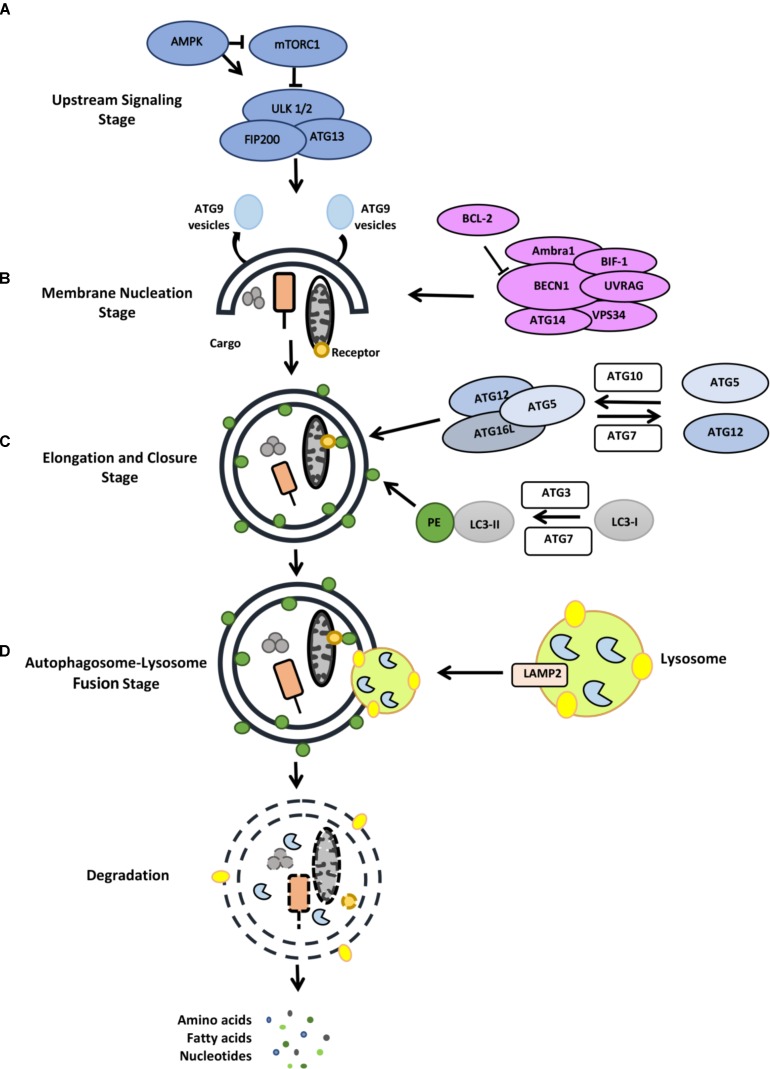
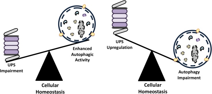
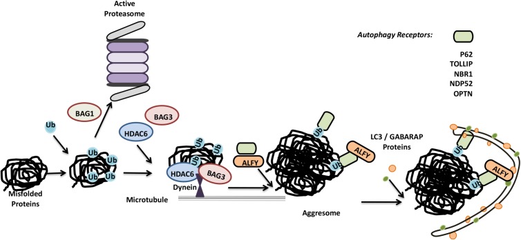
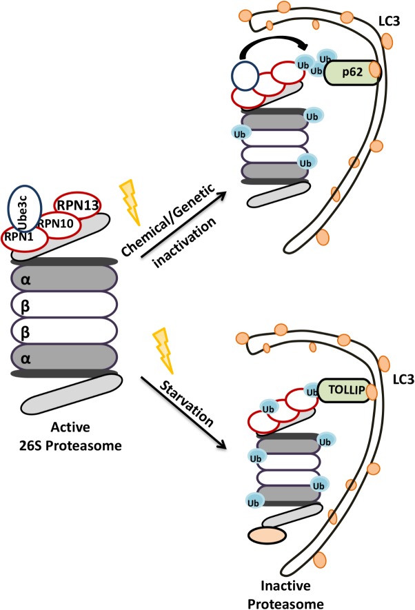
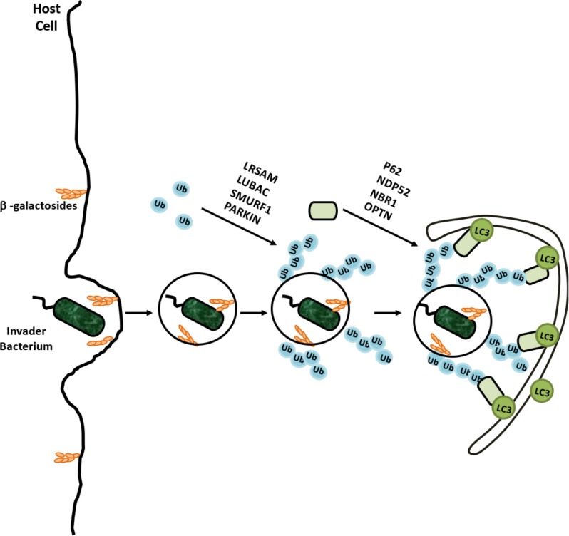
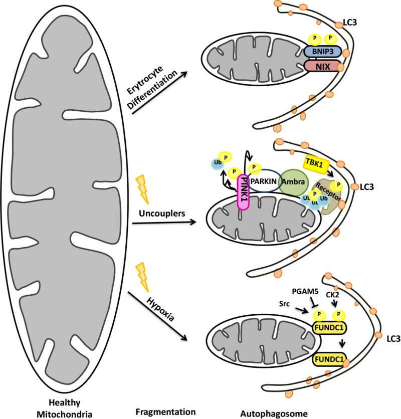
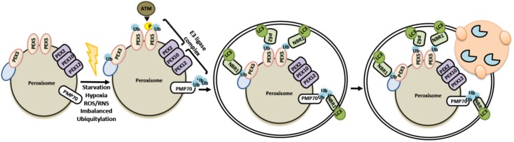
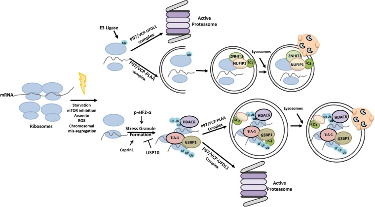
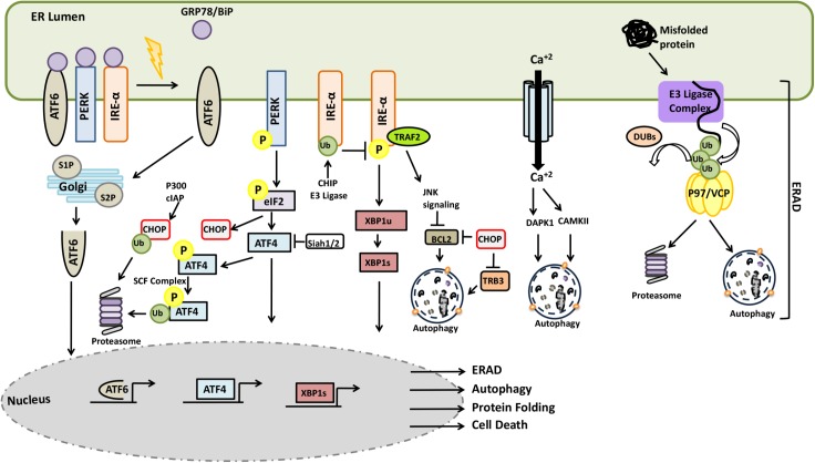
Autophagy and the ubiquitin-proteasome system (UPS) are the two major intracellular quality control and recycling mechanisms that are responsible for cellular homeostasis in eukaryotes. Ubiquitylation is utilized as a degradation signal by both systems, yet, different mechanisms are in play. The UPS is responsible for the degradation of short-lived proteins and soluble misfolded proteins whereas autophagy eliminates long-lived proteins, insoluble protein aggregates and even whole organelles (e.g., mitochondria, peroxisomes) and intracellular parasites (e.g., bacteria). Both the UPS and selective autophagy recognize their targets through their ubiquitin tags. In addition to an indirect connection between the two systems through ubiquitylated proteins, recent data indicate the presence of connections and reciprocal regulation mechanisms between these degradation pathways. In this review, we summarize these direct and indirect interactions and crosstalks between autophagy and the UPS, and their implications for cellular stress responses and homeostasis.

Keywords: UPS; autophagy; mitophagy; organelle homeostasis; proteasome; protein quality control; proteostasis; ubiquitylation.


Figures

FIGURE 1
FIGURE 1
The Ubiquitin-Proteasome System. Initially through C-terminal glycine, ubiquitin is attached to a cysteine residue of an activating enzyme, E1, in an ATP-dependent manner. The active ubiquitin is then associated with a cysteine residue of an ubiquitin conjugating enzyme, E2. Finally, specificity of ubiquitin transfer is ensured by E3 ubiquitin ligase family of proteins that bind to selected protein subsets (Hershko and Ciechanover, 1998). In the case of RING finger E3 ligases, the transfer of ubiquitin is direct from E2-ubiquitin to the substrate, even if the presence of E3 is required for substrate selection. At present, 2 genes are known to encode E1 isoforms, at least 40 genes encode E2’s, and over 600 E3 ubiquitin ligases were defined in the human genome (Pickart and Eddins, 2004; Clague et al., 2015). Each E1 isoform reveals a distinct preference for different E2 enzymes, while association of E2 and E3 depend on cellular context, generating extensive combinatorial complexity.
FIGURE 2
FIGURE 2
Stages of the autophagy pathway (for detail, see the text). (A) Upstream signaling, (B) membrane nucleation stage, (C) elongation and closure stage, (D) autophagosome-lysosome fusion stage.
FIGURE 3
FIGURE 3
The compensatory balance between the activities of autophagy and the UPS in order to maintain cellular homeostasis.
FIGURE 4
FIGURE 4
Misfolded proteins can be eliminated by both the UPS and autophagy system. Misfolded proteins are ubiquitylated and based on the differences in ubiquitin linkages and ubiquitin binding proteins, they are directed for proteasomal degradation or further accumulated in aggresomes. Aggresomes are selectively cleared by autophagy.
FIGURE 5
FIGURE 5
Schematic representation of the selective degradation of proteasomes by autophagy. Upon starvation and functional defects proteasomes become ubiquitylated and degraded by autophagic machinery.
FIGURE 6
FIGURE 6
Selective degradation of invaders by xenophagy is example of coregulation of the UPS and autophagy. Cellular degradation of invading bacterium was ubiquilated by various E3 ligases and recognized by adaptor proteins for recruitment autophagic membranes around bacterium.
FIGURE 7
FIGURE 7
Mitochondrial elimination by autophagy requires the activity of both the UPS and autophagy.
FIGURE 8
FIGURE 8
Selective removal of peroxisomes by autophagy utilizes ubiquitylation as signal.
FIGURE 9
FIGURE 9
Ubiquitylation primes ribosomes and stress granules for proteasomal degradation and autophagic elimination.
FIGURE 10
FIGURE 10
Crosstalk between the UPS and autophagy systems during ER stress and ERAD.

References

    1. Aerbajinai W., Giattina M., Lee Y. T., Raffeld M., Miller J. L. (2003). The proapoptotic factor Nix is coexpressed with Bcl-xL during terminal erythroid differentiation. Blood 102 712–717. 10.1182/blood-2002-11-3324 - DOI - PubMed
    1. Altieri D. C. (2010). Survivin and IAP proteins in cell-death mechanisms. Biochem. J. 430 199–205. 10.1042/BJ20100814 - DOI - PMC - PubMed
    1. Ambivero C. T., Cilenti L., Main S., Zervos A. S. (2014). Mulan E3 ubiquitin ligase interacts with multiple E2 conjugating enzymes and participates in mitophagy by recruiting GABARAP. Cell. Signal. 26 2921–2929. 10.1016/j.cellsig.2014.09.004 - DOI - PubMed
    1. Amodio N., Scrima M., Palaia L., Salman A. N., Quintiero A., Franco R., et al. (2010). Oncogenic role of the E3 ubiquitin ligase NEDD4-1, a PTEN negative regulator, in non-small-cell lung carcinomas. Am. J. Pathol. 177 2622–2634. 10.2353/ajpath.2010.091075 - DOI - PMC - PubMed
    1. An H., Harper J. W. (2018). Systematic analysis of ribophagy in human cells reveals bystander flux during selective autophagy. Nat. Cell Biol. 20 135–143. 10.1038/s41556-017-0007-x - DOI - PMC - PubMed

LinkOut - more resources