. 2018 Jun:3:71-81.
doi: 10.1016/j.cophys.2018.03.005. Epub 2018 Mar 20.

BH3-Dependent and Independent Activation of BAX and BAK in Mitochondrial Apoptosis

Affiliations

BH3-Dependent and Independent Activation of BAX and BAK in Mitochondrial Apoptosis

Paul S Jeng et al. Curr Opin Physiol. 2018 Jun.

Abstract

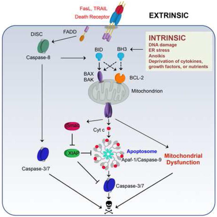
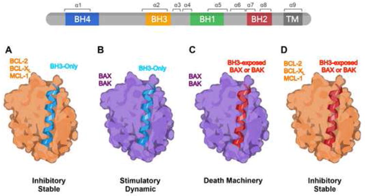
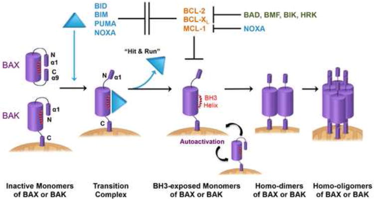
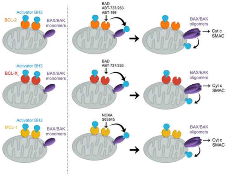
Mitochondria play key roles in mammalian apoptosis, a highly regulated genetic program of cell suicide. Multiple apoptotic signals culminate in mitochondrial outer membrane permeabilization (MOMP), which not only couples the mitochondria to the activation of caspases but also initiates caspase-independent mitochondrial dysfunction. The BCL-2 family proteins are central regulators of MOMP. Multidomain pro-apoptotic BAX and BAK are essential effectors responsible for MOMP, whereas anti-apoptotic BCL-2, BCL-XL, and MCL-1 preserve mitochondrial integrity. The third BCL-2 subfamily of proteins, BH3-only molecules, promotes apoptosis by either activating BAX and BAK or inactivating BCL-2, BCL-XL, and MCL-1. Through an interconnected hierarchical network of interactions, the BCL-2 family proteins integrate developmental and environmental cues to dictate the survival versus death decision of cells by regulating the integrity of the mitochondrial outer membrane. Over the past 30 years, research on the BCL-2-regulated apoptotic pathway has not only revealed its importance in both normal physiological and disease processes, but has also resulted in the first anti-cancer drug targeting protein-protein interactions.


Conflict of interest statement

The authors declare no conflict of interest.

Figures

Figure 1
Figure 1. The intrinsic and extrinsic pathways of apoptosis
The intrinsic pathway is initiated by death stimuli including DNA damage, ER stress, anoikis, and deprivation of cytokines, growth factors or nutrients, resulting in transcriptional or post-translational activation of BH3-only molecules (BH3s). Activator BH3s, including BID, BIM, PUMA, and NOXA, directly activate BAX and BAK to induce the homo-oligomerization of BAX and BAK, leading to mitochondrial outer member premeabilization (MOMP) and release of cytochrome c and SMAC from the mitochondrial intermembrane space to the cytosol. Upon binding to cytochrome c and dATP, APAF-1 oligomerizes into a heptameric complex known as the apoptosome, resulting in the recruitment and activation of caspase-9 and subsequent activation of effector caspase-3/7. The extrinsic pathway of apoptosis is initiated by engagement of cell surface death receptors, such as FAS or TRAIL receptors, resulting in the recruitment of adaptor proteins such as FAS-associated death domain (FADD). FADD then dimerizes with procaspase-8 to form the death-inducing signaling complex (DISC) and promote the auto-activation of procaspase-8. In ‘type I’ cells with low expression of the caspase inhibitor XIAP, such as T-lymphocytes, death receptor mediated caspase-8 activation is sufficient to activate effector caspase-3/-7. In ‘type II’ cells with high expression of XIAP, such as hepatocytes, effector caspase activation requires a mitochondrial amplification loop to alleviate XIAP-mediated caspase inhibition through mitochondrial release of SMAC. Caspase-8 mediated proteolytic cleavage of cytosolic BID into truncated BID (tBID) activates BAX and BAK-dependent MOMP, connecting the extrinsic pathway to the intrinsic mitochondrial apoptosis pathway.
Figure 2
Figure 2. BH3-in-groove: a structural basis of BCL-2 family interactions that control survival or death decisions
A schematic depicts conceptual modeling of the different interactions among the BCL-2 family proteins. The BCL-2 family proteins regulate mitochondrial apoptosis through protein-protein interactions, all involving the same BH3 helix-in-groove structure. Multidomain BCL-2 family members have four BCL-2 homology (BH) domains and a C-terminus hydrophobic transmembrane domain (α9 helix). The BH1, BH2, and BH3 domains of multidomain anti-apoptotic and pro-apoptotic members form a hydrophobic binding groove (or canonical dimerization groove) that accommodates the amphipathic alpha helical BH3 domain of BH3-only molecules as well as the BH3 domains of “BH3-exposed” BAX or BAK. The binding of BH3s to multidomain anti-apoptotic members is inhibitory and stable (A), whereas the binding of activator BH3s to BAX/BAK is stimulatory and dynamic (B). The BH3-in-groove interaction also forms the structural basis for the formation of symmetric BAX or BAK homo-dimers (C), the minimal unit for the assembly of higher-order homo-oligomers that permeabilize the mitochondrial outer membrane. The BH3 domain of most BAX or BAK present in viable cells is not exposed. Only partially activated BAX or BAK will expose the BH3 domain and as a result can bind to anti-apoptotic BCL-2 members (D).
Figure 3
Figure 3. Interconnected hierarchical regulation of BAX- and BAK-dependent mitochondrial apoptosis
Activator BH3s, including BID, BIM, PUMA, and NOXA, directly interact with BAX and BAK to induce the stepwise structural reorganization of BAX and BAK. In viable cells, BAX exists as a cytosolic monomer with its α1 helix keeping the C-terminal α9 helix engaged in the dimerization groove, while BAK is constitutively inserted in the MOM via its C-terminal α9 helix. The binding of activator BH3s drives the dissociation of an N-terminal α1 helix of BAX or BAK and mobilizes the C-terminal α9 of BAX for translocation to the MOM. Activator BH3s remain associated with the N-terminally exposed BAX or BAK through the canonical dimerization groove to drive the exposure of the BH3 domain. Partially activated, BH3-exposed BAX or BAK monomers then can bind to the hydrophobic dimerization groove of another BAX or BAK molecule to initiate homo-dimerization and subsequent homo-oligomerization. The interaction between activator BH3s and BAX or BAK is “hit-and-run” because the same binding interface of BAX and BAK is used for homo-dimerization. Anti-apoptotic BCL-2, BCL-XL, and MCL-1 sequester activator BH3s to prevent the initiation of BAX and BAK activation, providing frontline protection. As the second line of defense, anti-apoptotic BCL-2 members can also sequester “BH3-exposed” BAX and BAK monomers to prevent the homo-oligomerization of BAX and BAK. Autoactivation of BAX and BAK can occur independently of activator BH3s when BCL-2, BCL-XL, and MCL-1 are simultaneously downregulated, albeit with slower kinetics compared to BH3-mediated activation. BH3-exposed BAX or BAK monomers can serve as activators of BAX and BAK to induce a “feed-forward” amplification loop for the initiation of mitochondrial apoptosis, bypassing the need for activator BH3s.
Figure 4
Figure 4. Indirect activation of BAX or BAK by inactivator BH3s and BH3 mimetics
Anti-apoptotic BCL-2, BCL-XL, and MCL-1 preserve mitochondrial integrity by sequestering activator BH3s to prevent activation of BAX and BAK. Pro-apoptotic “inactivator” BH3s, including BAD, BMF, BIK, and HRK, displace sequestered BID/BIM/PUMA from anti-apoptotic BCL-2 and BCL-XL, thereby activating BAX and BAK indirectly. NOXA is unique among all BH3s in that it can prevent MCL-1 from sequestering BID, BIM, and PUMA due to its high binding affinity to MCL-1. Hence, NOXA is both an activator and inactivator BH3. The BH3-mimietic small molecules ABT-737 and ABT-263 (navitoclax) activate BAX and BAK indirectly through displacing activator BH3s from BCL-2 and BCL-XL, whereas ABT-199 (venetoclax) selectively targets BCL-2. Selective inhibitors for BCL-XL or MCL-1 (S63845) with preclinical activity have also been generated.

Similar articles

Cited by

References

    1. Danial NN, Korsmeyer SJ. Cell death: critical control points. Cell. 2004;116:205–219. - PubMed
    1. Wang X. The expanding role of mitochondria in apoptosis. Genes Dev. 2001;15:2922–2933. - PubMed
    1. Zhou M, Li Y, Hu Q, Bai Xc, Huang W, Yan C, Scheres SH, Shi Y. Atomic structure of the apoptosome: mechanism of cytochrome c-and dATP-mediated activation of Apaf-1. Genes & development. 2015;29:2349–2361. - PMC - PubMed
    1. Korsmeyer SJ. Bcl-2 initiates a new category of oncogenes: regulators of cell death. Blood. 1992;80:879–886. - PubMed
    1. Brem EA, Letai A. BOK: Oddball of the BCL-2 Family. Trends in cell biology. 2016;26:389–390. - PubMed

LinkOut - more resources