Comparative Study
. 2018 Oct;301(10):1628-1637.
doi: 10.1002/ar.23918. Epub 2018 Oct 17.

Developing an In Vitro Model to Screen Drugs for Nerve Regeneration

Affiliations
Comparative Study

Developing an In Vitro Model to Screen Drugs for Nerve Regeneration

Melissa L D Rayner et al. Anat Rec (Hoboken). 2018 Oct.

Abstract

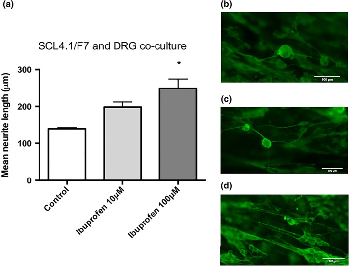
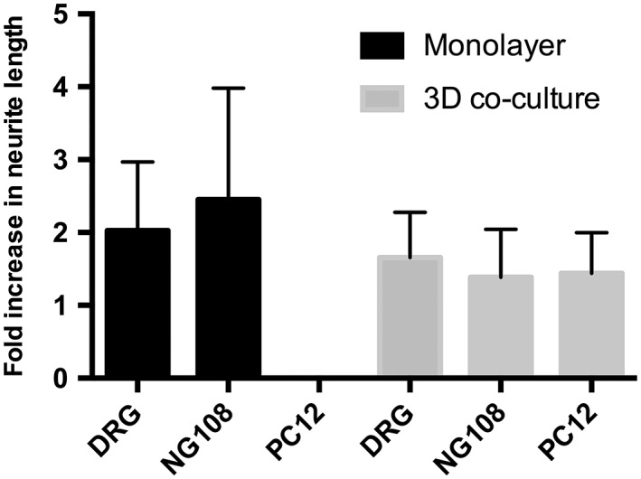
Peripheral nerve injuries (PNI) have a high prevalence and can be debilitating, resulting in life-long loss or disturbance in end-organ function, which compromises quality of life for patients. Current therapies use microsurgical approaches but there is the potential for enhancing recovery through other therapeutic modalities such as; cell-based conduits, gene therapy and small molecules. A number of molecular targets and drugs which have the potential to improve nerve regeneration have been identified, however, there are challenges associated with moving therapies toward clinical translation. Due to the lack of detailed knowledge about the pro-regenerative effect of potential drug treatments, there is a need for effective in vitro models to screen compounds to inform future pre-clinical and clinical studies. The interaction between regenerating neurites and supporting Schwann cells is a key feature of the nerve environment, therefore, in vitro models that mimic this cellular association are useful tools. In this study, we have investigated various cell culture models, including simple monolayer systems and more complex 3D-engineered co-cultures, as models for use in PNI drug development. Anat Rec, 301:1628-1637, 2018. © 2018 The Authors. The Anatomical Record published by Wiley Periodicals, Inc. on behalf of American Association of Anatomists.

Keywords: drug discovery; peripheral nerve; regeneration; therapies; tissue model.


Figures

Figure 1
Figure 1
Ibuprofen induces neurite growth in monolayer NG108–15 neurons. Significant increases in neurite length were seen in the presence of 100 μM and 200 μM ibuprofen when compared to the no‐drug treatment control, after 72 hr exposure. No neurite growth was seen with the growth inhibitor GW9662 (a). Fluorescence micrographs of the monolayer cultures show neurite length with (b) no drug treatment, (c) 100 μM GW9662, (d) 10 μM ibuprofen, (e) 100 μM ibuprofen and (f) 200 μM ibuprofen. Cultures were immunostained to detect β‐III tubulin (green). N = 6, mean ± SEM for each condition. One‐way ANOVA with Tukey's post hoc test, ***P < 0.001 and ****P < 0.0001.
Figure 2
Figure 2
Ibuprofen induces neurite growth on monolayer crude prep DRG neurons. Significant increases in neurite length were seen with 10 μM and 100 μM doses of ibuprofen, however, all doses increased neurite growth when compared to no drug control (shown by dotted line) and 100 μM GW9662 (shown in red) after 72 hr exposure (a). Fluorescence micrographs of the cultures show neurite length with no drug treatment (b), and 100 μM ibuprofen (c). Cultures were immunostained to detect β‐III tubulin (green). N = 6, mean ± SEM for each condition One‐way ANOVA with Dunnett's post hoc test, **P < 0.01.
Figure 3
Figure 3
Ibuprofen increases neurite growth in the 3D EngNT co‐culture model with both cell lines; (a) PC12 and (b) NG108–15. A significant increase in growth was seen with 100 μM ibuprofen but not with 10 μM with both cell lines. Additionally, no significant increase in neurite growth was seen with a 200 μM dose. Representation of 3D EngNT containing aligned Schwann cells (green) and neurons seeding on the gel surface (red) (c). Fluorescence micrographs of PC12 neuronal cells seeded on the gels without (d) and with (e) 100 μM drug treatment. Cultures were immunostained to detect β‐III tubulin (green). N = 6 of PC12 co‐culture and N = 3 of NG108–15 co‐culture gels, mean ± SEM for each condition. One‐way ANOVA with Dunnett's post hoc test, *P < 0.05, **P < 0.01, and ****P < 0.0001.
Figure 4
Figure 4
Ibuprofen induces neurite growth in the 3D EngNT co‐culture model with DRGs (a). A significant increase was seen with a dose of 100 μM ibuprofen but not 10 μM. Fluorescence micrographs of DRG neurons on EngNT gels without (b) and with (c) 10 μM drug treatment and (d) 100 μM drug treatment. Cultures were immunostained to detect β‐III tubulin (green). N = 6, mean ± SEM for each condition. One‐way ANOVA with Dunnett's post hoc test, *P < 0.05.
Figure 5
Figure 5
Ibuprofen increases the number of neurites detected in the distal stump 5 mm from the injury site following a local dose of 7 μg/day ibuprofen for 21 days. N = 3 animals per group. Mean ± SEM for each condition. T‐test, **P < 0.01.
Figure 6
Figure 6
The data sets for the 100 μM dose of ibuprofen in the various different in vitro models were normalized to their respective controls and presented as fold changes over the no drug treatment condition. Neurite growth increase was seen in all of the in vitro models except the monolayer PC12 cultures where no growth was seen. N = 6 (N = 3 for NG108–15 3D co‐culture). Mean ± SD.

References

    1. Ahmed I, Liu HY, Mamiya PC, Ponery AS, Babu AN, Weik T, Schindler M, Meiners S. 2006. Three‐dimensional nanofibrillar surfaces covalently modified with tenascin‐C‐derived peptides enhance neuronal growth in vitro. J Biomed Mater Res A 76:851–860. - PubMed
    1. Angius D, Wang H, Spinner RJ, Gutierrez‐Cotto Y, Yaszemski MJ, Windebank AJ. 2012. A systematic review of animal models used to study nerve regeneration in tissue‐engineered scaffolds. Biomaterials 33:8034–8039. - PMC - PubMed
    1. Backstrom E, Chambers BJ, Kristensson K, Ljunggren HG. 2000. Direct NK cell‐mediated lysis of syngenic dorsal root ganglia neurons in vitro. J Immunol 165:4895–4900. - PubMed
    1. Baldwin SP, Krewson CE, Saltzman WM. 1996. PC12 cell aggregation and neurite growth in gels of collagen, laminin and fibronectin. Int J Dev Neurosci 14:351–364. - PubMed
    1. Balgude AP, Yu X, Szymanski A, Bellamkonda RV. 2001. Agarose gel stiffness determines rate of DRG neurite extension in 3D cultures. Biomaterials 22:1077–1084. - PubMed

Publication types

MeSH terms

Substances