

Case Reports
doi: 10.4269/ajtmh.18-0431.
Case Report: Right Hemispheric Neuroimaging Abnormalities in a Patient with Dengue Encephalopathy
Affiliations
- PMID: 30334519
- PMCID: PMC6221225
- DOI: 10.4269/ajtmh.18-0431
Item in Clipboard
Case Reports
Case Report: Right Hemispheric Neuroimaging Abnormalities in a Patient with Dengue Encephalopathy
Am J Trop Med Hyg.
2018 Nov.
Abstract
Dengue encephalitis and dengue encephalopathy are frequent neurological complications of systemic dengue virus infection. Neuroimaging is normal in approximately 50% of patients. Common imaging abnormalities involve periventricular structures, including the basal ganglion, thalamus, and periventricular white matter. We describe an unusual case of dengue encephalopathy with unilateral imaging abnormalities involving the right cerebral hemisphere and mimicking the involvement of the right middle cerebral artery.
Figures
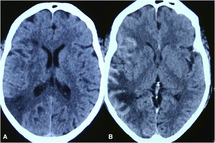
(A) Non-contrast computed tomography scan head showing sulcal effacement and ill-defined hypodensity on the right side (B) post contrast image showing gyral enhancement in the right temporoparietal region. This figure appears in color at www.ajtmh.org .
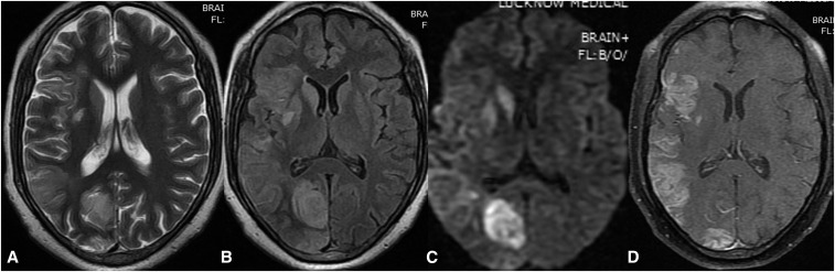
T2 and fluid-attenuated inversion recovery (FLAIR) (A and B) images showing signal intensity alteration involving right hemisphere and basal ganglia, respectively. (C) diffusion-weighted magnetic resonance imaging (DWI) image showing restricted diffusion in right basal ganglia and occipital region. (D) Post contrast T1 image showing gyral enhancement.
Similar articles
-
Dengue encephalopathy: very unusual neuroimaging findings.J Neurovirol. 2017 Oct;23(5):779-782. doi: 10.1007/s13365-017-0547-7. Epub 2017 Jul 17. J Neurovirol. 2017. PMID: 28718068
-
Encephalitis and myelitis associated with dengue viral infection clinical and neuroimaging features.Clin Neurol Neurosurg. 2008 Jun;110(6):635-40. doi: 10.1016/j.clineuro.2008.03.011. Epub 2008 May 7. Clin Neurol Neurosurg. 2008. PMID: 18467022
-
Acute disseminated encephalomyelitis complicating dengue infection with neuroimaging mimicking multiple sclerosis: A report of two cases.Mult Scler Relat Disord. 2016 Nov;10:112-115. doi: 10.1016/j.msard.2016.10.001. Epub 2016 Oct 4. Mult Scler Relat Disord. 2016. PMID: 27919476
-
Acute disseminated encephalomyelitis associated with dengue infection: a case report with literature review.J Neurol Sci. 2013 Dec 15;335(1-2):216-8. doi: 10.1016/j.jns.2013.08.029. Epub 2013 Sep 2. J Neurol Sci. 2013. PMID: 24035291 Review.
-
Encephalitis in the clinical spectrum of dengue infection.Neurol India. 2010 Jul-Aug;58(4):585-91. doi: 10.4103/0028-3886.68655. Neurol India. 2010. PMID: 20739797 Review.
Cited by
-
Viral Parkinsonism: An underdiagnosed neurological complication of Dengue virus infection.PLoS Negl Trop Dis. 2022 Feb 9;16(2):e0010118. doi: 10.1371/journal.pntd.0010118. eCollection 2022 Feb. PLoS Negl Trop Dis. 2022. PMID: 35139081 Free PMC article.
References
-
- Guzman MG, Gubler DJ, Izquierdo A, Martinez E, Halstead SB, 2016. Dengue infection. Nat Rev Dis Primers 2: 16055. - PubMed
-
- World Health Organization , 2018. Dengue and Severe Dengue: Fact Sheet. Available at: http://www.who.int/en/news-room/fact-sheets/detail/dengue-and-severe-dengue. Accessed July 9, 2018.
-
- Jain P, Prakash S, Khan DN, Garg RK, Kumar R, Bhagat A, Ramakrishna V, Jain A, 2017. Aetiology of acute encephalitis syndrome in Uttar Pradesh, India from 2014 to 2016. J Vector Borne Dis 54: 311–316. - PubMed
Publication types
MeSH terms
Substances
LinkOut - more resources
Full Text Sources
Medical
