

Applications of Isolated-Check Visual Evoked Potential in Early Stage of Open-Angle Glaucoma Patients
- PMID: 30334529
- PMCID: PMC6202595
- DOI: 10.4103/0366-6999.243564
Applications of Isolated-Check Visual Evoked Potential in Early Stage of Open-Angle Glaucoma Patients
Abstract
Background: Standard automated perimetry does not sufficiently detect early open-angle glaucoma (OAG) in the clinic. New visual function tests for early glaucoma damage are therefore needed. The present study evaluated whether an isolated-check visual evoked potential (icVEP) could be used to detect visual function abnormalities in early-stage OAG and to explore potential related factors.
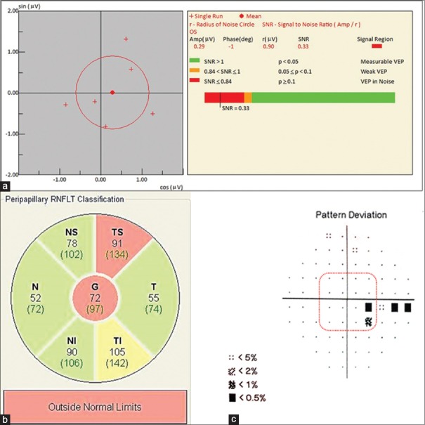
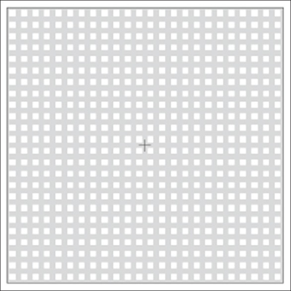
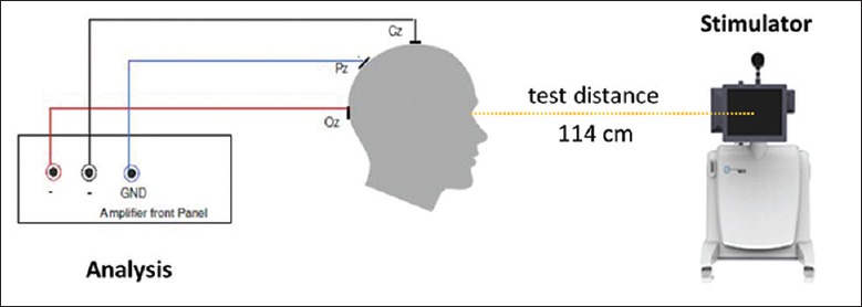
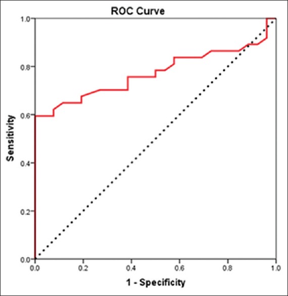
Methods: This was a cross-sectional study. Thirty-seven OAG patients with early-stage visual field loss (mean deviation ≥ -6.00 dB) detected by the Humphrey Field Analyzer (30-2 SITA program) and 26 controls were included in this study. Optical coherence tomography (OCT) was used to detect retinal nerve fiber layer (RNFL) defects. The icVEP preferentially evaluates the magnocellular-ON pathway. VEPs were recorded and signal-to-noise ratios (SNRs) were derived based on multivariate analysis. Eyes that yielded an SNR ≤1 were considered abnormal. Receiver operating characteristic (ROC) curve analysis was used to estimate the accuracy of group classification. Correlations between SNRs and related factors were analyzed.
Results: Based on an SNR criterion of 1, the icVEP had a sensitivity of 62.2% and a specificity of 92.3% for diagnosing early-stage OAG with 74.6% classification accuracy. The ROC curve analysis, however, suggested that an SNR criterion of 0.93 would produce the highest classification accuracy (77.3%). Both RNFL thinning in the temporal superior quadrant on OCT and number of abnormal test points in the central 11° visual field (pattern deviation, P < 0.5%) significantly correlated with the SNR (P < 0.05).
Conclusions: The icVEP detected visual function abnormalities in approximately 3/5 of eyes with early-stage OAG with greater than 90% specificity. SNR correlated with both a decrease in RNFL thickness and severity of central visual field loss.
分离格栅视觉诱发电位在早期开角型青光眼患者中的应用 摘要 背景: 标准自动视野计(SAP)在临床中无法充分地检测早期开角型青光眼。我们需要新型的视功能检查法来检测早期青光眼损伤。本研究将评估分离格栅视觉诱发电位(icVEP)在检测早期开角型青光眼(OAG)视功能异常中的能力,探索其潜力及相关因素。 方法: 这是一项横断面研究。37名结合标准自动视野计(Humphery视野分析仪,30-2 SITA程序)检测结果(MD≥-6dB)诊断为早期视野异常的早期OAG患者,以及26名健康志愿者被纳入该研究。光学相干断层扫描(OCT)用于检测OAG患者的视网膜神经纤维层(RNFL)缺损。icVEP能够特异性地刺激视网膜神经节细胞的大细胞(M细胞)ON通路。刺激产生视觉诱发电位被记录下来,通过多元统计方法得到信号噪声比(SNR)。SNR≤1被考虑为icVEP结果异常。受试者工作特征(ROC)曲线用于分析SNR分组的准确度。进一步分析相关因素与SNR的相关性。 结果: SNR判定值为1时,icVEP对早期OAG诊断的灵敏度为62.2%、特异度为92.3%,准确度为74.6%;而当SNR判定值为0.93时准确度达到最高值(77.3%),相应的灵敏度59.5%、特异度100%。颞上象限RNFL厚度的变薄以及中心11°视野的异常测试点数(模式偏差图,P<0.5%)均与SNR显著相关(P<0.05)。 结论: icVEP能够检测出早期OAG患者的近3/5存在青光眼性视功能异常,特异度超过90%;其评价指标SNR与OCT测得的RNFL厚度的下降以及中心11°视野缺损的程度均有相关性。.
Keywords: Cross-Sectional Study; Isolated-Check; Open-Angle Glaucoma; Signal-To-Noise Ratios; Visual Evoked Potential.
Conflict of interest statement
There are no conflicts of interest
Figures
References
-
- Quigley HA, Dunkelberger GR, Green WR. Chronic human glaucoma causing selectively greater loss of large optic nerve fibers. Ophthalmology. 1988;95:357–63. doi: 10.1016/S0161-6420(88)33176-3. - PubMed
-
- Quigley HA, Dunkelberger GR, Green WR. Retinal ganglion cell atrophy correlated with automated perimetry in human eyes with glaucoma. Am J Ophthalmol. 1989;107:453–64. doi: 10.1016/0002-9394(89)90488-1. - PubMed
-
- Bjerre A, Grigg JR, Parry NR, Henson DB. Test-retest variability of multifocal visual evoked potential and SITA standard perimetry in glaucoma. Invest Ophthalmol Vis Sci. 2004;45:4035–40. doi: 10.1167/iovs.04-0099. - PubMed
-
- Klistorner A, Graham SL. Objective perimetry in glaucoma. Ophthalmology. 2000;107:2283–99. doi: 10.1016/S0161-6420(00)00367-5. - PubMed
-
- Graham SL, Klistorner AI, Goldberg I. Clinical application of objective perimetry using multifocal visual evoked potentials in glaucoma practice. Arch Ophthalmol. 2005;123:729–39. doi: 10.1001/archopht.123.6.729. - PubMed
MeSH terms
LinkOut - more resources
Full Text Sources
Miscellaneous
