

Rickettsia typhi as Cause of Fatal Encephalitic Typhus in Hospitalized Patients, Hamburg, Germany, 1940-1944
- PMID: 30334722
- PMCID: PMC6200005
- DOI: 10.3201/eid2411.171373
Rickettsia typhi as Cause of Fatal Encephalitic Typhus in Hospitalized Patients, Hamburg, Germany, 1940-1944
Abstract
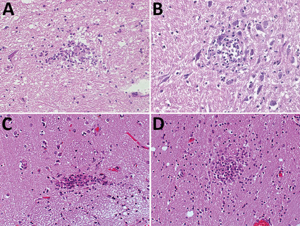
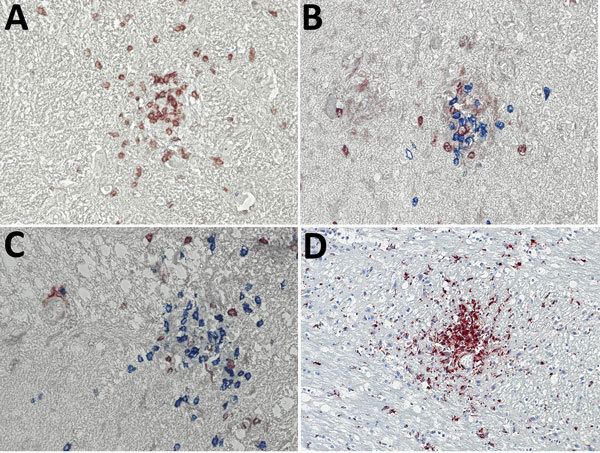
We evaluated formalin-fixed paraffin-embedded tissue specimens from 7 patients who died with encephalitic typhus in Hamburg, Germany, during World War II. The archived specimens included only central nervous system tissues >70 years old that had been stored at room temperature. We demonstrated successful detection of Rickettsia typhi DNA by a nested qPCR specific to prsA in 2 patients. These results indicate that R. typhi infections contributed to typhus outbreaks during World War II. Immunohistochemical analyses of brain tissue specimens of R. typhi DNA-positive and -negative specimens showed perivascular B-cell accumulation. Around blood vessels, nodular cell accumulations consisted of CD4-positive and CD8-positive T cells and CD68-positive microglia and macrophages; neutrophils were found rarely. These findings are similar to those of previously reported R. prowazekii tissue specimen testing. Because R. typhi and R. prowazekii infections can be clinically and histopathologically similar, molecular analyses should be performed to distinguish the 2 pathogens.
Keywords: CD4; CD8; Germany; Rickettsia prowazekii; Rickettsia typhi; T cells; World War II; bacteria; body louse; brain lesions; encephalitic typhus; endemic typhus; epidemic; fatality; flea; hospitalized patients; immunohistochemistry; murine typhus; nested PCR; outbreak; rat; rickettsiosis; typhus; typhus nodules; vector-borne infections.
Figures
References
-
- Keller C, Krüger A, Schwarz NG, Rakotozandrindrainy R, Rakotondrainiarivelo JP, Razafindrabe T, et al. High detection rate of Rickettsia africae in Amblyomma variegatum but low prevalence of anti-rickettsial antibodies in healthy pregnant women in Madagascar. Ticks Tick Borne Dis. 2016;7:60–5. 10.1016/j.ttbdis.2015.08.005 - DOI - PubMed
MeSH terms
LinkOut - more resources
Full Text Sources
Research Materials
