. 2019 Jan 2;129(1):192-208.
doi: 10.1172/JCI122819. Epub 2018 Nov 26.

Androgen receptor splice variant-7 expression emerges with castration resistance in prostate cancer

Affiliations

Androgen receptor splice variant-7 expression emerges with castration resistance in prostate cancer

Adam Sharp et al. J Clin Invest. .

Abstract

Background: Liquid biopsies have demonstrated that the constitutively active androgen receptor splice variant-7 (AR-V7) associates with reduced response and overall survival from endocrine therapies in castration-resistant prostate cancer (CRPC). However, these studies provide little information pertaining to AR-V7 expression in prostate cancer (PC) tissue.

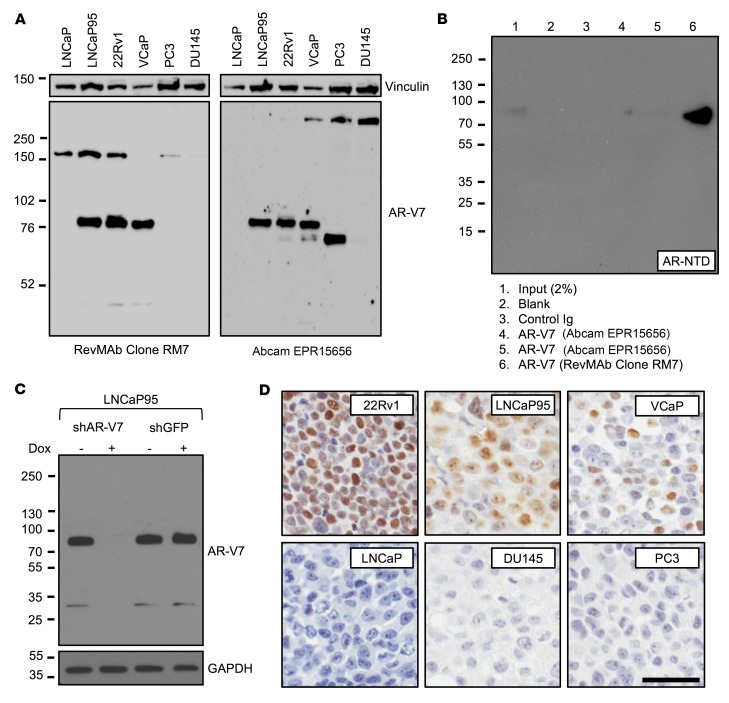
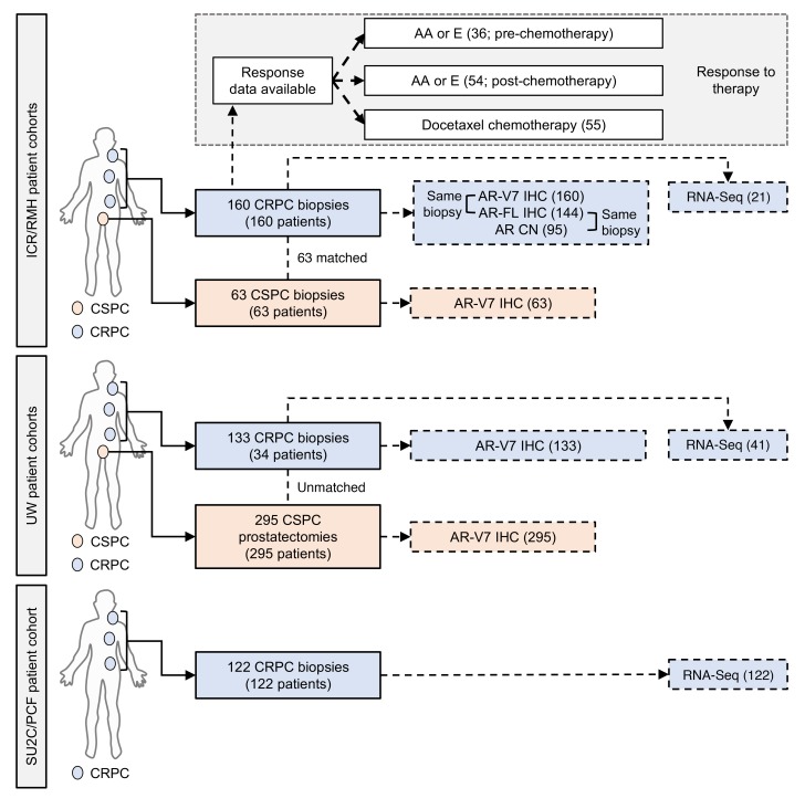
Methods: Following generation and validation of a potentially novel AR-V7 antibody for IHC, AR-V7 protein expression was determined for 358 primary prostate samples and 293 metastatic biopsies. Associations with disease progression, full-length androgen receptor (AR-FL) expression, response to therapy, and gene expression were determined.

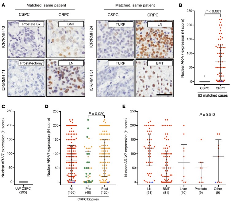
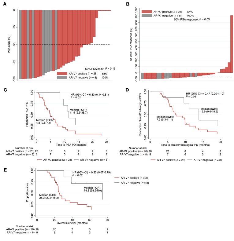
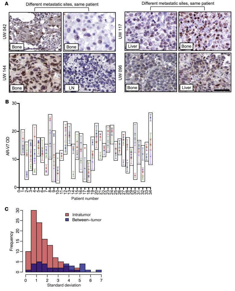
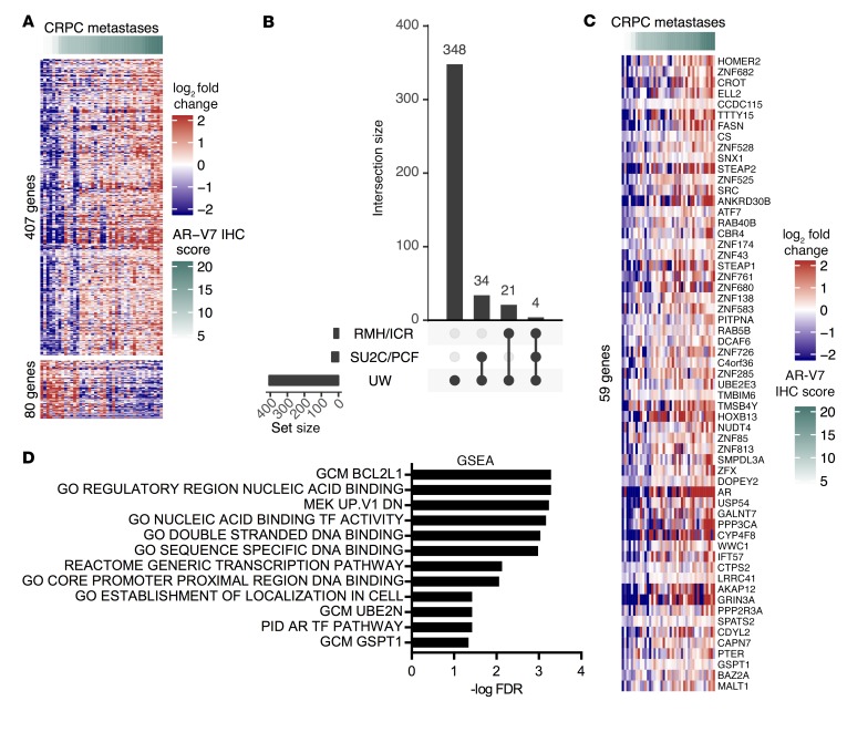
Results: We demonstrated that AR-V7 protein is rarely expressed (<1%) in primary PC but is frequently detected (75% of cases) following androgen deprivation therapy, with further significant (P = 0.020) increase in expression following abiraterone acetate or enzalutamide therapy. In CRPC, AR-V7 expression is predominantly (94% of cases) nuclear and correlates with AR-FL expression (P ≤ 0.001) and AR copy number (P = 0.026). However, dissociation of expression was observed, suggesting that mRNA splicing remains crucial for AR-V7 generation. AR-V7 expression was heterogeneous between different metastases from a patient, although AR-V7 expression was similar within a metastasis. Moreover, AR-V7 expression correlated with a unique 59-gene signature in CRPC, including HOXB13, a critical coregulator of AR-V7 function. Finally, AR-V7-negative disease associated with better prostate-specific antigen (PSA) responses (100% vs. 54%, P = 0.03) and overall survival (74.3 vs. 25.2 months, hazard ratio 0.23 [0.07-0.79], P = 0.02) from endocrine therapies (pre-chemotherapy).

Conclusion: This study provides impetus to develop therapies that abrogate AR-V7 signaling to improve our understanding of AR-V7 biology and to confirm the clinical significance of AR-V7.

Funding: Work at the University of Washington and in the Plymate and Nelson laboratories is supported by the Department of Defense Prostate Cancer Research Program (W81XWH-14-2-0183, W81XWH-12-PCRP-TIA, W81XWH-15-1-0430, and W81XWH-13-2-0070), the Pacific Northwest Prostate Cancer SPORE (P50CA97186), the Institute for Prostate Cancer Research, the Veterans Affairs Research Program, the NIH/National Cancer Institute (P01CA163227), and the Prostate Cancer Foundation. Work in the de Bono laboratory was supported by funding from the Movember Foundation/Prostate Cancer UK (CEO13-2-002), the US Department of Defense (W81XWH-13-2-0093), the Prostate Cancer Foundation (20131017 and 20131017-1), Stand Up To Cancer (SU2C-AACR-DT0712), Cancer Research UK (CRM108X-A25144), and the UK Department of Health through an Experimental Cancer Medicine Centre grant (ECMC-CRM064X).

Keywords: Oncology; Prostate cancer.


Conflict of interest statement

Conflict of interest: AS, WY, DD, DNR, IF, CB, GS, RR, AN, JW, SC, and JSDB are employees of The Institute of Cancer Research, which has a commercial interest in abiraterone. AS has served as an advisory member for Sanofi and Roche-Genentech. JL is an inventor of a relevant technology that has been licensed to A&G, Tokai, and Qiagen. PSN has served as an advisory member for Janssen, Astellas, and Roche. SPB has served as an advisory member for Janssen, Sanofi, and Astellas. JSDB has served as an advisory member for Astellas, Janssen, Taiho, Vertex, AstraZeneca, Bayer, Genmab, Genentech, GlaxoSmithKline, Merck, Pfizer, Roche, and Sanofi-Aventis. JSDB is a National Institute for Health Research (NIHR) Senior Investigator. The views expressed in this article are those of the author(s) and not necessarily those of the NHS, the NIHR, or the Department of Health.

Figures

Figure 1
Figure 1. Validation and optimization of a potentially novel AR-V7 antibody (Clone RM7) for immunohistochemistry.
(A) Western blot (long exposure) of AR-V7–positive (LNCaP95, 22Rv1, and VCaP) and –negative (LNCaP, PC3, and DU145) PC cell lines using a novel recombinant rabbit monoclonal anti–AR-V7 antibody (Clone RM7) and a previously reported anti–AR-V7 antibody (EPR15656). All cell lines except LNCaP95 (10% charcoal-stripped serum) were grown in 10% FBS. (B) Immunoprecipitation of AR-V7 from M12–cumate-inducible AR-V7 cells using the same concentration of AR-V7 antibodies and Western blot performed with AR N-terminal domain (AR-NTD) antibody. (C) LNCaP95 cells with doxycycline-inducible shRNA to AR-V7 were treated with (or without) doxycycline and Western blot performed with AR-V7 antibody (RM7). (D) Micrographs of AR-V7 detection by IHC using AR-V7 antibody (RM7) in cell line pellets positive (22Rv1, LNCaP95, and VCaP) and negative (LNCaP, DU145, and PC3) for AR-V7 (original magnification, ×200; scale bar: 50 μm).
Figure 2
Figure 2. Summary of clinical samples analyzed.
Overview of the ICR/RMH, UW, and SU2C/PCF patient cohorts utilized for this study. The ICR/RMH patient cohort included 63 CSPC biopsies and 160 CRPC biopsies stained for nuclear AR-V7 expression. Of the 160 biopsies with AR-V7 expression, AR-FL expression was available in 144 biopsies, AR copy number in 95, and RNA-Seq in 21. Response data were available for AA or E before chemotherapy in 36 patients, AA or E after chemotherapy in 54 patients, and docetaxel chemotherapy in 55 patients. The UW patient cohort included 295 CSPC tissues (from 295 patients) who had radical prostatectomy as primary therapy and 133 CRPC biopsies of metastases (from 34 patients). Of 133 CRPC biopsies from 34 patients with AR-V7 expression, RNA-Seq was available in 41 biopsies. The SU2C/PCF patient cohort included 122 CRPC biopsies with RNA-Seq analysis.
Figure 3
Figure 3. AR-V7 protein expression in PC.
(A) Representative micrographs of AR-V7 detection by IHC in 4 ICR/RMH patients with matched CSPC and CRPC biopsies (original magnification, ×200; scale bar: 50 μm). Prostate (Prostate Bx), prostatectomy, transurethral resection of the prostate (TURP), lymph node (LN), and bone marrow trephine (BMT) biopsies are shown. (B) Nuclear AR-V7 expression (H score [HS]) in 63 same-patient matched CSPC (gray) and CRPC (red) biopsies from the ICR/RMH cohort. Three AR-null CRPC cases with neuroendocrine features are shown (blue). Median HS and interquartile range are shown. P value was calculated using Wilcoxon signed-rank test. (C) Nuclear AR-V7 expression (HS) in 295 prostatectomy samples before any AR-targeted therapy. Median HS and interquartile range are shown. (D) Nuclear AR-V7 expression (HS) in 160 CRPC biopsies (red) and dichotomized (orange) by before (40 biopsies) and after (120 biopsies) AA or E treatment. Three AR-null CRPC cases with neuroendocrine features are shown (blue). Twenty biopsies taken after progression on primary ADT (with or without bicalutamide) and prior to standard therapy for CRPC are shown (green). Median HS and interquartile range are shown. P value was calculated using Mann-Whitney test. (E) Nuclear AR-V7 expression (HS) in 160 CRPC biopsies (red) from lymph node (LN), bone (BMT), liver, prostate, and other sites of metastases. Three AR-null CRPC cases with neuroendocrine features are shown (blue). Median HS and interquartile range are shown. P value was calculated using nonparametric equality-of-medians test.
Figure 4
Figure 4. AR-V7 status and response to AR-targeting therapies (AA or E) prior to chemotherapy in CRPC.
Thirty-six patients received AR-targeting therapies (AA or E) prior to chemotherapy for CRPC. (A) Percentage PSA nadir on AR-targeting therapies for AR-V7–negative (H score ≤ 10; gray) and AR-V7–positive (H score > 10; red) CRPC patients is shown. Fifty percent PSA nadir rate is shown. P value was calculated using Fisher’s exact test. (B) Percentage 12-week 50% PSA response rate on AR-targeting therapies for AR-V7–negative (gray) and AR-V7–positive (red) CRPC patients is shown. Twelve-week 50% PSA response rate is shown. P value was calculated using Fisher’s exact test. (CE) Kaplan-Meier curves show time to PSA progression (C), time to clinical/radiological progression (D), and overall survival (E) from start of AR-targeting therapy. Hazard ratios (HRs) with 95% confidence intervals (CIs) are shown. P value was calculated using univariate Cox proportional hazards model.
Figure 5
Figure 5. AR-FL and AR-V7 mRNA and protein quantification with AR copy number analysis in CRPC.
(A) AR-FL and AR-V7 mRNA expression in fragments per kilobase of transcript per million mapped reads (FPKM) for 122 CRPC transcriptomes from the SU2C/PCF cohort is shown. Spearman’s rank correlation is shown. (B) Expression (H score [HS]) for nuclear, cytoplasmic, and total (nuclear + cytoplasmic) AR-V7 (gray) and AR-FL N-terminal domain (NTD; red) is shown. Median HS and interquartile range are shown. (C) Expression (HS) of total AR-FL NTD protein and total AR-V7 protein in 144 CRPC biopsies from the ICR/RMH CRPC cohort is shown. Spearman’s rank correlation is shown. (D) Expression (HS) of total AR-FL NTD protein and AR copy number (log2) in 95 CRPC biopsies from the ICR/RMH CRPC cohort are shown. Cases with AR mutations are shown (L702H gray, T878A green, H875Y purple, K313E yellow). Spearman’s rank correlation is shown. (E) Expression (HS) of total AR-V7 protein and AR copy number (log2) in 95 CRPC biopsies from the ICR/RMH CRPC cohort are shown. Cases with AR mutations are shown. Spearman’s rank correlation is shown. (F) Expression (HS) of nuclear AR-FL NTD protein and nuclear AR-V7 protein in 144 CRPC biopsies from the ICR/RMH CRPC cohort is shown. Spearman’s rank correlation is shown.
Figure 6
Figure 6. AR-V7 protein expression variability within metastasis and between metastases from individual patients with CRPC.
(A) Representative micrographs of AR-V7 detection by IHC in 4 UW patients with multiple CRPC biopsies (original magnification, ×200; scale bar: 50 μm). (B) Nuclear AR-V7 expression (OD) in 133 metastases from 34 CRPC patients from the UW CRPC cohort. Mean OD and standard deviation (SD) for 3 measurements from each metastasis are shown. Each box encloses all metastases from a patient. Different colors for each patient represent an individual metastasis. (C) Frequency distribution of SD within a metastasis (Intratumor; comparison of triplicates in a metastasis; red) and between metastases (Between-tumor; comparison of multiple metastasis within a patient; blue) is shown. Median SD is 1.2 for intratumor measurements and 2.9 for between-tumor measurements.
Figure 7
Figure 7. AR-V7 protein expression is associated with a unique gene signature in metastatic CRPC.
(A) Expression (OD) of nuclear AR-V7 protein correlated (q < 0.05) with gene mRNA expression of 487 (407 upregulated and 80 downregulated) genes in 41 metastases from 24 patients from the UW CRPC cohort. Heatmap shows metastases ranked in order of nuclear AR-V7 expression (OD) and mean-centered log2 fold change in gene mRNA expression. (B) Fifty-nine of the 407 upregulated genes were validated in either 21 CRPC metastases from the ICR/RMH CRPC cohort or 122 CRPC transcriptomes from the SU2C/PCF cohort. Figure shows overlap of significantly correlated genes between the 3 cohorts. (C) Heatmap shows metastases ranked in order of nuclear AR-V7 expression (OD) and mean-centered log2 fold change in gene mRNA expression of the 59-gene signature in the UW CRPC cohort (n = 41). (D) Pathway overrepresentation analysis using MSigDB v6.2 (H, Hallmark; CP, Canonical Pathways; C4, Computational Gene Sets; C5, GO; and C6, Oncogenic Pathway) in the 59-gene set. Pathways with FDR less than 0.05 are shown. GSEA, gene set enrichment analysis.

References

    1. Siegel R, Naishadham D, Jemal A. Cancer statistics, 2013. CA Cancer J Clin. 2013;63(1):11–30. doi: 10.3322/caac.21166. - DOI - PubMed
    1. Visakorpi T, et al. In vivo amplification of the androgen receptor gene and progression of human prostate cancer. Nat Genet. 1995;9(4):401–406. doi: 10.1038/ng0495-401. - DOI - PubMed
    1. Robinson D, et al. Integrative clinical genomics of advanced prostate cancer. Cell. 2015;162(2):454. doi: 10.1016/j.cell.2015.06.053. - DOI - PubMed
    1. Chen CD, et al. Molecular determinants of resistance to antiandrogen therapy. Nat Med. 2004;10(1):33–39. doi: 10.1038/nm972. - DOI - PubMed
    1. Kumar A, et al. Substantial interindividual and limited intraindividual genomic diversity among tumors from men with metastatic prostate cancer. Nat Med. 2016;22(4):369–378. doi: 10.1038/nm.4053. - DOI - PMC - PubMed

Publication types

MeSH terms

Substances