

Sertoli-Leydig cell tumors of ovary: A case series
- PMID: 30334998
- PMCID: PMC6211859
- DOI: 10.1097/MD.0000000000012865
Sertoli-Leydig cell tumors of ovary: A case series
Abstract
Introduction: The purpose of this study was to report the clinical features, computed tomography (CT) and magnetic resonance imaging (MRI) findings, clinical management, and prognoses of 7 patients with Sertoli-Leydig cell tumors (SLCT) of ovary, and to review the literature of this rare condition.
Methods: Seven patients with pathologically confirmed ovarian SLCT were included. Their clinical, CT and MRI characteristics (CT images obtained from 6 patients and MR images from 4 patients), clinical management, and prognoses of 7 patients were retrospectively analyzed.
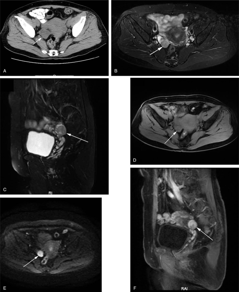
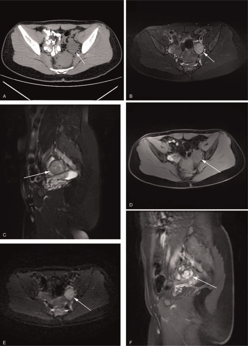
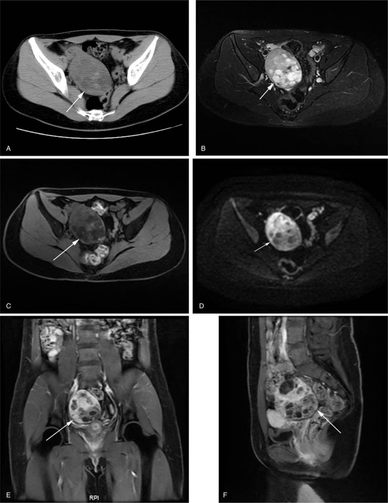
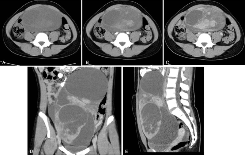
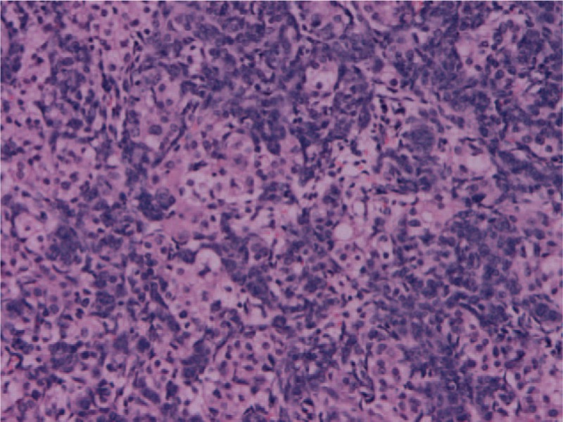
Results: Patients symptoms included irregular menstruation (n = 3), infertile (n = 1), vaginal bleeding after 7 years of menopause (n = 1), a palpable abdominal mass (n = 1), and abdominal pain (n = 1). Three patients had elevated alpha-fetoprotein (AFP), 1 had elevated cancer antigen 125 (CA125), and 2 had elevated Testosterone (T). The 7 tumors of 7 patients were solid or mixed solid-cystic mass with clear boundaries. The solid components of the tumors showed iso-dense on CT. On MRI, the solid components showed iso- or slightly low signal intensity (SI) on T1-weighted imaging (T1WI), high or slightly high SI on T2WI, and high on diffusion-weighted imaging (DWI) with low apparent diffusion coefficient (ADC) value. On contrast-enhanced CT and MRI, 1 tumor exhibited heterogeneous enhancement consisting of multiple nodules with relatively marked homogeneous enhancement, and other 6 tumors showed moderate or marked and constantly heterogeneous enhancements. All patients were treated with surgical excision. Only 3 had received postoperative chemotherapy. With the exception of 1 patient lost to follow-up, the other 6 patients exhibited tumor-free survival with a median follow-up time of 13.5 months, the longest follow-up time being 24 months.
Conclusion: The patients of SLCT can present with hormonal magnification and manifest high AFP, CA125, and T levels. SLCT is characterized by a solid or mixed solid-cystic mass on CT/MR scans, and shows marked or moderated heterogeneous and constantly enhancement upon postcontrast study. The clinical characteristics and imaging findings are features and appropriated imaging should be performed whenever an SLCT is suspected.
Conflict of interest statement
The authors have nothing to disclose and report no conflicts of interest.
Figures
Similar articles
-
Imaging, clinical, and pathologic findings of Sertoli-leydig cell tumors.Sci Prog. 2021 Apr-Jun;104(2):368504211009668. doi: 10.1177/00368504211009668. Sci Prog. 2021. PMID: 33848213 Free PMC article.
-
Contrast-enhanced ultrasound findings of ovarian Sertoli-Leydig cell tumor: a case report with ultrasonographic-pathological insights.BMC Womens Health. 2025 Jul 18;25(1):358. doi: 10.1186/s12905-025-03910-0. BMC Womens Health. 2025. PMID: 40682079 Free PMC article.
-
Sertoli-Leydig cell tumor of the ovary: Analysis of a single institution database and review of the literature.J Obstet Gynaecol Res. 2019 Jul;45(7):1311-1318. doi: 10.1111/jog.13977. Epub 2019 May 20. J Obstet Gynaecol Res. 2019. PMID: 31106943 Review.
-
Sertoli-Leydig cell tumor of ovary in children: A report of two cases, including retiform variant.Indian J Pathol Microbiol. 2021 Jul-Sep;64(3):559-562. doi: 10.4103/IJPM.IJPM_443_20. Indian J Pathol Microbiol. 2021. PMID: 34341273
-
Ovarian Sertoli-Leydig cell tumors: a single institution experience and review of the literature.Eur J Gynaecol Oncol. 2017;38(2):214-220. Eur J Gynaecol Oncol. 2017. PMID: 29953783 Review.
Cited by
-
Severe intrahepatic cholestasis of pregnancy due to a Sertoli-Leydig cell tumour in a woman with polycystic ovary syndrome: a case report.BMC Pregnancy Childbirth. 2022 Nov 2;22(1):807. doi: 10.1186/s12884-022-05159-z. BMC Pregnancy Childbirth. 2022. PMID: 36324123 Free PMC article.
-
Ovarian Sertoli-Leydig Tumour in a Postmenopausal Female With Virilisation: Magnetic Resonance Imaging Findings.Cureus. 2025 Jun 13;17(6):e85965. doi: 10.7759/cureus.85965. eCollection 2025 Jun. Cureus. 2025. PMID: 40662027 Free PMC article.
-
An Ovarian Sertoli-Leydig Cell Tumor with Elevated Alpha-Fetoprotein in an Adolescent: A Rare Case Report and Literature Review.Medicina (Kaunas). 2024 Sep 10;60(9):1477. doi: 10.3390/medicina60091477. Medicina (Kaunas). 2024. PMID: 39336518 Free PMC article. Review.
-
15-Year-Old Patient with an Unusual Alpha-Fetoprotein-Producing Sertoli-Leydig Cell Tumor of Ovary.Case Rep Obstet Gynecol. 2022 Apr 12;2022:4759826. doi: 10.1155/2022/4759826. eCollection 2022. Case Rep Obstet Gynecol. 2022. PMID: 35450124 Free PMC article.
-
Fertility-Sparing Surgery for Ovarian Cancer.J Clin Med. 2021 Sep 18;10(18):4235. doi: 10.3390/jcm10184235. J Clin Med. 2021. PMID: 34575345 Free PMC article. Review.
References
-
- Gui T, Cao D, Shen K, et al. A clinicopathological analysis of 40 cases of ovarian Sertoli-Leydig cell tumors. Gynecol Oncol 2012;127:384–9. - PubMed
-
- Akman L, Ertas IE, Gokcu M, et al. Ovarian Sertoli-Leydig cell tumors: a multicenter long-term clinicopathological analysis of 27 patients. J Cancer Res Ther 2016;12:290–4. - PubMed
-
- Edge SB, Compton CC. The American Joint Committee on Cancer: the 7th edition of the AJCC cancer staging manual and the future of TNM. Ann Surg Oncol 2010;17:1471–4. - PubMed
-
- Young RH, Scully RE. Ovarian Sertoli-Leydig cell tumors. A clinicopathological analysis of 207 cases. Am J Surg Pathol 1985;9:543–69. - PubMed
MeSH terms
Substances
LinkOut - more resources
Full Text Sources
Medical
Research Materials
Miscellaneous
