. 2018 Oct 18;16(10):e2005924.
doi: 10.1371/journal.pbio.2005924. eCollection 2018 Oct.

Preservation of myocardial contractility during acute hypoxia with OMX-CV, a novel oxygen delivery biotherapeutic

Affiliations

Preservation of myocardial contractility during acute hypoxia with OMX-CV, a novel oxygen delivery biotherapeutic

Jason Boehme et al. PLoS Biol. .

Erratum in

Abstract

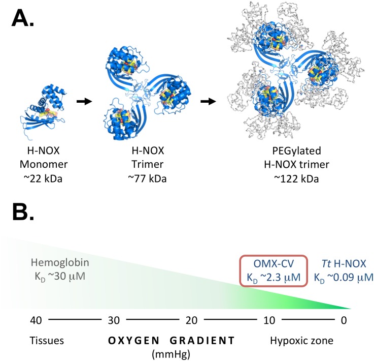
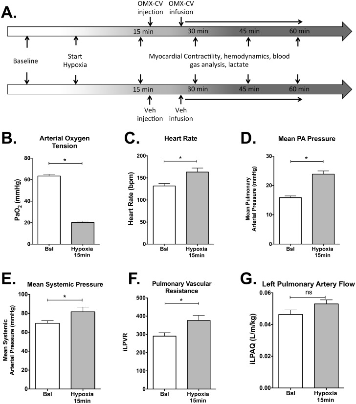
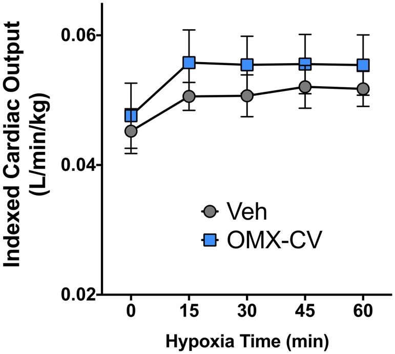
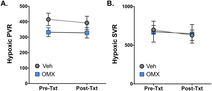
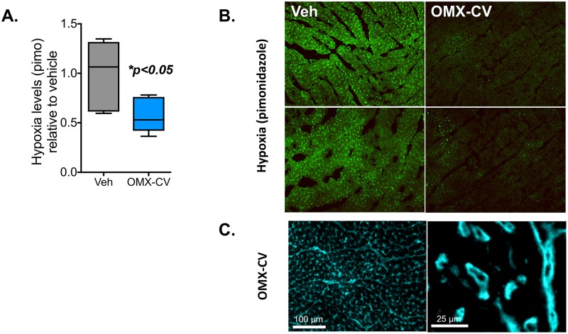
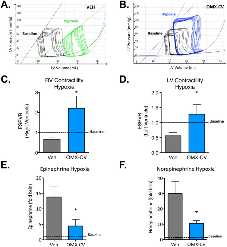
The heart exhibits the highest basal oxygen (O2) consumption per tissue mass of any organ in the body and is uniquely dependent on aerobic metabolism to sustain contractile function. During acute hypoxic states, the body responds with a compensatory increase in cardiac output that further increases myocardial O2 demand, predisposing the heart to ischemic stress and myocardial dysfunction. Here, we test the utility of a novel engineered protein derived from the heme-based nitric oxide (NO)/oxygen (H-NOX) family of bacterial proteins as an O2 delivery biotherapeutic (Omniox-cardiovascular [OMX-CV]) for the hypoxic myocardium. Because of their unique binding characteristics, H-NOX-based variants effectively deliver O2 to hypoxic tissues, but not those at physiologic O2 tension. Additionally, H-NOX-based variants exhibit tunable binding that is specific for O2 with subphysiologic reactivity towards NO, circumventing a significant toxicity exhibited by hemoglobin (Hb)-based O2 carriers (HBOCs). Juvenile lambs were sedated, mechanically ventilated, and instrumented to measure cardiovascular parameters. Biventricular admittance catheters were inserted to perform pressure-volume (PV) analyses. Systemic hypoxia was induced by ventilation with 10% O2. Following 15 minutes of hypoxia, the lambs were treated with OMX-CV (200 mg/kg IV) or vehicle. Acute hypoxia induced significant increases in heart rate (HR), pulmonary blood flow (PBF), and pulmonary vascular resistance (PVR) (p < 0.05). At 1 hour, vehicle-treated lambs exhibited severe hypoxia and a significant decrease in biventricular contractile function. However, in OMX-CV-treated animals, myocardial oxygenation was improved without negatively impacting systemic or PVR, and both right ventricle (RV) and left ventricle (LV) contractile function were maintained at pre-hypoxic baseline levels. These data suggest that OMX-CV is a promising and safe O2 delivery biotherapeutic for the preservation of myocardial contractility in the setting of acute hypoxia.


Conflict of interest statement

EM and JRF are consultants to Omniox, Inc.

Figures

Fig 1
Fig 1. Illustration of OMX-CV H-NOX protein and its oxygen-binding characteristics.
(A) Ribbon diagrams depicting an H-NOX protein monomer, H-NOX protein trimer, and PEGylated H-NOX protein trimer. The heme cofactor and the bound oxygen are depicted in yellow and red. Models were made using a Tt H-NOX structure (PDB ID 1U4H) and PyMOL [17]. (B) Illustration depicting the relative oxygen affinities of hemoglobin, Tt H-NOX, and OMX-CV overlaid on an oxygen gradient from normoxia to hypoxia. The oxygen affinity of hemoglobin facilitates release of oxygen in peripheral tissues (PO2 of about 40 mmHg), while the oxygen affinity of OMX-CV facilitates release of oxygen into hypoxic tissues (PO2 of about 10 mmHg). H-NOX, heme-based nitric oxide/oxygen; KD, dissociation constant; mmHg, millimeters mercury; OMX-CV, Omniox-cardiovascular; PEG, polyethylene glycol; PO2, partial pressure of oxygen; Tt, Thermoanaerobacter tengcongensis.
Fig 2
Fig 2. Physiologic responses of the cardiovascular system to acute alveolar hypoxia.
(A) Schematic of experimental protocol. Physiologic measurements were continuously recorded and logged every second for the duration of the study. At each designated time point, physiologic data were averaged over a 60-second period in 5-second intervals. (B) Average measured PaO2 in mmHg of all animals (n = 13) at baseline (Bsl) compared with 15 minutes following institution of hypoxic ventilation. (C) Average heart rate of all animals at Bsl compared with 15 minutes following institution of hypoxic ventilation. (D) Average mean pulmonary arterial pressure (in mmHg) of all animals at Bsl compared with 15 minutes following institution of hypoxic ventilation. (E) Average mean systemic arterial pressure (in mmHg) of all animals at Bsl compared with 15 minutes following institution of hypoxic ventilation. (F) Average indexed PVR of all animals at baseline (Bsl) compared with 15 minutes following institution of hypoxic ventilation. PVR of the left lung was calculated as the difference of mean pulmonary arterial pressure and left atrial pressure divided by the indexed LPA blood flow. (G) Average indexed left pulmonary arterial blood flow of all animals at Bsl compared with 15 minutes following institution of hypoxic ventilation. Flow was indexed to body size by dividing by the animal’s weight in kilograms. In all figures, “*” denotes significance with p < 0.05, while “ns” denotes p > 0.05. Error bars demonstrate standard error of the mean. Primary data can be found in S1 Table. bpm, beats per minute; Bsl, baseline; iLPAQ, indexed left pulmonary artery flow; iLPVR, indexed left pulmonary vascular resistance; LPA, left pulmonary artery; mmHg, millimeters mercury; OMX-CV, Omniox-cardiovascular; PA, pulmonary artery; PaO2, arterial oxygen tension; Veh, vehicle.
Fig 3
Fig 3. Cardiac output in control and OMX-CV–treated animals.
Indexed left pulmonary arterial blood flow in vehicle- versus OMX-CV–treated groups over the duration of the experimental protocol. Time 0 represents the physiologic baseline and other time points represent total duration of hypoxic ventilation. Error bars correspond to the standard error of the mean. There is a statistically significant interaction between time and iLPA flow (p <0.05) by two-way ANOVA. There is no significant difference between OMX-CV (n = 6) and vehicle (n = 7) groups. Primary data can be found in S1 Table. iLPA, indexed left pulmonary artery; OMX-CV, Omniox-cardiovascular; Veh, vehicle.
Fig 4
Fig 4. SVR and PVR before and after OMX-CV and vehicle administration.
(A) Indexed PVR in vehicle- (n = 7) and OMX-CV–treated (n = 6) animals during hypoxic ventilation immediately prior to (pre-txt) and following (post-txt) treatment administration. There are no statistically significant differences between groups or within groups pre- and posttreatment. Error bars represent the standard error of the mean. (B) Indexed SVR in vehicle- and OMX-CV–treated animals pre-txt and post-txt. There are no statistically significant differences between groups or within groups pre- and posttreatment. Error bars represent the standard error of the mean. Primary data can be found in S1 Table. OMX-CV, Omniox-cardiovascular; post-txt, immediately following treatment administration; pre-txt, immediately prior to treatment administration; PVR, pulmonary vascular resistance; SVR, systemic vascular resistance; Veh, vehicle.
Fig 5
Fig 5. Myocardial hypoxia in control and OMX-CV–treated animals.
In a subset of vehicle- and OMX-CV–treated animals (n = 3 each), following measurement of physiologic parameters, pimonidazole was administered intravenously and tissues were collected for analysis 30 minutes later. (A) Quantification of pimonidazole adducts in vehicle- and OMX-CV–treated myocardial tissue by pimonidazole ELISA. Values are ±SEM, *p < 0.05 by Student t test. (B) Representative images of vehicle- and OMX-CV–treated myocardium tissue sections immunostained with antibodies targeting pimonidazole adducts. (C) Representative images of OMX-CV–treated myocardial tissue sections immunostained with antibodies targeting the OMX-CV molecule. OMX-CV, Omniox-cardiovascular; pimo, pimonidazole; Veh, vehicle.
Fig 6
Fig 6. Ventricular contractility and circulating catecholamine levels in control and OMX-CV–treated animals.
(A) Representative Pressure-Volume loops obtained from the left ventricle of a vehicle-treated animal during transient IVC occlusion. LV pressure is measured on the y-axis and LV volume on the x-axis. The superimposed line tangential to the end systolic pressure volume points of each family of loops defines the ESPVR. The family of loops in black and their corresponding ESPVR were obtained during the physiologic baseline, while the green loops and ESPVR were obtained from the same animal following 1 hour of hypoxic ventilation. (B) Representative Pressure-Volume loops obtained from the LV of an OMX-CV–treated animal during transient IVC occlusion. The family of loops in black and their corresponding ESPVR were obtained during the physiologic baseline, while the blue loops and ESPVR were obtained from the same animal following 1 hour of hypoxic ventilation. (C) Mean right ventricular contractility (as assessed by slope of the ESPVR relative to baseline) in vehicle- (n = 7) and OMX-CV–treated (n = 6) animals after 1 hour of hypoxic ventilation. Error bars represent the standard error of the mean; “*” denotes a significant difference between groups with p < 0.05. (D) Mean left ventricular contractility (as assessed by slope of the ESPVR relative to baseline) in vehicle- (n = 7) and OMX-CV–treated (n = 6) animals after 1 hour of hypoxic ventilation. Error bars represent the standard error of the mean; “*” denotes a significant difference between groups with p < 0.05. (E) Mean serum epinephrine levels (expressed as fold change relative to physiologic baseline) after 1 hour of hypoxic ventilation in vehicle- (n = 7) and OMX-CV–treated (n = 6) animals. Error bars represent the standard error of the mean; “*” denotes a significant difference between groups with p < 0.05. (F) Mean serum norepinephrine levels (expressed as fold change relative to physiologic baseline) at 1 hour of hypoxic ventilation in vehicle- and OMX-CV–treated animals. Error bars represent the standard error of the mean; “*” denotes a significant difference between groups with p < 0.05. Primary data can be found in S1 Table. bsln, baseline; ESPVR, end systolic pressure-volume relationship; IVC, inferior vena cava; LV, left ventricle; mmHg, millimeters mercury; OMX-CV, Omniox-cardiovascular; RV, right ventricle; Veh, vehicle.

Similar articles

Cited by

References

    1. Kutko MC, Calarco MP, Flaherty MB, Helmrich RF, Ushay HM, et al. (2003) Mortality rates in pediatric septic shock with and without multiple organ system failure. Pediatr Crit Care Med 4: 333–337. 10.1097/01.PCC.0000074266.10576.9B - DOI - PubMed
    1. Martin GS (2012) Sepsis, severe sepsis and septic shock: changes in incidence, pathogens and outcomes. Expert Rev Anti Infect Ther 10: 701–706. 10.1586/eri.12.50 - DOI - PMC - PubMed
    1. Heckbert SR, Vedder NB, Hoffman W, Winn RK, Hudson LD, et al. (1998) Outcome after hemorrhagic shock in trauma patients. J Trauma 45: 545–549. - PubMed
    1. Reynolds HR, Hochman JS (2008) Cardiogenic shock: current concepts and improving outcomes. Circulation 117: 686–697. 10.1161/CIRCULATIONAHA.106.613596 - DOI - PubMed
    1. Taegtmeyer H (1994) Energy metabolism of the heart: from basic concepts to clinical applications. Curr Probl Cardiol 19: 59–113. - PubMed

Publication types

MeSH terms