. 2018 Oct 18;14(10):e1007643.
doi: 10.1371/journal.pgen.1007643. eCollection 2018 Oct.

Identification of UHRF2 as a novel DNA interstrand crosslink sensor protein

Affiliations

Identification of UHRF2 as a novel DNA interstrand crosslink sensor protein

Anna Motnenko et al. PLoS Genet. .

Abstract

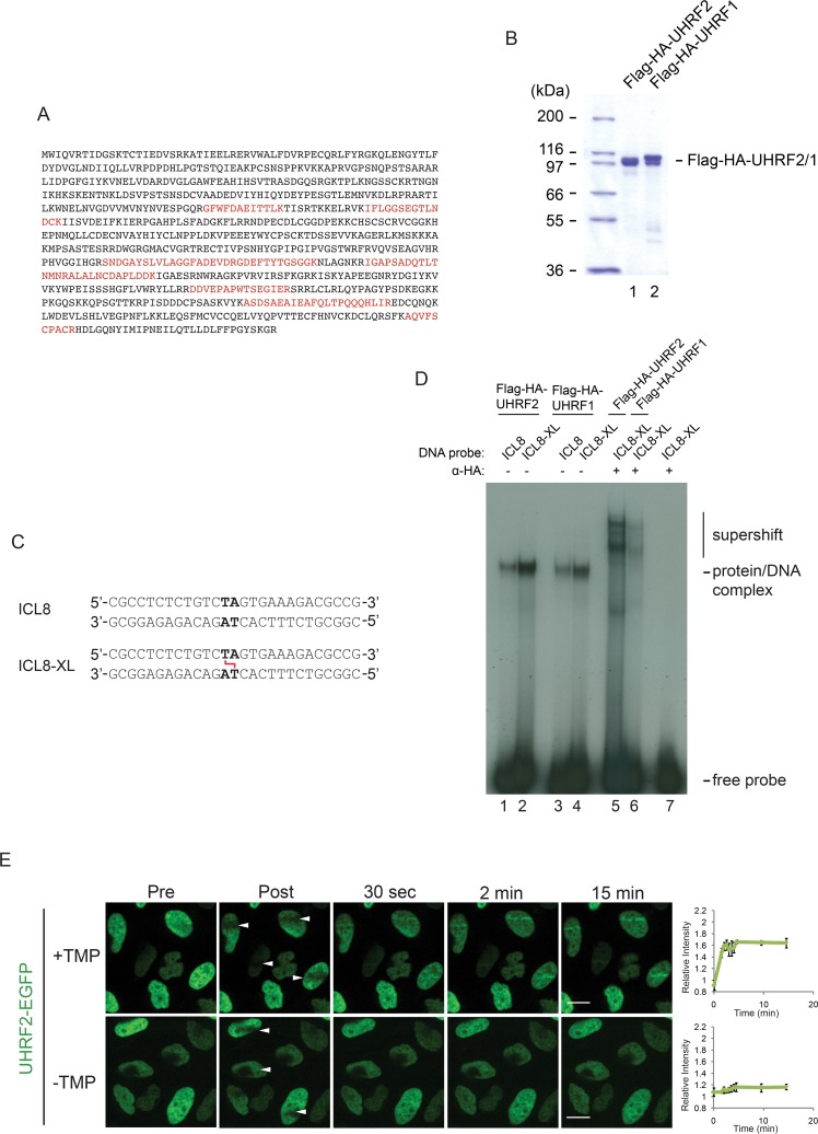
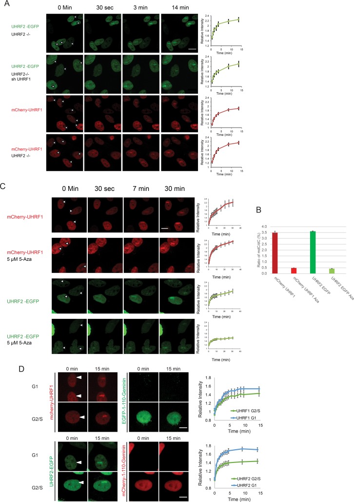
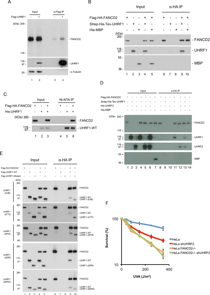
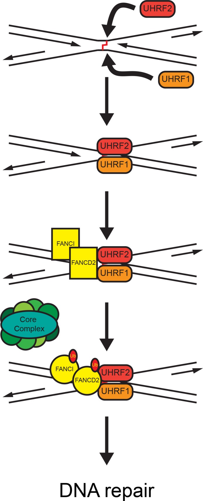
The Fanconi Anemia (FA) pathway is important for repairing interstrand crosslinks (ICLs) between the Watson-Crick strands of the DNA double helix. An initial and essential stage in the repair process is the detection of the ICL. Here, we report the identification of UHRF2, a paralogue of UHRF1, as an ICL sensor protein. UHRF2 is recruited to ICLs in the genome within seconds of their appearance. We show that UHRF2 cooperates with UHRF1, to ensure recruitment of FANCD2 to ICLs. A direct protein-protein interaction is formed between UHRF1 and UHRF2, and between either UHRF1 and UHRF2, and FANCD2. Importantly, we demonstrate that the essential monoubiquitination of FANCD2 is stimulated by UHRF1/UHRF2. The stimulation is mediating by a retention of FANCD2 on chromatin, allowing for its monoubiquitination by the FA core complex. Taken together, we uncover a mechanism of ICL sensing by UHRF2, leading to FANCD2 recruitment and retention at ICLs, in turn facilitating activation of FANCD2 by monoubiquitination.


Conflict of interest statement

The authors have declared that no competing interests exist.

Figures

Fig 1
Fig 1. UHRF2 binds ICLs in vitro and is recruited to ICLs in vivo.
A) Mass spectrometry identification of UHRF2. 11 peptides were identified. Amino acids present in identified peptides are indicated in red color. B) Coomassie blue stain of Flag-HA-tagged recombinant UHRF1 and UHRF2 proteins purified from Sf9 insect cells. C) Schematic of non-ICL and ICL-containing DNA probes used for EMSA in (D). D) EMSA showing the preferential binding of recombinant Flag-HA-tagged UHRF2 and UHRF1 to ICL-containing DNA probe (ICL8-XL) compared to control DNA probe (ICL8) DNA. A super-shift using HA antibody against Flag-HA-tagged UHRF2 and UHRF1 confirms that the protein/DNA complex is specifically formed by UHRF2 and UHRF1 binding to the DNA probe. E) Live-cell imaging of HeLa cells expressing exogenous EGFP-tagged UHRF2 with and without pre-treatment with TMP. Cells were micro irradiated at the sites indicated with white arrows. Scale bar indicates 10μm. Charts indicate quantification of relative intensity of signal at the irradiated sites. UHRF2 was recruited only the presence of TMP. Error bars show SEM n = 4/treatment.
Fig 2
Fig 2. Depletion of UHRF2 reduces cell survival in response to ICL-causing agents.
A) Clonogenic survival assay of HeLa and HeLa UHRF2-/- cells in response to MMC. Depletion of UHRF2 reduces cell survival. Experiment was repeated 3 times, each with a p-value <0.01 at 2 ng/ml MMC. B) Clonogenic survival assay of HeLa and HeLa UHRF2 -/- cells in response to TMP/UVA. Depletion of UHRF2 reduces cell survival. Experiment was repeated 2 times, each with a p-value <0.01 at 80 J/m2. C) Clonogenic survival assay of HeLa and HeLa UHRF2-/- cells in response to UVC. Experiment was performed once in triplicate. D) Clonogenic survival assay of HeLa and HeLa UHRF2-/- cells in response to HU. Experiment was performed once in triplicate.
Fig 3
Fig 3. The SRA domain of UHRF2 is required for its recruitment to ICLs.
A) Schematic representation of UHRF2 indicating positions of domains and deletions. B) Live-cell imaging of HeLa UHRF2 -/- cells (CRIPR/Cas9-mediated knockout) complemented with EGFP-tagged UHRF2 containing deletions as indicated in (A). Cells were pre-treated with TMP and micro irradiated at the sites indicated with white arrows. Scale bar indicates 10μm. Charts indicate quantification of relative intensity of signal at the irradiated sites. The p-values between EGFP-UHRF2 and EGFP-UHRF2-ΔTTD, EGFP-UHRF2-ΔPHD and EGFP-UHRF2-ΔSRA at the 20 min time-point are 0.001, 0.001, and 0.0000008, respectively. Error bars show SEM n = 8/treatment. C) Clonogenic survival assay of HeLa UHRF2 -/- cells complemented with the UHRF2 domain deletion mutants indicated in (A). Experiment was repeated 2 times and with p-values for EGFP-UHRF2-ΔSRA and EGFP-UHRF2-ΔTTD compared to EGFP-UHRF2 of 0.03 and 0.175, respectively. Error bars show SEM.
Fig 4
Fig 4. UHRF2 and UHRF1 mediate FANCD2 recruitment and ubiquitination in response to DNA damage.
A) HeLa cells expressing mCherry-tagged FANCD2 where subjected to depletion of UHRF1 by shRNA and/or depletion of UHRF2 by CRISPR/Cas9-mediated knockout, pre-treated with TMP, and then microirradiated at the sites indicated with white arrows. Charts indicate quantification of relative intensity of signal at the irradiated sites. Depletion of UHRF1 and UHRF2 impairs FANCD2 recruitment. Scale bar indicates 10μm n = 8/treatment. B) Western blot analysis of lysates from HeLa cells or HeLa cells where UHRF1 and/or UHRF2 were depleted using shRNA-mediated knockdown or CRISPR/Cas9-mediated knockout following treatment with TMP/UVA and harvested at 3 and 6 hours. Asterisk indicate unspecific band. Chart represents data from three independent experiments (two replicate experiments are shown in S5 Fig), and shows ratio of FANCD2-Ub to FANCD2. Strong accumulation of monoubiquitinated FANCD2 (FANCD2-Ub) occurs in HeLa cells but is reduced when UHRF1 and UHRF2 are depleted (p-value = 0.059 for the 6h timepoint). C) Flag-HA-tagged UHRF2 or Flag-HA-tagged UHRF1 were expressed in HeLa cells and immunoprecipitated from nuclear extracts using anti-Flag antibodies. Immunoprecipitates were analyzed by immunoblotting using anti-UHRF2 and anti-UHRF1 antibodies, as indicated. UHRF2 is co-immunoprecipitated with UHRF1 and vice versa. p300 is used as a loading control. D) In vitro binding assays of His-UHRF2 and wild type or deletion mutants of Flag-UHRF1 (purified from Sf9 cells). His-UHRF2 was immunoprecipitated and immunoblotting demonstrated that the SRA domain of UHRF2 is responsible for the protein-protein interaction between UHRF1 and UHRF2. E) Clonogenic survival assay of HeLa cells where UHRF1 and/or UHRF2 are depleted. Error bars show SEM. n = 3.
Fig 5
Fig 5
A) UHRF1 and UHRF2 are recruited to ICLs in vivo independently of each other. HeLa cells expressing either EGFP-tagged UHRF2 or mCherry-tagged UHRF1, were depleted of endogenous UHRF2 (CRISPR/Cas-9) and/or UHRF1 (shRNA). The cells were pre-treated with TMP and then microirradiated at the indicated sites. Charts indicate quantification of relative intensity of signal at the irradiated sites. Error bars show SEM n = 8/treatment. B) Genomic DNA from cells in (C) was assessed for the degree of DNA methylation. The percent of methylated cytosines vs. non-methylated cytosines is shown for each treatment. C) Cells expressing mCherry-tagged UHRF1 or EGFP-tagged UHRF2 were subjected to 5 μM 5-Aza-2'-deoxycytidine for 5 days. Cells were pre-treated with TMP and microirradiated at the sites indicated by the white arrows. The charts show the quantification of the stripe intensity. Error bars show SEM, n = 8 per treatment. The p-value for UHRF2 Aza compared to control UHRF2 is 0.005. D) Cells expressing either UHRF1-mCherry and EGFP-1-110-Geminin, or, UHRF2-EGFP and mCherry-1-110-Geminin, were imaged for 15 after the introduction of ICLs by microirradiation following treatment with TMP. The intensity of the fluorescent proteins in the microirradiated areas were quantified and plotted, in either G1 or S/G2-phase cells. Representative images shown. Error is shown as SEM, n = 16 per fluorophore. Scale bar indicates 10μm.
Fig 6
Fig 6. UHRF1 interacts directly with FANCD2.
A) Immunoprecipitation of Flag-UHRF1 from HeLa.shUHRF1 cells expressing exogenous Flag-UHRF1. HeLa.Scramble was used as a negative control. Cells were treated with TMP/UVA and allowed to recover for 1 hour before lysis, immunoprecipitation and immunoblotting. B) In vitro binding assay using Flag-HA-FANCD2, Strep-6xHis-Tev-UHRF1 (both purified from Sf9 cells) and 6xHis-MBP (purified from E. coli). HA-FANCD2 was immunoprecipitated and immunoblotting demonstrated co-immunoprecipitation of UHRF1 but not of MBP, which was used as a negative control. C) In vitro binding assay using Flag-HA-FANCD2 and 6xHis-UHRF1 (both purified from Sf9 cells). 6xHis-UHRF1 was purified using NTA-agarose, and immunoblotting demonstrated co-purification of FANCD2. D) In vitro binding assay using HA-FANCD2, Strep-6xHis-Tev-UHRF1, 6xHis-UHRF2 (all purified from Sf9 cells) and 6xHis-MBP (purified from E. coli). HA-FANCD2 was immunoprecipitated and immunoblotting demonstrated co-immunoprecipitation of UHRF1 and UHRF2 but not of MBP, which was used as a negative control. E) In vitro binding assays of Flag-HA-FANCD2 and wild type or deletion mutants of Flag-UHRF1 (purified from Sf9 cells). HA-FANCD2 was immunoprecipitated and immunoblotting demonstrated that the SRA domain of UHRF1 is responsible for the protein-protein interaction between UHRF1 and FANCD2. F) Clonogenic survival assay of HeLa, HeLa FANCD2 -/-, HeLa shUHRF2, and HeLa FANCD2 -/- shUHRF1 cells in response to TMP/UVA. Experiment was performed once in triplicate.
Fig 7
Fig 7
A proposed model of FANCD2 and FANCI recruitment and activation at ICLs via UHRF1 and UHRF2. UHRF1 and UHRF2 are rapidly and independently recruited to the site of the ICL. Recruitment of UHRF1 and UHRF2 facilitate recruitment/retention of the FANCD2/FANCI complex. When FANCD2 and FANCI are bound at the ICL a putative conformational change allows for monoubiquitination by the core complex. The now active FANCD2/FANCI complex activates downstream repair proteins, leading to repair of the ICL.

References

    1. Duxin JP, Walter JC. What is the DNA repair defect underlying Fanconi anemia? Curr Opin Cell Biol. 2015;37:49–60. 10.1016/j.ceb.2015.09.002 ; PubMed Central PMCID: PMCPMC4688103. - DOI - PMC - PubMed
    1. Auerbach AD. Fanconi anemia and its diagnosis. Mutat Res. 2009;668(1–2):4–10. Epub 2009/07/23. 10.1016/j.mrfmmm.2009.01.013 ; PubMed Central PMCID: PMCPMC2742943. - DOI - PMC - PubMed
    1. Lopez-Martinez D, Liang CC, Cohn MA. Cellular response to DNA interstrand crosslinks: the Fanconi anemia pathway. Cellular and molecular life sciences: CMLS. 2016;73(16):3097–114. 10.1007/s00018-016-2218-x ; PubMed Central PMCID: PMCPMC4951507. - DOI - PMC - PubMed
    1. Ceccaldi R, Sarangi P, D'Andrea AD. The Fanconi anaemia pathway: new players and new functions. Nat Rev Mol Cell Biol. 2016;17(6):337–49. 10.1038/nrm.2016.48 . - DOI - PubMed
    1. Bluteau D, Masliah-Planchon J, Clairmont C, Rousseau A, Ceccaldi R, Dubois d'Enghien C, et al. Biallelic inactivation of REV7 is associated with Fanconi anemia. J Clin Invest. 2016. 10.1172/JCI88010 . - DOI - PMC - PubMed

Publication types

MeSH terms