

Sonographic assessment of the prevalence and evolution of fluid collections as a complication of kidney transplantation
- PMID: 30335921
- PMCID: PMC6440513
- DOI: 10.15557/JoU.2018.0018
Sonographic assessment of the prevalence and evolution of fluid collections as a complication of kidney transplantation
Abstract
Aim of the study: The aim of this study is to assess the prevalence and evolution of perirenal fluid collections in a group of 488 patients who have undergone kidney transplantation.
Material and methods: Sonographic documentation of 488 deceased-donor kidney recipients was evaluated for the prevalence of perirenal fluid collections and their evolution in time, depending on selected demographic features of the patients, time of detection, initial dimensions and precise position of the collection relative to the kidney and the location of the transplanted organ in the right or left iliac fossa. The collected data were used for statistical analysis to determine the strength of the potential relationships.
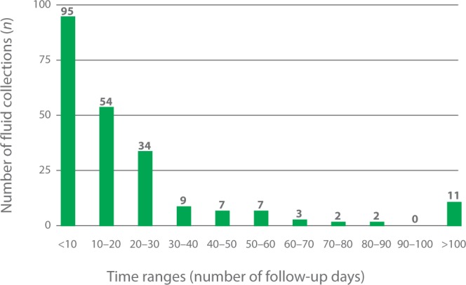
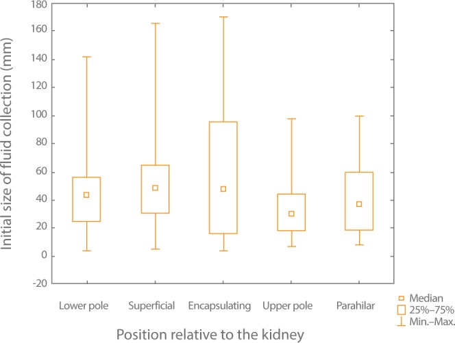
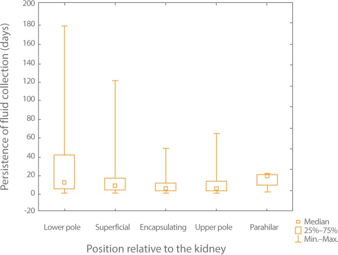
Results: In 146 out of 488 subjects perirenal fluid collections were found. In 1/3 of the patients more than one fluid collection was diagnosed. Over 40% of fluid collections were detected within 10 days from the date of the first scan and 24.11% were detected within 10-20 days from the date of the first scan. The majority of fluid collections were located near the lower pole of the kidney. Perihilar collections were the least common. Collections encapsulating the kidney and subcutaneous collections were the largest in size on average. A statistically significant difference between the size of collections located on the surface and the size of those located near the upper pole of the transplanted kidney was demonstrated. However, no correlation was proven to exist between the persistence of the fluid collection and its position relative to the transplanted kidney and its initial size.
Conclusions: The correct evaluation of a fluid collection's dynamics of development and nature requires periodic follow-up of the recipient, preferably in a single clinical center. Ultrasonography is an inexpensive, non-invasive and repeatable method for the determination of the presence of fluid collections. However, the decision whether treatment is necessary requires the sonographic image to be compared with the laboratory signs of inflammation and biochemical analysis of the contents of fluid collections.
Keywords: diagnostic imaging; kidney diseases; kidney transplantation; ultrasonography.
© Polish Ultrasound Society.
Figures


References
-
- Centrum Organizacyjno-Koordynacyjne ds. Transplantacji : http://www.poltransplant.pl/.
-
- Pollak R, Veremis SA, Maddux MS, Mozes MF: The natural history of and therapy for perirenal fluid collections following renal transplantation. J Urol 1988; 140: 716–720. - PubMed
-
- Friedewald SM, Molmenti EP, Friedewald JJ, DeJong MR, Hamper UM: Vascular and nonvascular complications of renal transplants: sonographic evaluation and correlation with other imaging modalities, surgery, and pathology. J Clin Ultrasound 2005; 33: 127–139. - PubMed
-
- Brown ED, Chen MY, Wolfman NT, Ott DJ, Watson NE Jr: Complications of renal transplantation: Evaluation with US and radionuclide imaging. Radiographics 2000; 20: 607–622. - PubMed
-
- Dubeaux VT, Oliveira RM, Moura VJ, Pereira JM, Henriques FP: Assessment of lymphocele incidence following 450 renal transplantations. Int Braz J Urol 2004; 30: 18–21. - PubMed
LinkOut - more resources
Full Text Sources
Research Materials