. 2018 Dec 15;444(2):71-82.
doi: 10.1016/j.ydbio.2018.10.002. Epub 2018 Oct 15.

FACT complex gene duplicates exhibit redundant and non-redundant functions in C. elegans

Affiliations

FACT complex gene duplicates exhibit redundant and non-redundant functions in C. elegans

Brittany Z Suggs et al. Dev Biol. .

Abstract

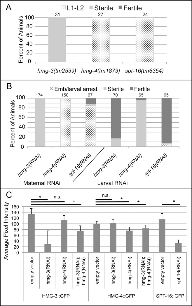
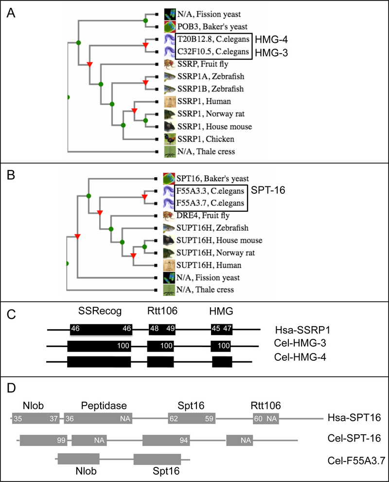
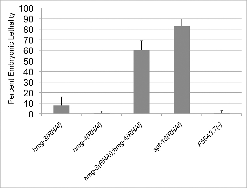
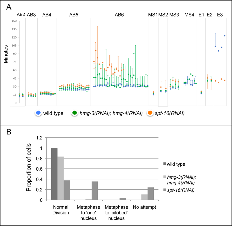
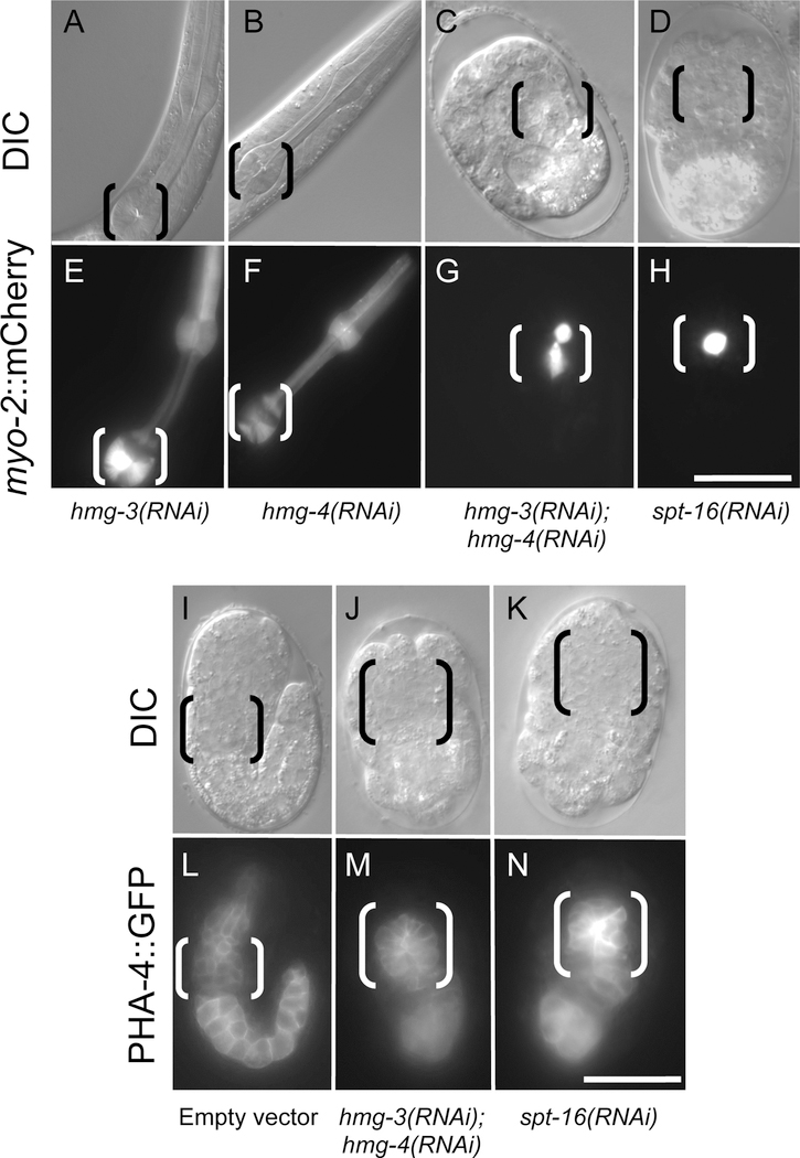
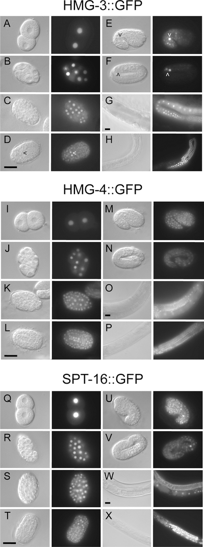
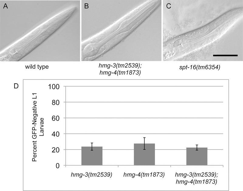
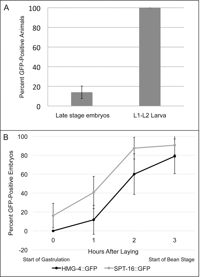
FACT (facilitates chromatin transcription) is a histone chaperone complex important in genomic processes including transcription, DNA replication, and DNA repair. FACT is composed of two proteins, SSRP1 and SPT16, which are highly conserved across eukaryotes. While the mechanisms for FACT in nucleosome reorganization and its relationship to DNA processes is well established, how these roles impact coordination in multicellular animal development are less well understood. Here we characterize the genes encoding FACT complex proteins in the nematode C. elegans. We show that whereas C. elegans includes one SPT16 gene (spt-16), two genes (hmg-3 and hmg-4) encode SSRP1 proteins. Depletion of FACT complex genes interferes with embryonic cell division and cell cycle timing generally, with anterior pharynx development especially sensitive to these defects. hmg-3 and hmg-4 exhibit redundancy for these maternally-provided embryonic functions, but are each uniquely required zygotically for normal germline development. This work provides a framework to study FACT gene function in developmental processes, and identifies that distinct functional requirements for gene duplicates can be manifest within a single tissue.

Keywords: Cell cycle timing; Gene duplication; Pharyngeal development; SPT16; SSRP1.


Conflict of interest statement

Declaration of Interest: none

Figures

Figure 1.
Figure 1.. Validation of RNAi knockdown.
A homozygous hmg-3(tm2539) mutants (GFP-negative) from heterozygous parents (hmg-3(tm2539)/hT2) exhibit a sterile phenotype, whereas homozygous hmg-4(tm1873) or spt-16(tm6354) mutants from heterozygous parents develop no further than L2 stage. Sample size indicated above the bar for each genotype. B hmg-3(RNAi) treatment of wild-type hermaphrodites (Maternal RNAi) results in sterile offspring, whereas treatment with hmg-4(RNAi) or spt-16(RNAi) results in animals that arrest as young larvae (hmg-4(RNAi)) or embryos (spt-16(RNAi); also see Figure 3, where each is evaluated directly for embryonic lethality). Seeding wild-type eggs onto RNAi plates (Larval RNAi) results in fertility for a majority of hmg-3(RNAi) and spt-16(RNAi) animals, and most hmg-4(RNAi) animals survive to sterile adulthood. Sample size indicated above the bar for each condition. C RNAi treatment of wild-type hermaphrodites (Maternal RNAi) reduces the intensity of fluorescent signal in hmg-3::GFP, hmg-4::GFP, and spt-16::GFP strains. The values represent average pixel signal intensity (arbitrary units) in the nucleus of the most mature oocyte in adult animals treated with RNAi for 24 hours (treatment starting in the L4 stage). * indicates p<0.005. n.s. indicates p>0.05; two tailed t-test. n>=8 for each condition.
Figure 2.
Figure 2.. Identification of FACT complex proteins encoded by the C. elegans genome.
A, B Phylogenetic trees for SSRP1 and SPT16 in model organisms generated by Treefam (Ruan et al., 2008) identify an inferred duplication of genes encoding SSRP1 and SPT16 in the C. elegans genome. C Comparison of amino acid similarities of the three major protein domains found in human SSRP1 (Hsa-SSRP1) to the predicted C. elegans orthologs. Domains are indicated by boxes; the name for each domain is noted above the topmost row. Numbers inside domain boxes indicate percent amino acid similarity between the protein and the one(s) below it; numbers on the left correspond to domain similarity with that of Cel-HMG −3, the right is similarity with Cel-HMG-4. Overall, Cel-HMG −3 and Cel -HMG-4 share 81% amino acid sequence identity, and the genes share 81% nucleotide sequence identity within the coding region. Genomic and RNA-seq data support two full length C. elegans SSRP1 orthologs. D Protein sequence comparison of the three major domains of human SPT16 (Hsa-SPT16) and C. elegans orthologs. Numbers inside the domain boxes indicate the amino acid similarity between the protein and the one(s) below it; numbers on the left correspond to domain similarity with Cel -SPT-16, the right is similarity with Cel-F55A3.7. Cel-SPT-16 has all four domains present in Hsa-SPT16, but F55A3.7 includes only the Nlob and Spt16 protein domains. NA (not applicable) is indicated in the boxes for these two domains for the comparison to F55A3.7. This gene structure, combined with RNA-seq data that identify limited transcript abundance of F55A3.7, has resulted in classification of F55A3.7 as a psuedogene. These data suggest that SPT-16 is the C. elegans SPT16 ortholog.
Figure 3.
Figure 3.. FACT complex genes are essential for normal embryonic development.
Embryonic lethality associated with single and double knockdown of FACT components, using RNA interference (RNAi). hmg-3(RNAi) or hmg-4(RNAi) treatment of hermaphrodites caused limited embryonic lethality among offspring but hmg-3(RNAi); hmg-4(RNAi) in combination resulted in high embryonic lethality, indicating an essential embryonic function that is redundant between these two genes. spt-16(RNAi) treatment resulted in a high level of embryonic lethality whereas embryos derived from mothers homozygous for the deletion allele F55A3.7(ok1829) are viable. This indicates spt-16 is essential for embryonic development. Data represent at least 100 offspring from 3 trials. Error bars correspond to +/− one standard deviation.
Figure 4.
Figure 4.. RNAi depletion of hmg-3; hmg-4 or spt-16 causes defects in the embryonic cell cycle.
A Cell cycle duration for all cells of the AB, MS and E lineages through the 6th division of AB. The graph shows medial, maximal and minimal cell cycle durations for control (blue), hmg-3(RNAi); hmg-4(RNAi) (green) and spt-16(RNAi) (orange). N=3 animals for each condition. Missing information (e.g. for spt-16(RNAi) at AB6, or hmg-3(RNAi); hmg-4(RNAi) at E3) indicates some cells failed to divide. Average (and standard deviation) of cell cycle length at AB6 for wild type, hmg-3(RNAi); hmg-4(RNAi), and spt-16(RNAi) in minutes was 33.4 (2.3; n=96), 47.8 (17.4; n=82), and 69.3 (19.0; n=36), respectively, with both hmg-3(RNAi); hmg-4(RNAi) and spt-16(RNAi) statistically different from wild type (two tailed t-test, p<<0.01). B Cell division defects observed at the 6th division of AB. Cells were classified based on whether they executed a relatively normal mitosis (Normal Division), did not initiate a division (No attempt), or initiated mitosis but resulted in morphological defects (Metaphase to either a single apparent nucleus, or a bilobed nucleus rather than the normal two distinct nuclei). Sample size is 96 cells each condition (32 cells each of three animals).
Figure 5.
Figure 5.. Depletion of hmg-3; hmg-4 or spt-16 results in a loss of anterior pharynx.
A-D Comparison of the terminal phenotype of hmg-3(RNAi); hmg-4(RNAi) or spt-16(RNAi) embryos with late stage control embryos (hmg-3(RNAi) or hmg-4(RNAi)). Black brackets indicate the posterior bulb of the pharynx. In hmg-3(RNAi); hmg-4(RNAi) or spt-16(RNAi) embryos animals, no anterior pharynx is apparent, and embryos do not elongate. E-H Fluorescence from myo-2::mCherry (a marker for differentiated pharyngeal cells) is seen in the pharynx in single hmg-3(RNAi) or hmg-4(RNAi) larvae but only in posterior pharynx cells corresponding to the grinder (marked by brackets) in hmg-3(RNAi); hmg-4(RNAi) or spt-16(RNAi) embryos. Thirty-five of 38 transgene-bearing hmg-3(RNAi); hmg-4(RNAi) and 19 of 19 spt-16(RNAi) animals exhibited expression in a region consistent with the posterior pharynx similar to that in the image. I-N Expression of the early pharyngeal-intestinal marker PHA-4::GFP is observed only in intestine and posterior pharynx in hmg-3(RNAi); hmg-4(RNAi) or spt-16(RNAi) embryos, whereas it identifies the anterior pharynx in control animals. Scale = 20 µm.
Figure 6.
Figure 6.. HMG-4::GFP and SPT-16::GFP are present in both germline and soma, whereas HMG-3::GFP is germline-restricted.
A-H HMG-3::GFP is nuclear-localized and present broadly in early embryos (A, two cell embryo; B-D, gastrulation through ventral cleft closure) Over developmental time, the somatic protein intensity is reduced compared to that in the germ cells, and in late embryonic stages it is absent from somatic cells (D, ventral cleft closure; E, comma stage; F, elongated embryo; arrowhead indicates germ cell(s)). Presence of the protein in the germline persists through adulthood in both sexes (G, adult hermaphrodite, H, adult male). I-P HMG-4::GFP is present broadly and in both somatic and germ cells from embryonic (I, two cell embryo; J-L, gastrulation through ventral cleft closure; M, comma stage; N, elongated embryo) through adult stages in both hermaphrodites (O) and males (P). Q-X Like HMG-4::GFP, SPT-16::GFP is seen in both somatic and germ cells from early embryonic stages (Q, two cell embryo; R-T, gastrulation through ventral cleft closure; U, comma stage; V, elongated embryo) through adult stages (W, adult hermaphrodite; X, adult male). Scale = 20 µm. Scale in D, L, and T is for all embryonic images (A-F, I-N, Q-V, taken with 100x objective) and scale in G, O, and W is for all adult images (G-H, O-P, W-X, taken with 40x objective).
Figure 7.
Figure 7.. The pharynx develops normally in hmg-3; hmg-4 or spt-16 mutant embryos derived from heterozygous mothers
A-C Homozygous hmg-3(tm2539); hmg-4(tm1873) and spt-16(tm6354) mutant larvae derived from heterozygous mothers have normal pharynges, indicating that maternal gene activity is sufficient for normal pharyngeal development. Scale = 20 µm. D. hmg-3(tm2539); hmg-4(tm1873) double mutants hatch at the same frequency as single mutants. Both genes are balanced by hT2, which is marked with a myo-2::GFP transgene. The percent of GFP-negative L1 larvae derived from heterozygous mother was evaluated, and no significant difference in survivability was observed. Data represent at least 125 offspring from at least 2 trials. Error bars correspond to +/− one standard deviation.
Figure 8.
Figure 8.. Zygotic expression of FACT complex proteins
Males homozygous for HMG-3::GFP, HMG-4::GFP or SPT-16::GFP were mated to spe −9(eb19) hermaphrodites to evaluate the onset of zygotic protein expression. A A comparison of the percent of HMG-3::GFP-positive animals at late embryonic and early larval stages indicates that zygotic expression initiates only in late embryonic/early larval stages, coincident with the initiation of germline cell division. B Offspring from spe-9 hermaphrodites mated with either HMG-4::GFP- or SPT-16::GFP-bearing males were evaluated at different timepoints after the parent had laid eggs for one hour. Zygotic expression for HMG-4::GFP and SPT-16::GFP begins much earlier than for HMG-3::GFP, and is apparent in somatic cells as well as germline. Data represent at least 30 offspring at each timepoint for both panels. Error bars correspond to the 95% confidence interval.
Figure 9.
Figure 9.. hmg-3 and hmg-4 are each required for normal germline development.
Germline of a wild-type L4 lethargus (A) and adult (B) hermaphrodite, with inset of sperm cells. Germline of hmg-3(tm2539) (C) and zygotic (L1 larval) hmg-4(RNAi) (D) adult animals exhibit evidence of germline proliferation and sperm cells (inset), but lack oocytes. Scale = 20 µm. Inset images are 3x the primary image. Quantification of sterile phenotype for hmg-3(tm2539) and hmg-4(RNAi) is in Figure 1.

Similar articles

Cited by

References

    1. Abe T, Sugimura K, Hosono Y, Takami Y, Akita M, Yoshimura A, Tada S, Nakayama T, Murofushi H, Okumura K, Takeda S, Horikoshi M, Seki M, Enomoto T, 2011. The histone chaperone facilitates chromatin transcription (FACT) protein maintains normal replication fork rates. J. Biol. Chem 286, 30504–30512. 10.1074/jbc.M111.264721 - DOI - PMC - PubMed
    1. Atlasi Y, Stunnenberg HG, 2017. The interplay of epigenetic marks during stem cell differentiation and development. Nat. Rev. Genet 18, 643–658. 10.1038/nrg.2017.57 - DOI - PubMed
    1. Attwood K, Fleyshman D, Prendergast L, Paszkiewicz G, Omilian AR, Bshara W, Gurova K, 2017. Prognostic value of histone chaperone FACT subunits expression in breast cancer. Breast Cancer Dove Med. Press 9, 301–311. 10.2147/BCTT.S126390 - DOI - PMC - PubMed
    1. Baugh LR, Hill AA, Slonim DK, Brown EL, Hunter CP, 2003. Composition and dynamics of the Caenorhabditis elegans early embryonic transcriptome. Dev. Camb. Engl 130, 889–900. - PubMed
    1. Birchler JA, Veitia RA, 2012. Gene balance hypothesis: connecting issues of dosage sensitivity across biological disciplines. Proc. Natl. Acad. Sci. U. S. A 109, 14746– 14753. 10.1073/pnas.1207726109 - DOI - PMC - PubMed

Publication types

LinkOut - more resources