. 2018 Oct 18;11(1):90.
doi: 10.1186/s13048-018-0462-4.

DAXX promotes ovarian cancer ascites cell proliferation and migration by activating the ERK signaling pathway

Affiliations

DAXX promotes ovarian cancer ascites cell proliferation and migration by activating the ERK signaling pathway

Sheng-Bing Liu et al. J Ovarian Res. .

Abstract

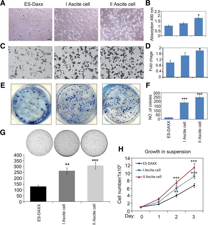
Background: The death-domain-associated protein (DAXX) was originally identified as a protein that binds to the transmembrane death receptor FAS and enhances both FAS-induced and transforming growth factor-β-dependent apoptosis. In a previous study, we found that nude mice injected with DAXX-overexpressing cells (ES-2-DAXX) accumulated large concentrations of first-generation ascites cells (I ascites cells). The role of DAXX in the development of ascites is unknown. The aim of this study was to analyze the effect of DAXX on proliferation and migration of ascites cells in ovarian cancer in vitro and in vivo.

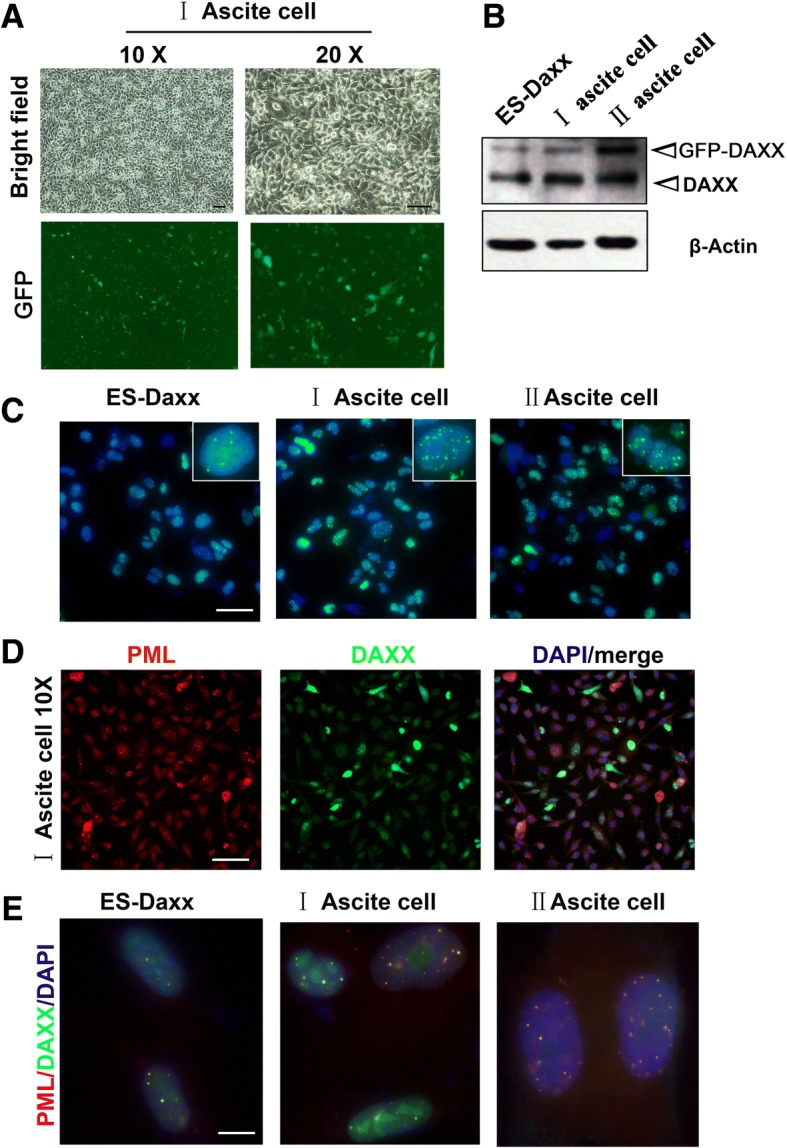
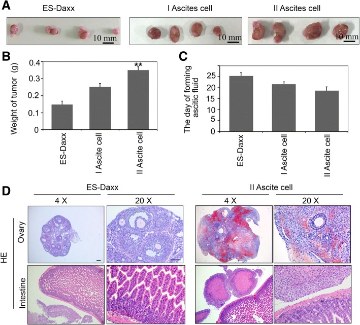
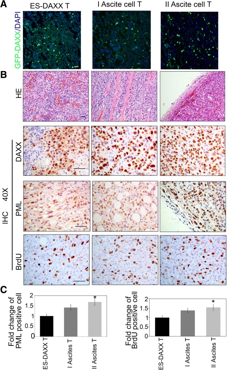
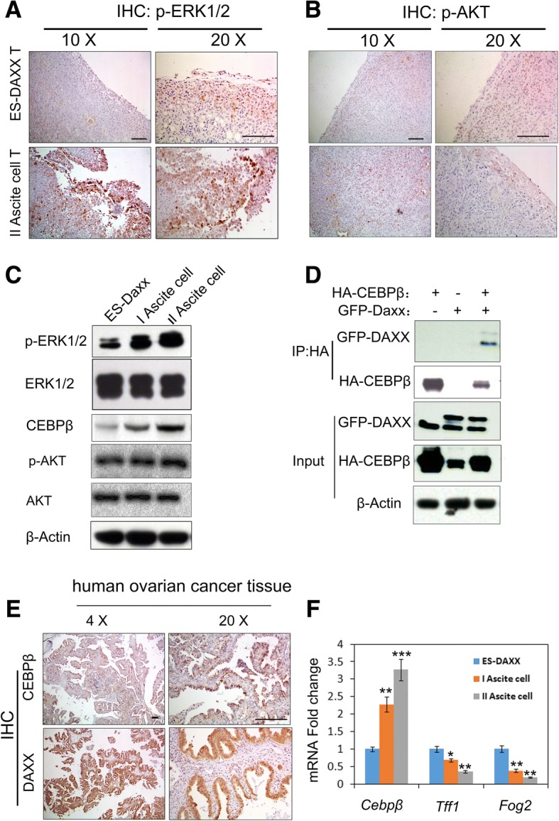
Methods: Nude mice were housed in cages with a 14:10 h light:dark cycle; water and food were provided ad libitum. ES-2-DAXX cells (1×10⁶) were injected intraperitoneally into athymic nude mice (8-week-old female mice). After 4 weeks, I ascites cells were collected. The I ascites cells were injected intraperitoneally into athymic nude mice (8-week-old female mice). After 4 weeks, II ascites cells were collected and cultured. Ascites cell survival, migration, and colony formation were measured using colony formation and cell growth assays. Immunofluorescent staining revealed the co-localization of DAXX and promyelocytic leukemia protein (PML) in ascites cell nuclei. Western blotting and immunohistochemistry showed that extracellular signal-related kinase (p-ERK) 1/2 and CEBP-β were highly expressed in tumor tissues formed by II ascites cells. Through immunoprecipitation, we also found that DAXX can interact with CEBP-β.

Results: DAXX enhanced ascites cell survival, migration, and colony formation. DAXX and PML nuclear foci dramatically increased in a passage-dependent manner in ascites cells, DAXX promoted the tumor growth of ascites cells in vivo, increased ascites cell proliferation in vivo, and enhanced ascites cell survival and migration by activating the ERK signalling pathway and integrating with CEBP-β.

Conclusions: DAXX can interact with CEBP-β. DAXX can induce ovarian cancer ascites formation by activating the ERK signal pathway and binding to CEBP-β.

Keywords: Ascites cell; Cell migration; Cell proliferation; DAXX.


Conflict of interest statement

Authors’ information

Where the work was performed: Laboratory of molecular biology, college of medicine, Jiaxing University.

Ethics approval

Normal ovaries were provided by the the Jiaxing Maternity and Child Health Care Hospital, China. The use of archived samples in this study was approved by the Jiaxing University Institutional Review Board. The NIH Guides for the Care and Use of Laboratory Animals were used as all animal protocols.

Consent for publication

Not applicable.

Competing interests

The authors declare that they have no competing interests.

Publisher’s Note

Springer Nature remains neutral with regard to jurisdictional claims in published maps and institutional affiliations.

Figures

Fig. 1
Fig. 1
DAXX enhances ascites cell colony formation and migration. a–b DAXX-induced ascites cell proliferation. Ascites cells were seeded into 96-well plates (3,000 cells/well) overnight, and then assessed by an MTT assay. Scale bar, 50 μm. cd Transwell experiment to determine the migration capability of ascites cells and their derivatives (GFP-DAXX-overexpressing ascites cells). Cells were added to transwells and allowed to migrate for 12 h. Cells at the upper surface of the membrane were removed with cotton swabs, and cells on the bottom surface were stained with hematoxylin and eosin. Scale bar, 100 μm. ef Colony formation assay for the growth of ascites cells with DAXX overexpression. Colony numbers were counted after 10–14 days. * p < 0.05; ***, p < 0.0001. g DAXX overexpression promotes anchorage-independent growth of ascite cells in vitro. Soft-agar colony-formation assay was performed and the colonies were stained with crystal violet for quantification. h DAXX overexpression promoted cell proliferation in suspension (detachment) conditions. Cells (1 × 105) were plated in 6-well culture dishes and cell number was determined with trypan blue staining. Three replicates were included. The error bars represent s.d. ns (p > 0.05); *** p < 0.001. Student’s t-test was applied
Fig. 2
Fig. 2
DAXX and PML nuclear localization in ascites cells. a Ascites cell morphology using brightfield and GFP microscopy (10 × and 20× magnification, respectively). b Western blot results for DAXX overexpression in ascites cells. c Ascites cells were harvested from nude mice and cultured in vitro. Fluorescence microscopy showing the localization of overexpressed DAXX (green) in ascites cells stably transfected with GFP-DAXX. Nuclei were stained with 4′,6-diamidino-2-phenylindole (DAPI; blue). Scale bar, 50 μm. d Immunofluorescence results for promyelocytic leukemia protein (PML) foci formation in ascites cells. Scale bar, 50 μm. e Immunofluorescence results for PML, DAXX and DAPI in ES-Daxx, I and II ascites cells. Scale bar, 10 μm
Fig. 3
Fig. 3
DAXX promotes ascites cell metastasis in vivo. a–b Ovarian cancer cells (ES-Daxx) and I and II ascites cells (106cells for each) were implanted intraperitoneally into different nude mice. After 20 days, tumors were removed and weighed (n = 5). c Quantify ascites formation time (d). d ES-Daxx and II ascites cells were injected intraperitoneally into nude mice (106 cells/mouse). HE staining showing cells metastasis to the intestine and ovary 3 weeks later. Scale bar, 250 μm
Fig. 4
Fig. 4
DAXX promotes ascites cell proliferation in vivo. a Cryosections were prepared from tumor tissues derived from GFP-DAXX-overexpressing ascites cells. b HE and Immunohistochemistry staining for DAXX, PML, and BrdU in representative DAXX-overexpressingascites cells. Scale bar, 50 μm. c Quantify PML and BrdU positive cells in B. *, p < 0.05
Fig. 5
Fig. 5
DAXX binds to CEBP-β and promotes ascites cell proliferation in vivo. a–b Immunohistochemistry staining for p-ERK1/2 and p-AKT in mouse tumor tissue. Scale bar, 200 μm. c DAXX-overexpressing ascites cells were subjected to immunoblotting analysis with antibodies against the indicated proteins. d DAXX-overexpressing ascites cells were transfected with the indicated combinations of plasmids encoding HA-CEBP-β. Cells were lysed after 24 h, and protein extracts were immunoprecipitated with anti-HA antibodies, followed by SDS-PAGE and western blotting. e Immunohistochemical analysis of DAXX and CEBP-β levels in human ovarian tumor tissue samples (n = 10). Sections were counterstained with hematoxylin and eosin. Scale bar, 200 μm. f Cebpβ, Tff1 and p16 mRNA expressions in ES-Daxx, I and II ascites cells were determined by Q-PCR

Similar articles

Cited by

References

    1. Ozols RF, Bookman MA, Connolly DC, Daly MB, Godwin AK, Schilder RJ, Xu X, Hamilton TC. Focus on epithelial ovarian cancer. Cancer Cell. 2004;5:19–24. doi: 10.1016/S1535-6108(04)00002-9. - DOI - PubMed
    1. Bast RC, Jr, Hennessy B, Mills GB. The biology of ovarian cancer: new opportunities for translation. Nat Rev Cancer. 2009;9:415–428. doi: 10.1038/nrc2644. - DOI - PMC - PubMed
    1. Lane D, Robert V, Grondin R, Rancourt C, Piché A. Malignant ascites protect against TRAIL-induced apoptosis by activating the PI3K/Akt pathway in human ovarian carcinoma cells. Int J Cancer. 2007;121:1227–1237. doi: 10.1002/ijc.22840. - DOI - PubMed
    1. Lane D, Goncharenko-Khaider N, Rancourt C, Piché A. Ovarian cancer ascites protects from TRAIL-induced cell death through αvβ5 integrin-mediated focal adhesion kinase and Akt activation. Oncogene. 2010;29:3519–3531. doi: 10.1038/onc.2010.107. - DOI - PubMed
    1. Ahmed N, Riley C, Oliva K, Rice G, Quinn M. Ascites induces modulation of 6β1 integrin and urokinase plasminogen activator receptor expression and associated functions in ovarian carcinoma. Br J Cancer. 2005;92:1475–1485. doi: 10.1038/sj.bjc.6602495. - DOI - PMC - PubMed