. 2018 Nov:37:428-441.
doi: 10.1016/j.ebiom.2018.10.033. Epub 2018 Oct 15.

vWA proteins of Leptospira interrogans induce hemorrhage in leptospirosis by competitive inhibition of vWF/GPIb-mediated platelet aggregation

Affiliations

vWA proteins of Leptospira interrogans induce hemorrhage in leptospirosis by competitive inhibition of vWF/GPIb-mediated platelet aggregation

Jia-Qi Fang et al. EBioMedicine. 2018 Nov.

Abstract

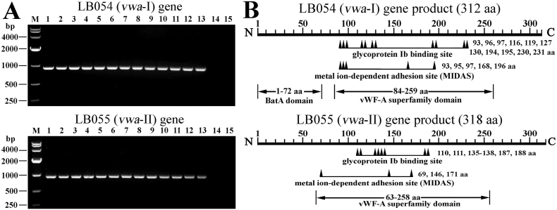
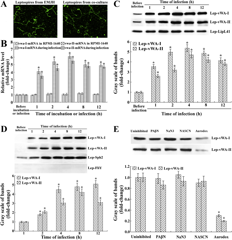
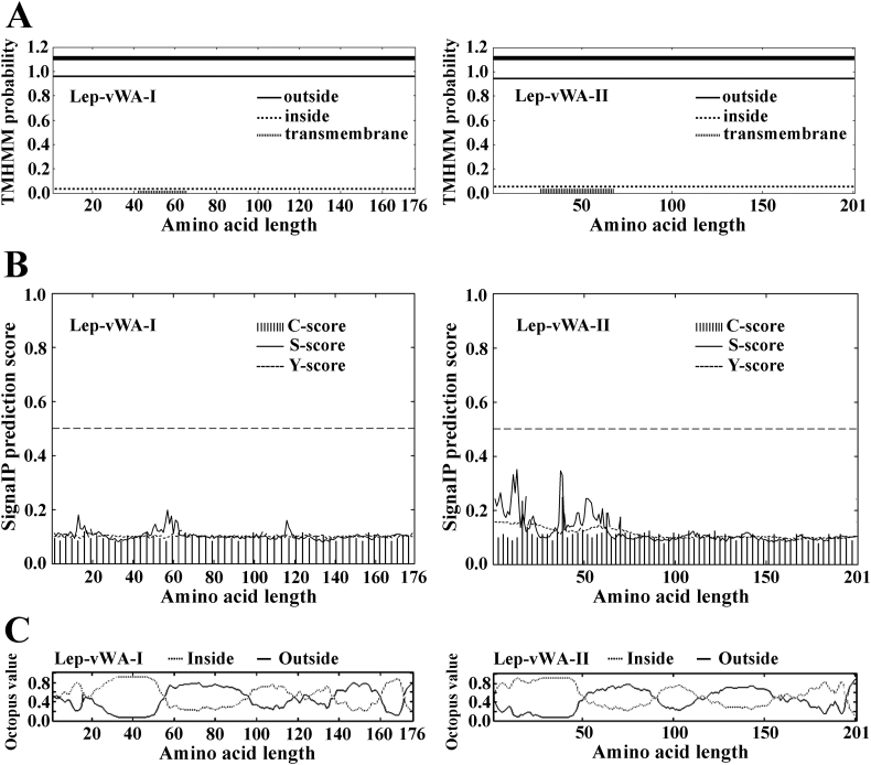
Backgroud: Leptospira interrogans is the major causative agent of leptospirosis, a worldwide zoonotic disease. Hemorrhage is a typical pathological feature of leptospirosis. Binding of von Willebrand factor (vWF) to platelet glycoprotein-Ibα (GPIbα) is a crucial step in initiation of platelet aggregation. The products of L. interrogans vwa-I and vwa-II genes contain vWF-A domains, but their ability to induce hemorrhage has not been determined.

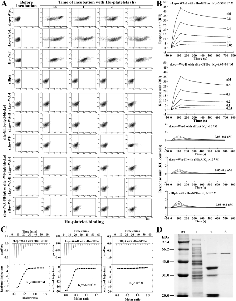
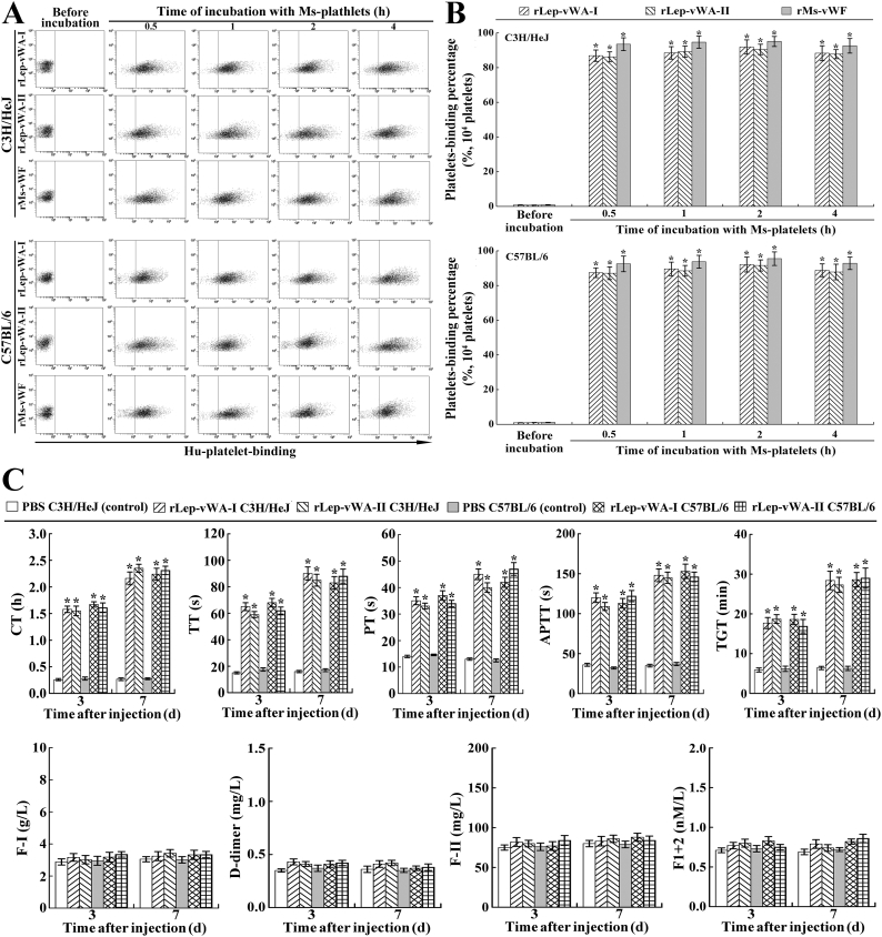
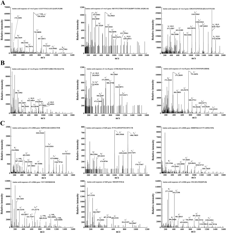
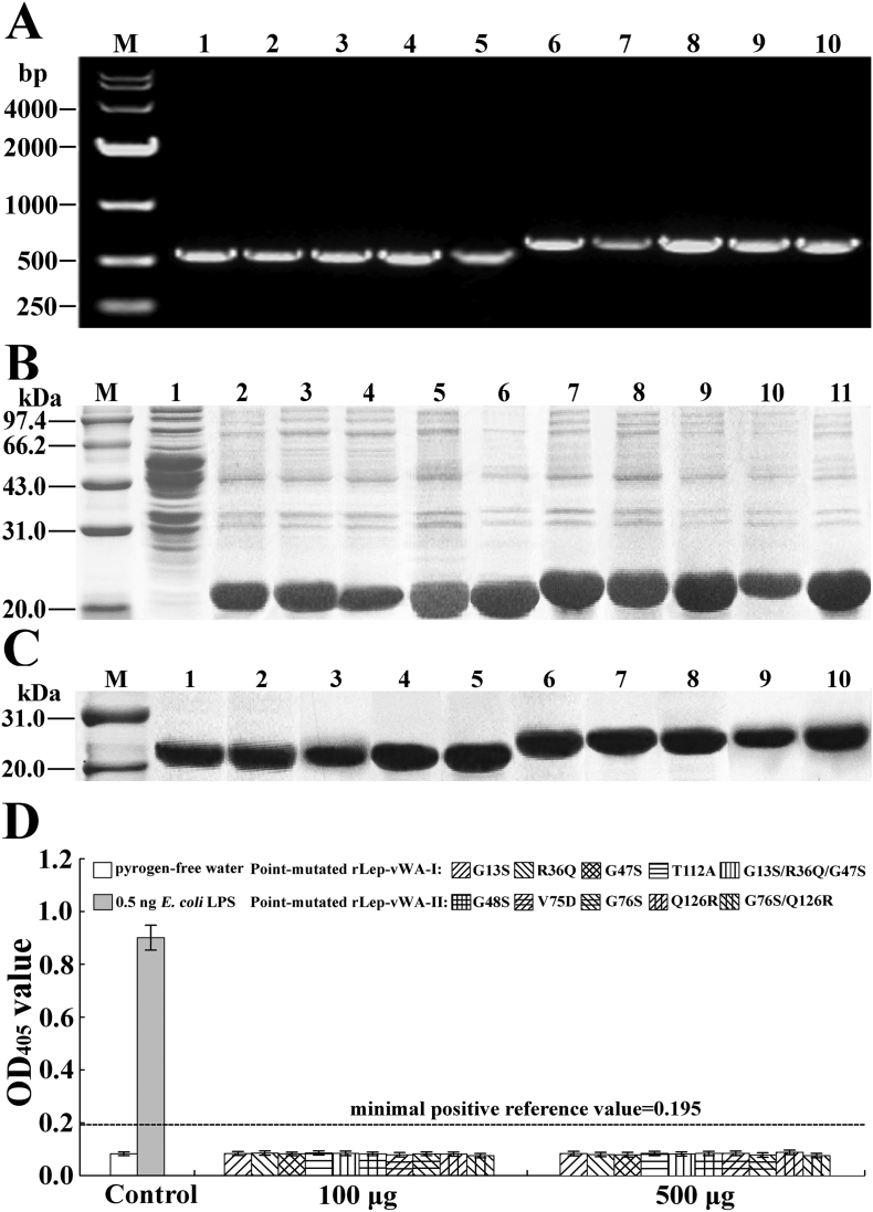
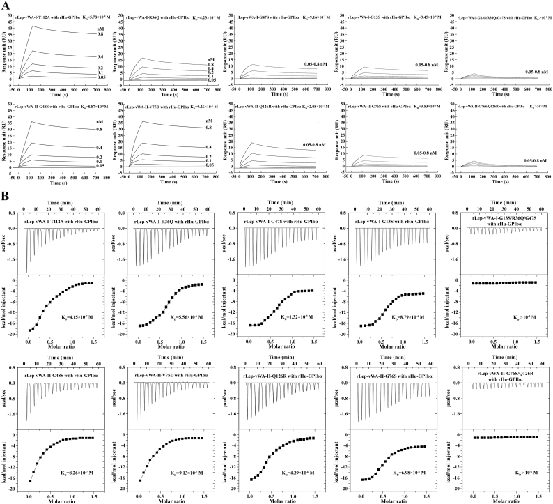
Methods: Human (Hu)-platelet- and Hu-GPIbα-binding abilities of the recombinant proteins expressed by L. interrogans strain Lai vwa-I and vwa-II genes (rLep-vWA-I and rLep-vWA-II) were detected by flowcytometry, surface plasmon resonance (SPR) and isothermal titration calorimetry (ITC). Hu-platelet aggregation and its signaling kinases and active components were detected by lumiaggregometry, Western analysis, spectrophotometry and confocal microscopy. Hu-GPIbα-binding sites in rLep-vWA-I and rLep-vWA-II were identified by SPR/ITC measurements.

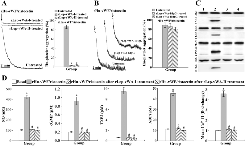
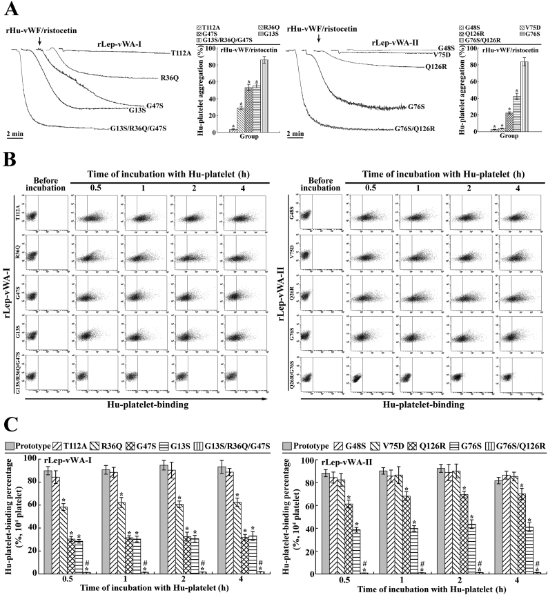
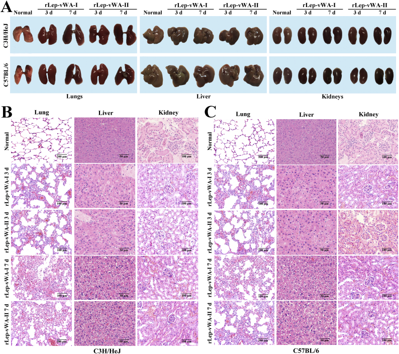
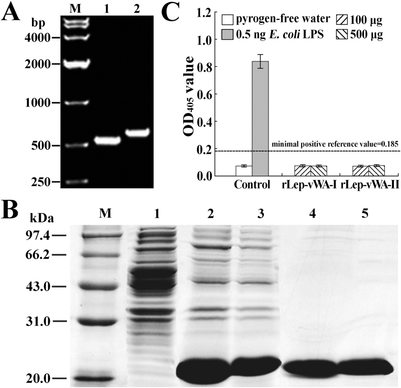
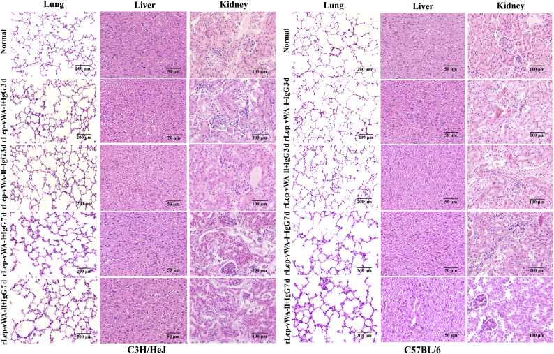
Findings: Both rLep-vWA-I and rLep-vWA-II were able to bind to Hu-platelets and inhibit rHu-vWF/ristocetin-induced Hu-platelet aggregation, but Hu-GPIbα-IgG, rLep-vWA-I-IgG and rLep-vWA-II-IgG blocked this binding or inhibition. SPR and ITC revealed a tight interaction between Hu-GPIbα and rLep-vWA-I/rLep-vWA-II with KD values of 3.87 × 10⁻⁷-8.65 × 10⁻⁸ M. Hu-GPIbα-binding of rL-vWA-I/rL-vWA-II neither activated the PI3K/AKT-ERK and PLC/PKC kinases nor affected the NO, cGMP, ADP, Ca2+ and TXA2 levels in Hu-platelets. G13/R36/G47 in Lep-vWA-I and G76/Q126 in Lep-vWA-II were confirmed as the Hu-GPIbα-binding sites. Injection of rLep-vWA-I or rLep-vWA-II in mice resulted in diffuse pulmonary and focal renal hemorrhage but this hemorrhage was blocked by rLep-vWA-I-IgG or rLep-vWA-II-IgG.

Interpretation: The products of L. interrogans vwa-I and vwa-II genes induce hemorrhage by competitive inhibition of vWF-mediated Hu-platelet aggregation.

Keywords: Competitive inhibition; Hemorrhage; Leptospira interrogans; Leptospirosis; Platelet aggregation; von Willebrand factor; vwa-I and vwa-II genes.


Figures

Fig. 1
Fig. 1
Distribution and domains of LB054 and LB055 genes in leptospiral strains. (A). The PCR products of leptospiral LB054 (vwa-I) and LB055 (vwa-II) genes, determined by PCR. Lane M: DNA marker. Lanes 1–13: amplicoms of the vwa-I or vwa-II genes from thirteen pathogenic strains belonging to L. interrogans, L. borgpetersenii or L. weilii. Lanes 14–15: no amplicoms of vwa-I or vwa-II genes from two saprophytic strains of L. biflexa. (B). Predicted functional domains in LB054 (vwa—I) and LB055 (vwa-II) genes of L. interrogans strain Lai.
Fig. 2
Fig. 2
Platelet- and GPIbα-binding ability of rLep-vWA-I and rLep-vWA-II. (A). Hu-platelet-binding ability of rLep-vWA-I and rLep-vWA-II, determined by flow cytometry. rHu-vWF and rHlpA, a commercial recombinant human vWF and a recombinant hemolysin of L. interrrogans, were used as the controls. (B). rHu-GPIbα-binding ability of rLep-vWA-I and rLep-vWA-II, determined by SPR. rHlpA, a recombinant hemolysin of L. interrrogans, was used as the control. (C). rHu-GPIbα-binding ability of rLep-vWA-I and rLep-vWA-II, determined by ITC. The legend is the same as shown in C. (D). rHu-GPIbα-captured leptospiral proteins, determined by co-precipitation test. Lane M: protein marker. Lane 1: total proteins from L. interrogans strain Lai. Lane 2: proteins released from protein-A-GPIbα-IgG beads. Lane 3: rHu-GPIbα control.
Fig. 3
Fig. 3
Inhibition of rLep-vWA-I and rLep-vWA-II on vWF-mediated Hu-platelet aggregation. (A). Inhibition of rLep-vWA-I and rLep-vWA-II on vWF/ristocetin-induced Hu-platelet aggregation, determined by platelet aggregation test. (B). Reversed inhibition of rLep-vWA-I-IgG and rLep-vWA-II-IgG on rLep-vWA-I and rLep-vWA-II blockage of vWF/ristocetin-induced Hu-platelet aggregation, determined by platelet aggregation test. (C). No phosphorylation of the AKT, ERK, PLC and PKC in rLep-vWA-I- or rLep-vWA-II-treated Hu-platelets, determined by Western Blot assay. Lane 1: normal Hu-platelets. Lane 2: vWF/ristocetin-treated Hu-platelets. Lanes 3 and 4: rLep-vWA-I- or rLep-vWA-II-treated Hu-platelets. (D). No increase of the NO, cGMP, TXA2, free ADP and Ca2+ levels in rLep-vWA-I- or rLep-vWA-II-treated Hu-platelets, determined by spectrophotometry and confocal microscopy.
Fig. 4
Fig. 4
GPIbα-binding sites in products of vwa-I528 and vwa-II603 mutants. (A). Decreased inhibition of point-mutated rLep-vWA-I and rLep-vWA-II on vWF/ristocetin-induced Hu-platelet aggregation, determined by platelet aggregation test. (B). Decreased Hu-platelet-binding ability of point-mutated rLep-vWA-I and rLep-vWA-II, determined by flow cytometry. (C). Statistical summary of the Hu-platelet-binding percentages of point-mutated rLep-vWA-I and rLep-vWA-II. Statistical data from experiments such as shown in B. Bars show the means ± SD of three independent experiments. *: p < .05 vs the Hu-platelet-binding percentages of prototypic rLep-vWA-I or rLep-vWA-II. #: p < .05 vs the Hu-platelet-binding percentages of single point-mutated rLep-vWA-I or rLep-vWA-II.
Fig. 5
Fig. 5
Increase of vwa-I and vwa-II gene expression and secretion during infection. (A) The integrity of leptospires from the lysates of Leptospira-cell co-cultures, examined by cofocal microscopy. (B) Increase of vwa-I- or vwa-II-mRNA levels in L. interrogans strain Lai during infection of HUVEC, determined by qRT-PCR. Bars show the mean ± SD of three independent experiments. The vwa-I- or vwa-II-mRNA levels in the spirochete from EMJH medium (before infection) were set as 1.0. *: p < .05 vs the vwa-I- or vwa-II-mRNA levels in the spirochete before infection or in incubation with RPMI-1640 medium at 37 °C. (C). Increase of Lep-vWA-I and Lep-vWA-II expression in L. interrogans strain Lai during infection of HUVEC, determined by Western Blot assay. LipL41, a leptospiral outer membrane lipoprotein, was used as the control. The immunoblotting bands reflecting Lep-vWA-I and Lep-vWA-II expression levels during infection were quantified by gray scales. Bars show the mean ± SD of three independent experiments. The gray scale values of immunoblotting bands from the spirochete in EMJH medium (before infection) were set as 1.0. *: p < .05 vs the gray scale values reflecting the expression levels of Lep-vWA-I and Lep-vWA-II of the spirochete before infection. (D). Secretion of Lep-vWA-I and Lep-vWA-II of L. interrogans strain Lai during infection of HUVEC, determined by Western Blot assay. Sph2, a secreted leptospiral hemolysin, and FliY, a leptospiral cytosolic protein, were used as the controls. The rest of legend is the same as in B but for detection of Lep-vWA-I and Lep-vWA-II secretion. *: p < .05 vs the gray scale values reflecting the secreted Lep-vWA-I and Lep-vWA-II levels of the spirochete before infection. (E). Lep-vWA-I and Lep-vWA-II secretion through T3SS during infection of HUVEC, determined by Western Blot assay. PAβN or Aurodox is the T1SS or T3SS inhibitor while NaN3 and NaSCN are T2SS inhibitors. The rest of legend is the same as in B but for determination of Lep-vWA-I and Lep-vWA-II secretion pathways. *: p < .05 vs the gray scale values reflecting the secreted Lep-vWA-I and Lep-vWA-II levels of the spirochete before infection.
Fig. 6
Fig. 6
rLep-vWA-I- and rLep-vWA-II-induced hemorrhage in mice. (A). The gross pathological changes of lungs, livers and kidneys of rLep-vWA-I- or rLep-vWA-II-injected C3H/HeJ or C57BL/6 mice. (B). rLep-vWA-I- or rLep-vWA-II-induced hemorrhage in C3H/HeJ mice, observed by microscopy after HE-staining. (C). rLep-vWA-I- or rLep-vWA-II-induced hemorrhage in C57BL/6 mice, observed by microscopy after HE-staining.
Fig. 7
Fig. 7
Ms-platelet-binding ability of rLep-vWA-I and blood coagulation dysfunction. (A). Ms-platelet-binding ability of rLep-vWA-I and rLep-vWA-II, determined by flow cytometry. rHu-vWF, a commercial recombinant mouse vWF, was used as the control. (B). Statistical summary of the Ms-platelet-binding percentages of rLep-vWA-I and rLep-vWA-II. Statistical data from experiments such as shown in A. Bars show the means ± SD of three independent experiments. *: p < .05 vs the normal Ms-platelets. (C). Extension of the CT, PT, TT, APTT and TGT as well as no change of the F—I, D-dimer, F-II and F1 + 2 levels of peripheral blood specimens from rLep-vWA-I- or rLep-vWA-II-injected mice, determined by using an Auto-Blood Coagulation Analyzer and ELISA. *: p < .05 vs the CT, PT, TT, APTT and TGT of peripheral blood specimens from normal mice.
Fig. S1
Fig. S1
Predicted characteristics of vwa-I and vwa-II gene products. (A). Structure and location of vwa-I and vwa-II gene products, predicted using TMHMM software. (B). No signal peptide sequence in vwa-I or vwa-II gene product, predicted using SignalP-4.1 software. (C). Position of vwa-I or vwa-II gene product, predicted using Octopus software.
Fig. S2
Fig. S2
Expression and extraction of rLep-vWA-I and rLep-vWA-II. (A). Amplification fragments of vwa-I and vwa-II gene segments (vwa-I528 and vwa-II603) from L. interrogans strain Lai, detected by PCR. Lane M: DNA marker. Lanes 2 and 3: amplicons of vwa-I and vwa-II gene segments (528 and 603 bp). (B). Expression and extraction effects of rLep-vWA-I and rLep-vWA-II, detected by SDS-PAGE. Lane M: protein marker. Lane 1: wild-type E. coli BL21DE3. Lanes 2 and 3 or 4 and 5: the expressed or extracted rLep-vWA-I (~20.2 kDa) and rLep-vWA-II (~23.1 kDa). (C). No LPS in rLep-vWA-I and rLep-vWA-II extracts, determined by spectrophotometeric limulus test.
Fig. S3
Fig. S3
GPIbα-captured proteins as the products of vwa-I and vwa-II genes. (A).The cleaved peptide sequences from GPIbα-captured proteins matching to the vwa-I gene product, determined by LC-MS/MS. (B).The cleaved peptide sequences from GPIbα-captured proteins matching to the vwa-II gene product, determined by LC-MS/MS. (C).The cleaved peptide sequences from GPIbα-captured proteins matching to the product of LA2066, folE or LA2836 genes, determined by LC-MS/MS.
Fig. S4
Fig. S4
Expression and product extraction of point-mutated vwa-I528 and vwa-II603 segments. (A). Amplification fragments of point-mutated vwa-I528 and vwa-II603 segments, detected by PCR. Lane M: DNA marker. Lanes 1-5: amplicons of vwa-I-G13S, vwa-I-R36Q, vwa-I-G47S, vwa-I-T112A and vwa-I-G13S/R36Q/G47S segments (528 bp). Lanes 6-10: amplicons of vwa-II-G48S, vwa-II-G76S, vwa-II-V75D, vwa-II-Q126R and vwa-II-G76S/Q126R segments (603 bp). (B). Expression of point-mutated vwa-I528 and vwa-II603 segments, detected by SDS-PAGE. Lane M: protein marker. Lane 1: wild-type E. coli BL21DE3. Lanes 2-6: the expressed rLep-vWA-I-G13S, rLep-vWA-I-R36Q, rLep-vWA-I-G47S, rLep-vWA-I-T112A and rLep-vWA-I-G13S/R36Q/G47S (~20.2 kDa). Lanes 7-11: the expressed rLep-vWA-II-G48S, rLep-vWA-II-G76S, rLep-vWA-II-V75D, rLep-vWA-II-Q126R and rLep-vWA-II-G76S/Q126R (~23.1 kDa). (C). Extraction of point-mutated vwa-I528 and vwa-II603 segment products, detected by SDS-PAGE. The legend is the same as shown in B but for detection of the product extraction by Ni-NTA affinity chromatography. (D). No LPS in the point-mutated rLep-vWA-I and rLep-vWA-II extracts, determined by spectrophotometric limulus test.
Fig. S5
Fig. S5
Decreased GPIbα-binding ability of the point-mutated rLep-vWA-I and rLep-vWA-II. (A). Decreased Hu-platelet-binding ability of point-mutated rLep-vWA-I and rLep-vWA-II proteins, determined by SPR. (B). Decreased Hu-platelet-binding ability of point-mutated rLep-vWA-I and rLep-vWA-II proteins, determined by ITC.
Fig. S6
Fig. S6
Blockage of hemorrhage by rLep-vWA-I-IgG and rLep-vWA-II-IgG in mice. The histopathological examination showed no visible hemorrhage in the IgG-blocked rLep-vWA-I- or rLep-vWA-II-injected mice.

Similar articles

Cited by

References

    1. Bharti A.R., Nally J.E., Ricaldi J.N. Leptospirosis: a zoonotic disease of global importance. Lancet Infect Dis. 2003;3(12):757–771. - PubMed
    1. Ko A.I., Goarant C., Picardeau M. Leptospira: the dawn of the molecular genetics era for an emerging zoonotic pathogen. Nat Rev Microbiol. 2009;7(10):736–747. - PMC - PubMed
    1. Zhang C.L., Wang H., Yan J. Leptospirosis prevalence in Chinese populations in the last two decades. Microbes Infect. 2012;14(4):317–323. - PubMed
    1. Smith J.K., Young M.M., Wilson K.L., Craig S.B. Leptospirosis following a major flood in Central Queensland, Australia. Epidemiol Infect. 2013;141(3):585–590. - PMC - PubMed
    1. Miraglia F., Matsuo M., Morais Z.M. Molecular characterization, serotyping, and antibiotic susceptibility profile of Leptospira interrogans serovar Copenhageni isolates from Brazil. Diagn Microbiol Infect Dis. 2013;77(3):195–199. - PubMed

MeSH terms