. 2018 Dec 1;24(23):5948-5962.
doi: 10.1158/1078-0432.CCR-18-1277. Epub 2018 Oct 18.

STAT3 Inhibition Combined with CpG Immunostimulation Activates Antitumor Immunity to Eradicate Genetically Distinct Castration-Resistant Prostate Cancers

Affiliations

STAT3 Inhibition Combined with CpG Immunostimulation Activates Antitumor Immunity to Eradicate Genetically Distinct Castration-Resistant Prostate Cancers

Dayson Moreira et al. Clin Cancer Res. .

Abstract

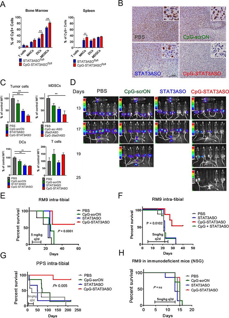
Purpose: Prostate cancers show remarkable resistance to emerging immunotherapies, partly due to tolerogenic STAT3 signaling in tumor-associated myeloid cells. Here, we describe a novel strategy combining STAT3 inhibition with Toll-like Receptor 9 (TLR9) stimulation to unleash immune response against prostate cancers regardless of the genetic background.

Experimental design: We developed and validated a conjugate of the STAT3 antisense oligonucleotide (ASO) tethered to immunostimulatory TLR9 agonist (CpG oligonucleotide) to improve targeting of human and mouse prostate cancer and myeloid immune cells, such as myeloid-derived suppressor cells (MDSC).

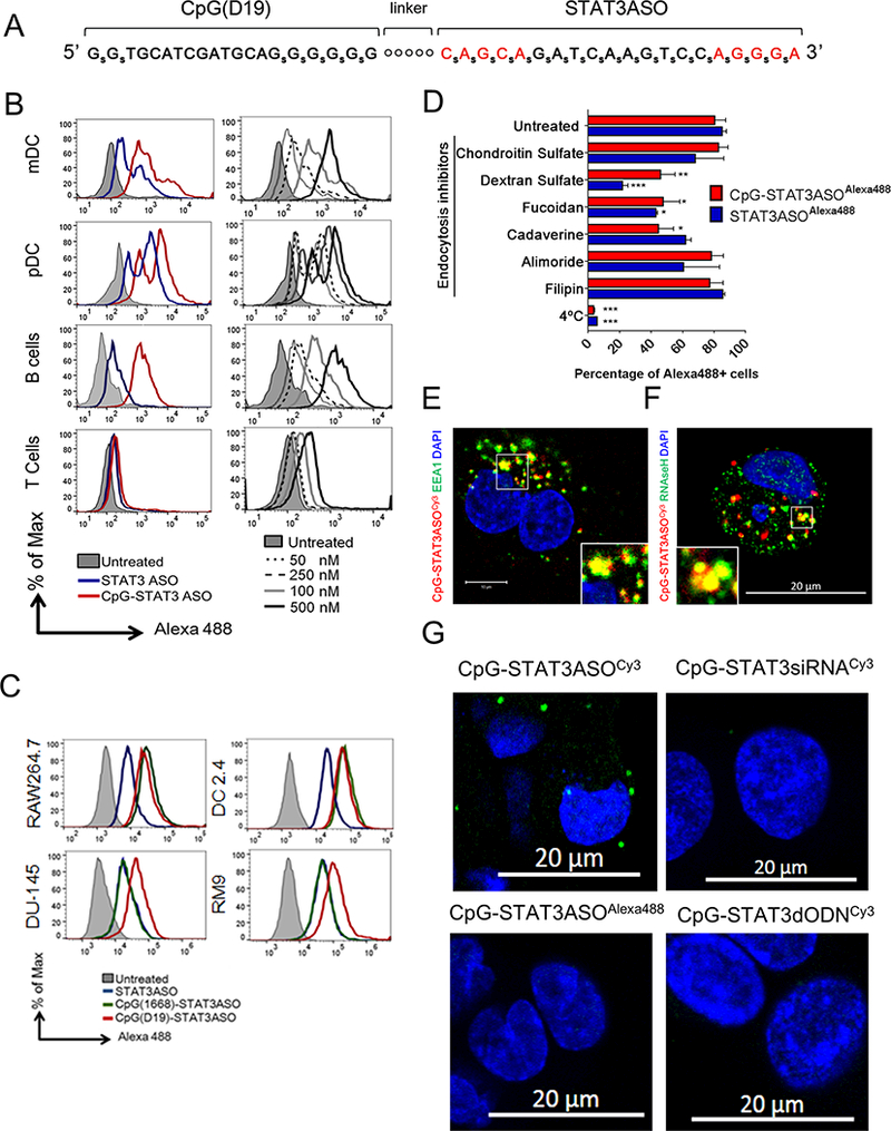
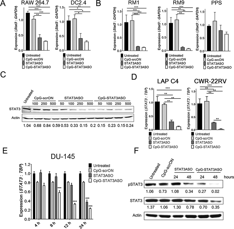
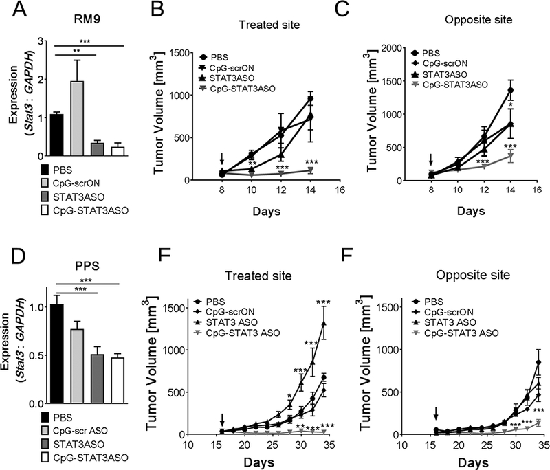
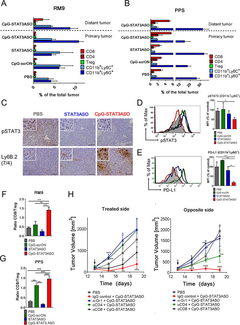
Results: CpG-STAT3ASO conjugates showed improved biodistribution and potency of STAT3 knockdown in target cells in vitro and in vivo. Systemic administration of CpG-STAT3ASO (5 mg/kg) eradicated bone-localized, Ras/Myc-driven, and Ptenpc -/- Smad4pc -/- Trp53c -/- prostate tumors in the majority of treated mice. These antitumor effects were primarily immune-mediated and correlated with an increased ratio of CD8+ to regulatory T cells and reduced pSTAT3+/PD-L1+ MDSCs. Both innate and adaptive immunity contributed to systemic antitumor responses as verified by the depletion of Gr1+ myeloid cells and CD8+ and CD4+ T cells, respectively. Importantly, only the bifunctional CpG-STAT3ASO, but not control CpG oligonucleotides, STAT3ASO alone, or the coinjection of both oligonucleotides, succeeded in recruiting neutrophils and CD8+ T cells into tumors. Thus, the concurrence of TLR9 activation with STAT3 inhibition in the same cellular compartment is indispensable for overcoming tumor immune tolerance and effective antitumor immunity against prostate cancer.

Conclusions: The bifunctional, immunostimulatory, and tolerance-breaking design of CpG-STAT3ASO offers a blueprint for the development of effective and safer oligonucleotide strategies for treatment of immunologically "cold" human cancers.


Conflict of interest statement

DISCLOSURE OF POTENTIAL CONFLICT OF INTEREST

M.K., P.S., D.M. and S.K.P. are inventors on patent application U.S. Provisional Application No.: 62/264,026 submitted by COH that covers the design of oligonucleotides presented in this report. All other authors declare no potential conflict of interest.

Figures

Figure 1.
Figure 1.. CpG-STAT3ASO conjugate design and cell-selective uptake.
(A) Single-stranded CpG-STAT3ASO design; subscript “S” = phosphothioated nucleotides; “o” = C3 units of the carbon linker; red = 2’-O-methyl-modified nucleotides. (B, C) The in vitro uptake of CpG-STAT3ASOAlexa488 compared to STAT3ASOAlexa488 by: (B) primary human immune cells (pDC: CD303+, mDCs: CD1c+, B cells: CD19+, and T-cells: CD3+); (C) mouse dendritic (DC2.4) and macrophage (RAW264.7) cells, and prostate cancer cells (DU145 and RM9). Cells were incubated for 1 h with 500 nM (B-left panel, C) or with various concentrations (B-right panel) of CpG-STAT3ASOAlexa488 and STAT3ASOAlexa488 without any transfection reagents. Oligonucleotide uptake was measured cytofluorimetrically. (D) CpG-STAT3ASO is internalized by prostate cancer cells via scavenger receptor- and clathrin-dependent endocytosis. DU145 cells were pretreated using various endocytosis inhibitors or placed in 4°C for 1 h before incubation with CpG-STAT3ASOAlexa488 (250 nM) or STAT3ASOAlexa488 (750 nM) for another hour. The percentage of Alexa488-positive cells was assessed by flow cytometry; shown are means+SEM from three independent experiments. (E, F) Partial colocalization of CpG-STAT3ASO with early endosomes and with RNase H1 after cellular uptake. The confocal microscopy to visualize Cy3-labeled oligonucleotides and (E) early endosomal antigen 1 (EEA1) or (F) RNase H1 in prostate cancer cells (DU145) after 15 min and 4 h of incubation with 250 nM CpG-STAT3ASOCy3, respectively. (G) The direct interaction of CpG-STAT3ASOCy3 with RNAse H1 as measured by in situ proximity ligation assay and confocal microscopy. Cells were incubated with 250 nM CpG-STAT3ASOCy3 or other labeled control oligonucleotides for 4 h before the analysis; shown are representative images from one of three independent experiments.
Figure 2.
Figure 2.. STAT3 knockdown in human and mouse target cells in vitro after treatment with CpG-STAT3ASO or STAT3ASO alone.
(A-B) CpG-STAT3ASO induces STAT3 knockdown in mouse myeloid (A) and prostate cancer (B) cells. Cells were treated using 500 nM CpG-STAT3ASO, STAT3ASO or control CpG-scrON for 18 h. The STAT3 mRNA levels were assessed using qPCR; means+SEM from one of three independent experiments performed in triplicates. (C) Dose-dependent STAT3 inhibition in mouse splenocytes derived from RM9-tumor bearing mice. Splenocytes were treated ex vivo with the indicated dose of CpG-STAT3ASO, STAT3ASO or CpG-scrON for 48 h and evaluated by Western blotting, with normalization to β-actin. Shown are the representative results from one of three independent experiments. (D) CpG-STAT3ASO reduces STAT3 expression in human CRW-22rv1 and LAPC4 prostate cancer cells. Cells were treated using 500 nM CpG-STAT3ASO, STAT3ASO or control CpG-scrON for 18 h. The STAT3 mRNA levels were assessed using qPCR; shown are means+SEM from one of three independent experiments performed in triplicates. (E, F) Time-dependent STAT3 knockdown by CpG-STAT3ASO vs. STAT3ASO in human DU145 cells at mRNA (E) or protein levels (F) as assessed using qPCR or Western blot, respectively; shown are means+SEM from one of three independent experiments. The relative STAT3 band intensities normalized to β-actin are indicated.
Figure 3.
Figure 3.. Local administration of CpG-STAT3ASO triggers systemic antitumor immunity against two genetically distinct mouse prostate cancer models.
C57BL/6 mice were injected subcutaneously at two sites with mouse syngeneic RM9 (A-C) or PPS (D-F) prostate cancer cells to generate dual tumor models. After tumors were established, one site was injected every other day intratumorally using 5 mg/kg of indicated oligonucleotides; the arrows indicate treatment initiation. (A, D) STAT3 mRNA levels were assessed using real-time qPCR at the end of the experiment; means+SEM (n = 6). RM9 and PPS tumor growth kinetics measured at the treated (B, E) and at the distant (C, F) tumor sites; means±SEM (n = 12). Shown are results combined from three independent experiments.
Figure 4.
Figure 4.. STAT3-inhibition combined with TLR9-stimulation is crucial for disrupting tolerogenic prostate tumor microenvironment and for immune cell recruitment.
Dual tumor models were established as described in Fig. 3. The left tumor site was injected intratumorally using 5 mg/kg of CpG-STAT3ASO, STAT3ASO or CpG-srcON every other day. (A, B) Immunophenotypic analysis showing differences in composition of the tumor microenvironment in RM9 (A) and PPS (B) tumors in both locations during the experiment. The percentages of immune cell populations, such as granulocytic (CD11b+Ly6G+Ly6CLO) and monocytic (CD11b+Ly6GLy6C+) myeloid cells, CD3+CD8+ T-cells, CD3+CD4+FoxP3 T-cells or CD3+CD4+FoxP3+ Tregs infiltrating tumors were measured using flow cytometry; means+SEM (n = 6/each treatment group). The detailed gating strategy is presented in the Supplemental Fig.S7. (C) STAT3 inhibition (top row) and recruitment of activated neutrophils (Ly6B.2+; clone 7/4) (bottom row) were assessed using immunohistochemical staining in treated tumors. (D) Activation of pSTAT3 and expression of PD-L1 (E) in CD11b+Ly6G+Ly6CLO cells isolated from RM9 tumors after oligonucleotide treatments. pSTAT3 and PD-L1 expression levels in the tumor and in the tumor-associated CD11b+Ly6G+Ly6CLO were assessed using flow cytometry; means+SEM. (n = 6). (F, G) Ratio of CD8 T-cell (CD3+CD8+) to Tregs (CD3+CD4+FOXP3+) in treated RM9 (F) and PPS (G) tumors as assessed using flow cytometry. Shown are ratios of CD8+ T-cells to Tregs; means+SEM (n = 6).
Figure 5.
Figure 5.. Systemic administration of CpG-STAT3ASO induces regression of bone-localized mouse prostate tumors in immunocompetent mice.
C57BL/6 mice were injected intratibially using RM9 or PPS prostate cancer cells. (A) Biodistribution of systemically injected CpG-STAT3ASOCy3 and STAT3ASOCy3 in RM9 tumor-bearing mice. Mice were injected IV using 2.5 mg/kg of either oligonucleotide and euthanized 3 h later. Percentages of Cy3+ T-cells (CD3+), macrophages (CD11b+F4/80+), DCs (CD11b+CD11c+) and MDSCs (CD11b+/Gr1+) were assessed using flow cytometry in single-cell suspensions of bone marrow or spleen. Results of two independent experiments using a total of 6 mice analyzed individually; means+SEM. (B-C) Systemic administration of CpG-STAT3ASO reduces STAT3 activation in bone-localized prostate tumors and in the tumor-associated immune cells. After tumors were established, mice were treated using IV injections (q2d) of 5 mg/kg of indicated oligonucleotides. After the third treatment, mice were euthanized and pSTAT3 activation was assessed in the tumors using immunohistochemistry (B) and flow cytometry (C) in tumor cells (LSCHISCCHICD11b-CD3-), MDSCs (CD11b+/Gr1+), DCs (CD11b+CD11c+), and T-cells (CD3+). C57BL/6 (D-G) or NSG (H) mice were intratibially injected using RM9-Luc or PPS-Luc prostate cancer cells. After tumors were established mice were treated using IV injections (q2d) of 5 mg/kg of indicated oligonucleotides. (D) Tumor progression was monitored using bioluminescent imaging on the AmiX (Spectral Instruments). (E) Repeated systemic administration of CpG-STAT3ASO induces regression of bone-localized tumors and increases the overall survival of mice. Shown are combined results from two independent experiments (n = 12 mice/each group). (F) Co-injection of CpG ODN and STAT3ASO fails to reproduce the efficacy of the bi-functional CpG-STAT3ASO conjugate against bone-localized RM9-Luc tumors (n = 6 mice/each group). (G) Systemic administration of CpG-STAT3ASO induced tumor regression in the bone-localized Pten-deficient tumor model (PPS-Luc). Results were combined from two independent experiments (n = 12 mice/each group). (H) The antitumor effect of CpG-STAT3ASO depended on the presence of an intact immune system and cannot be achieved in immunodeficient NSG mice (n = 6 mice/each group).
Figure 6.
Figure 6.. TLR9-stimulation combined with STAT3-inhibition alleviates tolerogenic activity of PMN-MDSCs from prostate cancer patients.
(A) CpG(D19)-STAT3ASO is efficiently internalized by human HLA-DRCD16CD15+ prostate cancer-associated PMN-MDSCs. Peripheral blood monocytes isolated from patients with advanced prostate cancers were incubated with 250 nM of fluorescently-labeled CpG(D19)-STAT3ASOAlexa488 conjugate or unconjugated STAT3ASOAlexa488 for 1 h without any transfection reagents. Percentages of Alexa488+ PMN-MDSCs were assessed using flow cytometry. The gating strategy (left panels) and bar graph (right) combining results from 6 individual patient’s samples; means+SD. (B) STAT3 knockdown in PMN-MDSCs treated using CpG(D19)-STAT3ASO or STAT3ASO alone. The CD15+ PMN-MDSCs enriched from prostate cancer patients’ PBMCs were treated using 500 nM of CpG-STAT3ASO, STAT3ASO or CpG-scrON. The levels of STAT3 mRNA were assessed at 18 h using real-time qPCR; shown are means+SD (n = 6). (C-E) Prostate cancer-associated PMN-MDSCs were treated with 500 nM of CpG-STAT3ASO, STAT3ASO or control CpG-scrON for 72 h and then co-cultured with allogeneic CD3+ T-cells at 3:1 ratio with anti-CD3/CD28 co-stimulation. Flow cytometry was used to determine T-cell proliferation using CFSE dilution assay (C), and percentages of IFNγ- (E) or granzyme B-producing (F) CD8+ T-cells. Dot plots from a representative sample (left) and bar graphs with combined results from all tested patients’ samples (right) are shown as means+SD (n = 12).

Similar articles

Cited by

References

    1. Pardoll DM. The blockade of immune checkpoints in cancer immunotherapy. Nat Rev Cancer. 2012;12:252–64. - PMC - PubMed
    1. Schweizer MT, Drake CG. Immunotherapy for prostate cancer: recent developments and future challenges. Cancer Metastasis Rev. 2014;33:641–55. - PMC - PubMed
    1. McNeel DG, Bander NH, Beer TM, Drake CG, Fong L, Harrelson S, et al. The Society for Immunotherapy of Cancer consensus statement on immunotherapy for the treatment of prostate carcinoma. J Immunother Cancer. 2016;4:92. - PMC - PubMed
    1. De Marzo AM, Platz EA, Sutcliffe S, Xu J, Grönberg H, Drake CG, et al. Inflammation in prostate carcinogenesis. Nat Rev Cancer. 2007;7:256–69. - PMC - PubMed
    1. Miyahira AK, Kissick HT, Bishop JL, Takeda DY, Barbieri CE, Simons JW, et al. Beyond immune checkpoint blockade: new approaches to targeting host-tumor interactions in prostate cancer: report from the 2014 Coffey-Holden prostate cancer academy meeting. Prostate. 2015;75:337–47. - PubMed

Publication types

MeSH terms