. 2019 Jan 4;294(1):50-62.
doi: 10.1074/jbc.RA118.005233. Epub 2018 Oct 18.

Evidence that a respiratory shield in Escherichia coli protects a low-molecular-mass FeII pool from O2-dependent oxidation

Affiliations

Evidence that a respiratory shield in Escherichia coli protects a low-molecular-mass FeII pool from O2-dependent oxidation

Joshua D Wofford et al. J Biol Chem. .

Abstract

Iron is critical for virtually all organisms, yet major questions remain regarding the systems-level understanding of iron in whole cells. Here, we obtained Mössbauer and EPR spectra of Escherichia coli cells prepared under different nutrient iron concentrations, carbon sources, growth phases, and O2 concentrations to better understand their global iron content. We investigated WT cells and those lacking Fur, FtnA, Bfr, and Dps proteins. The coarse-grain iron content of exponentially growing cells consisted of iron-sulfur clusters, variable amounts of nonheme high-spin FeII species, and an unassigned residual quadrupole doublet. The iron in stationary-phase cells was dominated by magnetically ordered FeIII ions due to oxyhydroxide nanoparticles. Analysis of cytosolic extracts by size-exclusion chromatography detected by an online inductively coupled plasma mass spectrometer revealed a low-molecular-mass (LMM) FeII pool consisting of two iron complexes with masses of ∼500 (major) and ∼1300 (minor) Da. They appeared to be high-spin FeII species with mostly oxygen donor ligands, perhaps a few nitrogen donors, and probably no sulfur donors. Surprisingly, the iron content of E. coli and its reactivity with O2 were remarkably similar to those of mitochondria. In both cases, a "respiratory shield" composed of membrane-bound iron-rich respiratory complexes may protect the LMM FeII pool from reacting with O2 When exponentially growing cells transition to stationary phase, the shield deactivates as metabolic activity declines. Given the universality of oxidative phosphorylation in aerobic biology, the iron content and respiratory shield in other aerobic prokaryotes might be similar to those of E. coli and mitochondria.

Keywords: Mössbauer spectroscopy; chemiosmotic coupling; cyanide; electron paramagnetic resonance (EPR); ferric uptake regulator; ferritin; iron homeostasis; iron metabolism; labile iron pool; metal homeostasis; mitochondria.


Conflict of interest statement

The authors declare that they have no conflicts of interest with the contents of this article

Figures
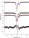
Figure 1.
Figure 1.
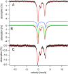
Low-temperature low-field (5 K, 0.05 T) Mössbauer spectra of WT E. coli harvested during exponential growth. A, glucose medium supplemented with 100 μm 57FeIII citrate; B, acetate medium supplemented with 1 (top) and 100 μm 57FeIII citrate (bottom). Gold, blue, green, and pink lines simulate FeIIRET, ISC, FeIILMM, and residual doublets, respectively. The red lines in this and other figures represent composite simulations assuming components, parameters, and percentages given in Table 1. The presence of the FeIILMM doublet in the spectrum from acetate-grown cells with 1 μm iron added is evident from the shift in the high-energy line of the NHHS FeII doublet. The spectrum in C is the sum of all six spectra in Fig. 1 and Fig. S1, after removing contributions from the four major doublets. The red line in C is a simulation assuming parameters typical of S = 52 FeIII or ferritins (the two cannot be distinguished) with 5% of overall spectral intensity. Unless specified otherwise, the magnetic field was applied parallel to the γ radiation.
Figure 2.
Figure 2.
Low-temperature X-band EPR spectra of whole packed E. coli cells grown on glucose (G100E) or acetate (A100E) and harvested under exponential growth conditions. Samples were grown in media supplemented with 100 μm FeIII citrate. Composite simulations are the red lines overlaying the data (black lines). Individual simulations are shown in Fig. S3. Temperature was 10 K, microwave frequency was 9.38 GHz, microwave power was 0.2 milliwatt, time constant was 0.293 s, and modulation amplitude was 10 G.
Figure 3.
Figure 3.
Mössbauer spectra (5 K, 0.05 T) of three separate batches (A, B, and C) of WT E. coli cells grown in glucose under reduced O2 conditions. Blue and green lines, simulations of the FeIILMM and FeIIRET species, respectively.
Figure 4.
Figure 4.
Mössbauer spectra (5 K, 0.05 T) of whole Δfur cells grown on glucose medium and harvested during exponential phase. The concentration of 57FeIII citrate was 1 (A), 10 (B), or 100 μm (C).
Figure 5.
Figure 5.
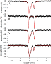
Mössbauer spectra (0.05 T) of whole E. coli cells grown on minimal medium, supplemented with 100 μm57Fe citrate, and harvested during exponential and stationary phases. A, WT cells harvested during exponential growth. B, same as A but harvested in stationary phase. C, ΔftnA cells harvested during exponential growth. D, same as C except collected at 100 K. E, same as C and D except harvested in stationary phase. Vertical dashed lines between C and D highlight the FeIII sextet extending slightly from the baseline. F, ΔbfrΔdps cells harvested during exponential phase. G, same as F but harvested during stationary phase. Spectra A, B, C, E, F, and G were collected at 5 K.
Figure 6.
Figure 6.
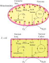
Respiratory shield model for mitochondria (top) and E. coli (bottom). The respiratory shield consists of the ISC- and heme-containing respiratory complexes located in the inner membrane of mitochondria and the cytoplasmic membrane of E. coli and other prokaryotes. The shield is operational when cells are metabolically active, oxidizing nutrient carbon and passing electrons through the respiratory electron-transfer chain and reducing some diffusing O2 to water. With the shield operational, the cytosolic regions become microaerobic. This protects the labile FeII pool in the cell from reaction with O2. When cells transition to stationary phase, they become metabolically less active, and the shield deactivates. Then additional O2 diffuses into the cytosol, where it reacts more rapidly with the labile FeII pool, forming FeIII oxyhydroxide nanoparticles. A similar deactivation of the shield occurs when respiratory complex IV is inhibited by cyanide.
Figure 7.
Figure 7.
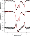
Mössbauer spectra (5 K, 0.05 T) of cyanide-treated E. coli cells. A, before treatment; B, after treatment. C, a difference spectrum of BA. The solid red line in C is a simulation in which the FeIILMM doublet in A is replaced by the nanoparticle doublet in B (13% of spectral intensity for each).
Figure 8.
Figure 8.
Mössbauer spectra (5 K, 0.05 T) of whole E. coli cells and associated retentate and flow-through solutions. A, sum of the spectra obtained of the three samples used in the experiment; B, FTS; C, retentate; D, same as C except after removing the FeIILMM contribution.
Figure 9.
Figure 9.
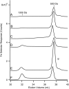
57Fe-detected LC-ICP-MS chromatograms of flow-through solutions from exponentially grown E. coli cells. Traces A, B, and C were for glucose-grown cells in which media were supplemented with 1, 10, and 100 μm 57FeIII citrate, respectively. D, E, and F, the same respective iron concentrations for acetate-grown cells.

Similar articles

Cited by

References

    1. Mettert E. L., and Kiley P. J. (2015) How Is Fe-S Cluster Formation Regulated? Annu. Rev. Microbiol. 69, 505–526 10.1146/annurev-micro-091014-104457 - DOI - PMC - PubMed
    1. Rouault T. A., and Tong W. H. (2005) Iron–sulphur cluster biogenesis and mitochondrial iron homeostasis. Nat. Rev. 6, 345–351 10.1038/nrm1620 - DOI - PubMed
    1. Braymer J. J., and Lill R. (2017) Iron-sulfur cluster biogenesis and trafficking in mitochondria. J. Biol. Chem. 292, 12754–12763 10.1074/jbc.R117.787101 - DOI - PMC - PubMed
    1. Outten F. W., and Theil E. C. (2009) Iron-based redox switches in biology. Antioxid. Redox Signal. 11, 1029–1046 10.1089/ars.2008.2296 - DOI - PMC - PubMed
    1. Gütlich P., Bill E., and Trautwein A. X. (2011) Mössbauer Spectroscopy and Transition Metal Chemistry, Springer, New York

Publication types

MeSH terms