. 2018 Dec 14;293(50):19429-19440.
doi: 10.1074/jbc.RA118.005173. Epub 2018 Oct 18.

The molecular basis of protein toxin HicA-dependent binding of the protein antitoxin HicB to DNA

Affiliations

The molecular basis of protein toxin HicA-dependent binding of the protein antitoxin HicB to DNA

Ashley J Winter et al. J Biol Chem. .

Abstract

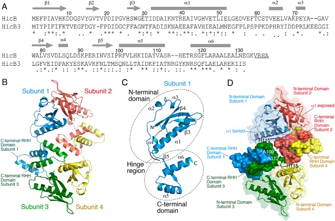
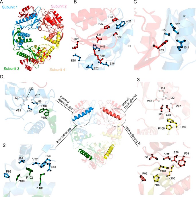
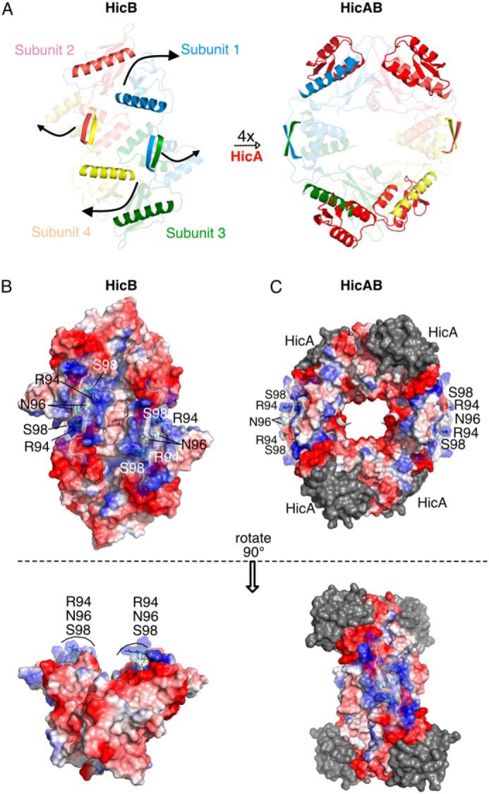
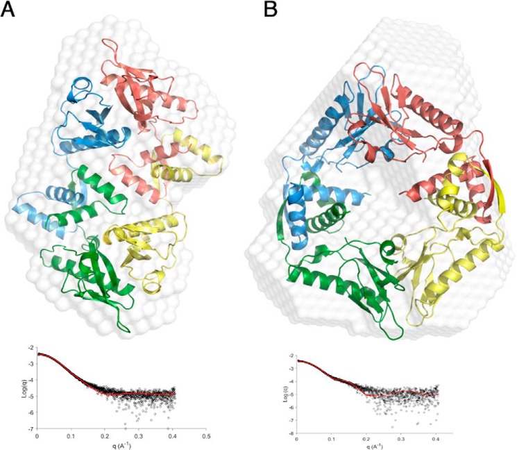
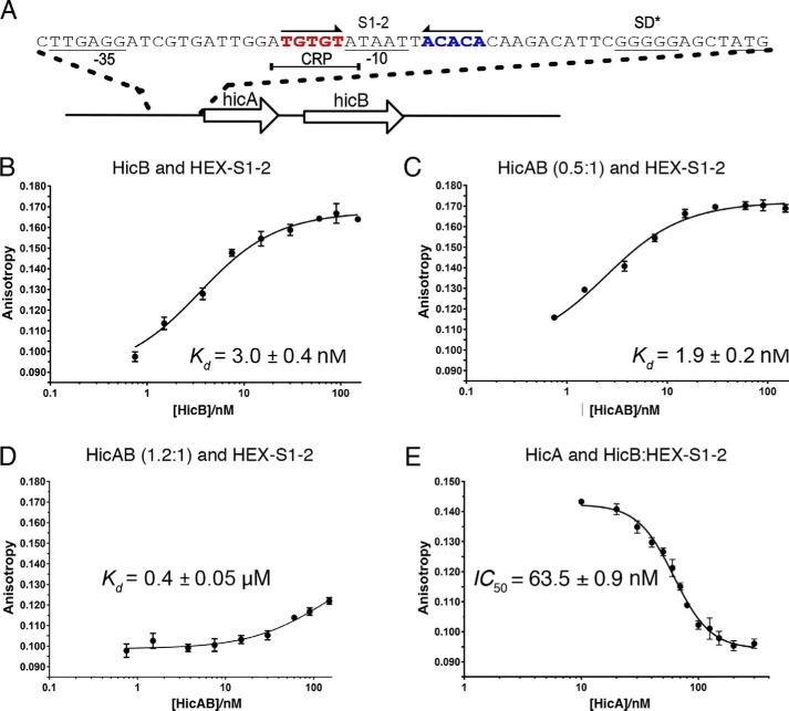
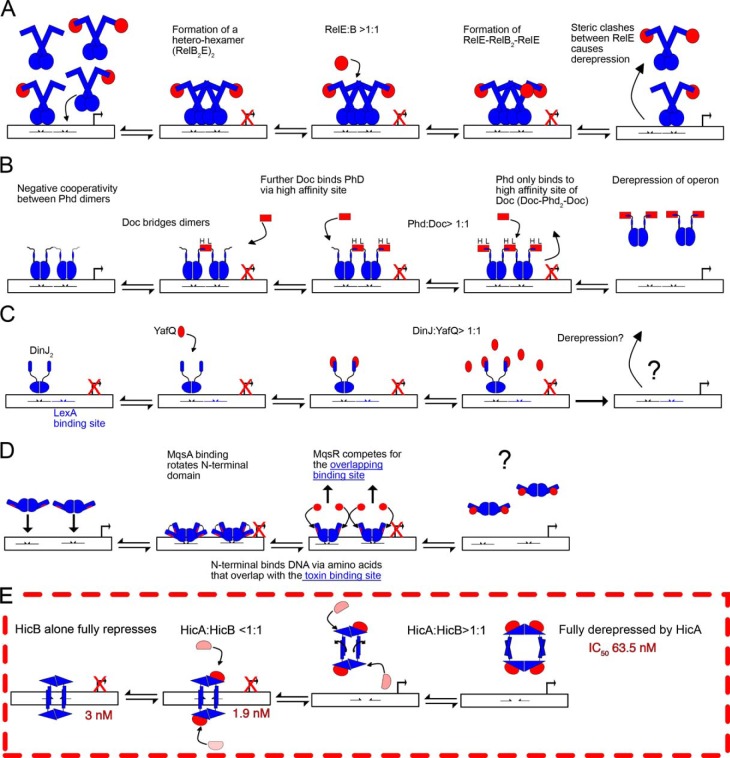
Toxin-antitoxin (TA) systems are present in many bacteria and play important roles in bacterial growth, physiology, and pathogenicity. Those that are best studied are the type II TA systems, in which both toxins and antitoxins are proteins. The HicAB system is one of the prototypic TA systems, found in many bacterial species. Complex interactions between the protein toxin (HicA), the protein antitoxin (HicB), and the DNA upstream of the encoding genes regulate the activity of this system, but few structural details are available about how HicA destabilizes the HicB-DNA complex. Here, we determined the X-ray structures of HicB and the HicAB complex to 1.8 and 2.5 Å resolution, respectively, and characterized their DNA interactions. This revealed that HicB forms a tetramer and HicA and HicB form a heterooctameric complex that involves structural reorganization of the C-terminal (DNA-binding) region of HicB. Our observations indicated that HicA has a profound impact on binding of HicB to DNA sequences upstream of hicAB in a stoichiometric-dependent way. At low ratios of HicA:HicB, there was no effect on DNA binding, but at higher ratios, the affinity for DNA declined cooperatively, driving dissociation of the HicA:HicB:DNA complex. These results reveal the structural mechanisms by which HicA de-represses the HicB-DNA complex.

Keywords: DNA-binding protein; HicAB; X-ray crystallography; antibiotic resistance; bacterial toxin; conditional cooperativity; persistence; protein-protein interaction; structural biology; toxin–antitoxin system; type II TA system.


Conflict of interest statement

The authors declare that they have no conflicts of interest with the contents of this article

Figures

Figure 1.
Figure 1.
Crystal structure of HicB. A, sequence of B. pseudomallei HicB and comparison to the closest structural homologue, HicB3 from Y. pestis. Symbols indicate a conserved residue (*), conservative mutation (:), and a semi-conservative mutation (.). Underlined C-terminal residues were deleted in the His6-tagged construct of HicB used in this study. B, the tetrameric crystal structure of HicB with subunits highlighted and dimerization interfaces at both the N-terminal and C-terminal across adjacent subunits. C, single subunit of HicB with secondary structure elements labeled. D, HicB tetramer with the C-terminal domains rendered with a solid surface and residues at the interfaces between C-terminal pairs are annotated.
Figure 2.
Figure 2.
The HicAB heterooctameric complex. A, cartoon representation of the HicAB heterooctamer where HicA (red) binds to the N-terminal domain of each HicB subunit (blue, pink, green, and yellow). B, HicA (red) has a triple stranded β-sheet that interacts with the α1 helix of HicB (blue). C, the functionally important residue His-24 (H24A in the crystal structure) is predicted to project into a polar pocket formed by Ser-27, Glu-41, and Asn-37 of HicB. D, cartoon representation of new interaction sites across the HicB tetramer. Box 1 highlights the new internal hydrophobic network formed because of the rotation of Leu-85 and Leu-88 to interact with Ile-22, Ile-43, Val-47, and Val-83 within subunit 1 of HicB (blue). Box 2 indicates new intersubunit interactions between adjacent subunits (1 and 3) of HicB (blue and green). Pro-100 and Phe-102 form hydrophobic contacts to Ile-22, Ile-43, Val-47, Val-57, and Val-83, whereas Glu-58 interacts with Lys-106. Because of the symmetrical nature of HicB within the heterooctamer, these interactions sites are conserved between subunits of HicB. This is highlighted for HicB subunit 2 (Boxes 3 and 4).
Figure 3.
Figure 3.
Conformation rearrangements induced by HicA binding and forming the complex HicAB. A, HicA (red) interacts with the α1 helix of each subunit of HicB (highlighted blue, pink, green, and yellow) in the unbound conformation to form the heterooctameric HicAB complex (PDB ID: 6G26), with β5 strands of HicB also highlighted to illustrate their rotation upon complexation. B, surface representation of HicB showing clustering of positive charges on one face mapped to Arg-94, Asn-96, and Ser-98 of the C-terminal domain of HicB. C, surface representation of HicAB highlighting perturbation of the positively charged patch of the C-terminal domain (Arg-94, Asn-96, and Ser-98) because of rotation of the two RHH domains. HicA is represented as gray to emphasize the surface charge of HicB.
Figure 4.
Figure 4.
Small angle X-ray scattering of HicB and HicAB. A, ab initio modeling of the crystal structure of the tetrameric form of HicB into the shape envelope of HicB (white), with each subunit of HicB appropriately colored blue, pink, green, and yellow. The FoXS profile of the proposed scattering profile for the crystal structure (red) against the experimental raw scattering data (black) is underneath (χ2 = 1.98). B, ab initio modeling of the HicB component of the HicAB crystal structure into the shape envelope of HicAB (white), with the corresponding FoXS profile underneath (χ2 = 2.89).
Figure 5.
Figure 5.
Overview of HicB binding to S1–2. A, overview of the hicAB operon. The palindromic region within the upstream region of hicAB that binds HicB. S1 and S2 are highlighted in red and blue. CRP, cAMP receptor protein. B, quantification of HicB binding to HEX–S1–2. Samples contained 7.5 nm HEX–S1–2 in DNA-binding buffer. The proportion of HEX–S1–2 bound by increasing concentration of HicB was followed (n = 1). Data of three independent repeats were fit to Equation 2. C, quantification of HicA/HicB binding to HEX–S1–2 (n = 1) at HicA:B 0.5:1. Again three repeats were fit to Equation 2. D, quantification of HEX–S1–2 binding to HicAB at 1.2:1 for three independent repeats (n = 1). Data were fit to Equation 2. E, quantification of HicA binding to a preformed complex of HicBM (40 nm) with HEX–S1–2 (7.5 nm) corresponding to 80% bound, HicB refers to the monomer concentration of HicB. The proportion of substrate displaced by increasing concentrations of HicA was calculated for three independent repeats via Equation 3 with a Hill coefficient of 2.6. For each experiment the mean value is plotted with error bars representing the S.E. Standard errors of Kd values were calculated in GraphPad Prism.
Figure 6.
Figure 6.
Overview of de-repression models for RelBE, Phd–Doc, DinJ–YafQ, MqsRA, and HicAB. The toxin is highlighted in red and the antitoxin in blue, whereas DNA-binding sites are represented by half arrows. A, the RelB2 dimer or RelB2E complex can bind to one of the two adjacent DNA operator sites, but it is proposed that the formation of a W-shaped heterohexameric complex (RelB2E)2 may occupy both adjacent DNA operator sites simultaneously to confer full transcriptional repression of the relBE operon. Excess toxin (RelE) binds a second site of a RelB dimer forming a rigid RelB2E2 heterotetramer, two heterotetramers that cannot simultaneously bind both operator sites because of steric hindrance resulting in transcriptional de-repression. Transcription of the relBE operon returns RelB and RelE to stoichiometric levels (≠ 1:1). B, Doc forms a heteropentameric complex with Phd (Phd2-Doc-Phd2) by binding to Phd2 via low (L) and high (H) affinity sites to confer full repression of the phd-doc operon by binding two operator sites. Doc allosterically regulates Phd to form a structured DNA-binding domain to ensure full transcription repression only occurs upon formation of a Phd–Doc complex. An excess of Doc preferentially binds Phd solely through H sites resulting in the formation of a rigid heterotetramer (Doc-Phd2-Doc) that cannot occupy both operator sites because of steric clashes between adjacent heterotetramers. C and D, in contrast both DinJ–YafQ (C) and MqsRA (D) do not follow the model of conditional cooperativity, as both toxins act as de-repressors rather than co-repressors. C, DinJ2 fully represses its operator via a single palindromic site. Addition of YafQ forms a YafQ-DinJ2-YafQ heterotetrameric complex, but an excess does not result in de-repression of the DinJ/YafQ–DNA complex and the de-repression mechanism is unknown. D, likewise, MqsA fully represses in the absence of MqsR. Excess MqsR competes with an overlapping DNA-binding site of MqsA (highlighted in red) and formation of a proposed heterotetrameric MqsRA complex results in de-repression (the published MqsRA complex is a partial model). MqsA cannot simultaneously bind both MqsA and DNA. E, like MqsA and DinJ, HicB alone results in saturation of the palindromic sequences (S1–2). HicA binds the surface exposed α1 helices of subunit 2 and 3 to form an intermediate HicA2HicB complex that does not result in an increase of affinity to S1–2. At concentrations of HicA >HicB, there is binding of a further two HicA molecules that results in the 90° rotation of the ribbon-helix-helix motifs and prevents binding to the palindromic sequences and dissociation of HicB from DNA. The intermediate steps of this pathway are unknown and either route, or an equilibrium between the two, cannot be discounted as of present.

Similar articles

Cited by

References

    1. Hall A. M. J., Gollan B., and Helaine S. (2017) Toxin-antitoxin systems: Reversible toxicity. Curr. Opin. Microbiol. 36, 102–110 10.1016/j.mib.2017.02.003 - DOI - PubMed
    1. Page R., and Peti W. (2016) Toxin-antitoxin systems in bacterial growth arrest and persistence. Nat. Chem. Biol. 12, 208–214 10.1038/nchembio.2044 - DOI - PubMed
    1. Lobato-Márquez D., Díaz-Orejas R., and García-Del Portillo F. (2016) Toxin-antitoxins and bacterial virulence. FEMS Microbiol. Rev. 40, 592–609 10.1093/femsre/fuw022 - DOI - PubMed
    1. Lewis K. (2010) Persister cells. Annu. Rev. Microbiol. 64, 357–372 10.1146/annurev.micro.112408.134306 - DOI - PubMed
    1. Gerdes K., and Maisonneuve E. (2012) Bacterial persistence and toxin-antitoxin loci. Annu. Rev. Microbiol. 66, 103–123 10.1146/annurev-micro-092611-150159 - DOI - PubMed

Publication types

LinkOut - more resources