. 2018 Nov 9;145(21):dev169698.
doi: 10.1242/dev.169698.

Notch signalling patterns retinal composition by regulating atoh7 during post-embryonic growth

Affiliations

Notch signalling patterns retinal composition by regulating atoh7 during post-embryonic growth

Alicia Pérez Saturnino et al. Development. .

Abstract

Patterning of a continuously growing naive field in the context of a life-long growing organ such as the teleost eye is of high functional relevance. Intrinsic and extrinsic signals have been proposed to regulate lineage specification in progenitors that exit the stem cell niche in the ciliary marginal zone (CMZ). The proper cell-type composition arising from those progenitors is a prerequisite for retinal function. Our findings in the teleost medaka (Oryzias latipes) uncover that the Notch-Atoh7 axis continuously patterns the CMZ. The complement of cell types originating from the two juxtaposed progenitors marked by Notch or Atoh7 activity contains all constituents of a retinal column. Modulation of Notch signalling specifically in Atoh7-expressing cells demonstrates the crucial role of this axis in generating the correct cell-type proportions. After transiently blocking Notch signalling, retinal patterning and differentiation is re-initiated de novo Taken together, our data show that Notch activity in the CMZ continuously structures the growing retina by juxtaposing Notch and Atoh7 progenitors that give rise to distinct complementary lineages, revealing coupling of de novo patterning and cell-type specification in the respective lineages.

Keywords: Atoh7; Cell specification; Medaka; Notch; Post-embryonic growth; Retinal progenitors.


Conflict of interest statement

Competing interestsThe authors declare no competing or financial interests.

Figures
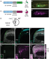
Fig. 1.
Fig. 1.
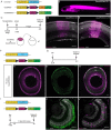
Notch signalling is active in a subset of retinal progenitors, which give rise to MG cells, ACs and BCs during retinal post-embryonic growth in medaka. (A) The tp1-MmHbb::d2GFP (tp1::d2GFP) Notch signalling reporter contains six copies of the tp1 promoter, a Notch-responsive promoter (blue striped boxes). Each tp1 promoter contains two RBP-Jk-binding sites (dark blue stripes). The tp1 promoter is followed by a minimal promoter (mouse β globin) and a destabilized GFP (d2GFP), which has a short half-life. (B) The tp1::d2GFP reporter line shows Notch signalling activation in various tissues such as brain (B), thymus (T) and intestine (I) in hatch medaka. (C) The tp1-MmHbb::tagRFP (tp1::tagRFP) Notch signalling reporter contains the tp1 Notch-responsive promoter followed by a tagRFP, a very stable red fluorescent protein with a long half-life. (D) The tp1::tagRFP reporter line shows Notch signalling activation in hatch medaka in the same tissues as the previously described green reporter line: brain (B), thymus (T) and intestine (I). (E) Schematic of the retina from a lateral view (90°). The CMZ is indicated with a square, which corresponds to the area that is shown in F-G″. (F-F″) Notch signalling (green) is active in a subset of retinal progenitors but it is active in neither RSCs nor differentiated cells (PRCs and MG cells), here labelled with Rx2 (cyan). Nuclear labelling with DAPI is in grey. n=10 fish. (G-G″) The two Notch reporter lines (tp1::d2GFP in green and tp1::tagRFP in magenta) show overlapping activation only in the progenitor area (bottom left insets); 66.14±18.07% (s.d.) of the GFP-positive cells are also tagRFP positive (n=8 retinae, 41 Notch reporter-positive cells). Owing to label retention, the tp1::tagRFP reporter line is also visible in differentiated cells (asterisks) derived from Notch-positive progenitors: MG cells, ACs and BCs. Scale bar: 20 μm.
Fig. 2.
Fig. 2.
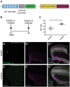
Notch signalling activation and atoh7 expression show a mutually exclusive pattern in the progenitor area of the post-embryonic medaka retina. (A) The atoh7 reporter line containing the atoh7 promoter followed by a GFP was crossed to the tp1::tagRFP Notch reporter. (B,B′) Notch signalling activation (magenta) and atoh7 expression (green) show mutually exclusive patterns in the progenitor area of the CMZ. DAPI is shown in grey in B. Orthogonal views (xz, yz) of the progenitor area in B′ are shown. (C-C″) Higher magnification of the progenitor area indicated with a square in B′. Notch signalling activation is shown in magenta (C), atoh7 in green (C′) and the merge is shown in C″. n=8 fish. Scale bars: 20 μm (B′); 10 μm (C-C″).
Fig. 3.
Fig. 3.
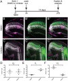
The GaudíRSNICD line recombines ubiquitously and is functional. (A) Specific promoters (X promoter) can be used to drive expression of an inducible Cre recombinase in specific cell populations to achieve targeted recombination of the GaudíRSG construct and the GaudíRSNICD. The GaudíRSG construct is the control construct: it switches from mCherry to H2B-GFP upon recombination. Black triangles represent LoxP sites. The GaudíRSNICD construct is the experimental construct: it switches from mCherry to Notch intracellular domain (NICD) fused to GFP upon recombination. (B) GaudíRSNICD fish after hatching showing ubiquitous mCherry expression (magenta). (C) Transplantations were performed at blastula stage (day 0, d0) from GaudíRSG or GaudíRSNICD embryos into wild-type (WT) embryos (see scheme below). The fish were screened for mCherry expression at hatch (d8) and grown for 14 days. After that, they were fixed and analysed (d22). (D,E) The transplanted cells from both lines (GaudíRSG in D and GaudíRSNICD in E) integrated in the host retina and differentiated into all cell types (Centanin et al., 2011). mCherry-positive cells are shown in magenta and nuclear labelling with DAPI in grey. The retinal cellular layers are also indicated. (F) GaudíRSNICD fish were crossed to a heatshock (HS)::Cre line. (G) The double-positive fish were heatshocked (HS!) at stage 34 (st 34) and fixed and analysed 10 days later. (H-H″) Recombined retinae show mCherry expression (magenta, H) and recombination in all three cellular layers (GFP-positive cells, green, H′). H″ shows the overlay of H and H′. The three retinal layers are indicated. (I) Recombination of the GaudíRSNICD construct was induced with an inducible Cre recombinase under the control of rx2 promoter. After overnight tamoxifen induction, the fish were incubated for 3 days in BrdU and then fixed and analysed. (J) Recombination in rx2-expressing cells such as PRCs, which are located in the ONL, could be observed. (J′) Massive proliferation in the central retina was detected by immunostaining against BrdU (magenta) (n=7 fish). This result recapitulated the previously reported phenotype observed upon Notch activation in Rx2-positive cells (Lust et al., 2016). Scale bars: 40 µm.
Fig. 4.
Fig. 4.
Notch signalling activation in Atoh7-positive progenitors shifts cell-fate ratios during post-embryonic growth. (A) The atoh7 promoter was used to drive an inducible Cre recombinase exclusively in Atoh7-positive cells to achieve targeted recombination of the GaudíRSG construct (control construct: switch from red to green upon recombination) and the GaudíRSNICD construct (functional construct: switch from red to Notch intracellular domain with a GFP upon recombination). (B) Recombination was triggered with an overnight induction with tamoxifen at hatch. The fish were incubated in BrdU overnight to label the induction time point and grown for 14 days afterwards. At day 16 the fish were fixed and the lineage was analysed. (C-H′) GaudíRSG fish (C-E′) and GaudíRSNICD fish (F-H′) both show recombined cells (GFP positive, green) belonging to the Atoh7 lineage. These recombined cells are located at the BrdU (magenta) stripe or more peripherally. (I) Stacked column graph showing the percentage of cells in each layer derived from Atoh7-positive progenitor in control (GaudíRSG) and experimental (GaudíRSNICD) conditions. (J) Quantification of the percentage of GFP-positive (Atoh7-derived) cells shows a decrease in the GCL (***P=0.0003, n=1755 cells in 4 GaudíRSG retinae and 1428 cells in 8 GaudíRSNICD retinae). (K) Quantification of the percentage of GFP-positive (Atoh7-derived) cells shows an increase in the INL (****P<0.0001, n=100 cells in 4 GaudíRSG retinae and 522 cells in 8 GaudíRSNICD retinae). (L) Quantification of the percentage of GFP-positive (Atoh7-derived) cells remained constant in the ONL (P=0.2412, n=164 cells in 4 GaudíRSG retinae and 268 cells in 8 GaudíRSNICD retinae). ns, not significant. Error bars represent s.d. Scale bars: 20 μm.
Fig. 5.
Fig. 5.
Notch signalling inhibition increases the number of Atoh7-positive progenitors. (A) Fish at hatching stage from a double reporter line carrying the tp1::d2GFP reporter and the atoh7::lyntdTomato reporter were used for this experiment. (B) The fish were treated with BrdU and DMSO (control) or Notch inhibitor (LY-411575) for 4 days. At day 4, the fish were fixed and analysed. (C) Quantification of Atoh7-positive cells (counted as lyntdTomato-positive cells) shows an increase upon Notch signalling inhibition (**P=0.001, n=1116 cells in 5 control fish and 1507 cells in 4 inhibited fish). (D-D″) Control fish show Notch signalling activation (GFP, green) and a restricted domain of Atoh7-positive cells (lyntdTomato, magenta). (E-E″) Notch-inhibited fish do not show Notch signalling activation and the atoh7 expression domain is expanded. Error bars represent s.d. Scale bars: 20 μm.
Fig. 6.
Fig. 6.
Notch signalling inhibition shifts cell-fate ratios within the Atoh7 lineage. (A) Wild-type fish were treated at hatch with BrdU and DMSO (control) or Notch inhibitor (LY-411575) for 4 days. At day 4, the BrdU and the DMSO or the inhibitor were washed out and the fish were grown for 14 days. At day 18, the fish were fixed and analysed. (B-B″) Control retinae display a normal morphology and the BrdU (magenta) stripe corresponding to the treatment time contains cells in the three cellular layers. The PRCs labelled with Zpr-1 (green) also display a normal morphology. (C-C″) Treated retinae display a disrupted morphology at the BrdU stripe, lacking the PRCs. (D-F) Quantification of BrdU-positive cells located in each layer in control conditions as well as in Notch-inhibited conditions reveals an increase in cells added to the INL and a decrease in photoreceptors located in the ONL (GCL: P=0.9372, n=710 cells in 3 control retinae and 1303 cells in 3 inhibited retinae; INL: **P=0.0042, n=1104 cells in 3 control retinae and 2443 cells in 3 inhibited retinae; ONL: **P=0.0013, n=334 cells in 3 control retinae and 169 cells in 3 inhibited retinae). Error bars represent s.d. Scale bars: 20 μm.
Fig. 7.
Fig. 7.
Notch signalling regulates the expression of atoh7 by lateral inhibition. The cells that exhibit Notch signalling activation will repress atoh7 expression and give rise to the cell types of the Notch lineage, namely MG cells, BCs and ACs (left). In the neighbouring cell, Notch signalling will be inactive and therefore atoh7 can be expressed. Those cells will give rise to cell types of the Atoh7 lineage: PRCs, ACs, HCs and RGCs (right). Scheme adapted from Centanin et al. (2014).

Similar articles

Cited by

References

    1. Andreazzoli M. (2009). Molecular regulation of vertebrate retina cell fate. Birth Defects Res. Part C - Embryo Today 87, 284-295. 10.1002/bdrc.20161 - DOI - PubMed
    1. Atkinson P. J., Huarcaya Najarro E., Sayyid Z. N. and Cheng A. G. (2015). Sensory hair cell development and regeneration: similarities and differences. Development 142, 1561-1571. 10.1242/dev.114926 - DOI - PMC - PubMed
    1. Austin C. P., Feldman D. E., Ida J. A. and Cepko C. L. (1995). Vertebrate retinal ganglion cells are selected from competent progenitors by the action of Notch. Development 121, 3637-3650. - PubMed
    1. Bajoghli B., Aghaallaei N., Hess I., Rode I., Netuschil N., Tay B.-H., Venkatesh B., Yu J.-K., Kaltenbach S. L., Holland N. D. et al. (2009). Evolution of genetic networks underlying the emergence of thymopoiesis in vertebrates. Cell 138, 186-197. 10.1016/j.cell.2009.04.017 - DOI - PubMed
    1. Baker N. E., Yu S. and Han D. (1996). Evolution of proneural atonal expression during distinct regulatory phases in the developing Drosophila eye. Curr. Biol. 6, 1290-1302. 10.1016/S0960-9822(02)70715-X - DOI - PubMed

Publication types

MeSH terms

Substances

LinkOut - more resources