

Patterned Deposition of Xylan and Lignin is Independent from that of the Secondary Wall Cellulose of Arabidopsis Xylem Vessels
- PMID: 30337427
- PMCID: PMC6305973
- DOI: 10.1105/tpc.18.00292
Patterned Deposition of Xylan and Lignin is Independent from that of the Secondary Wall Cellulose of Arabidopsis Xylem Vessels
Abstract
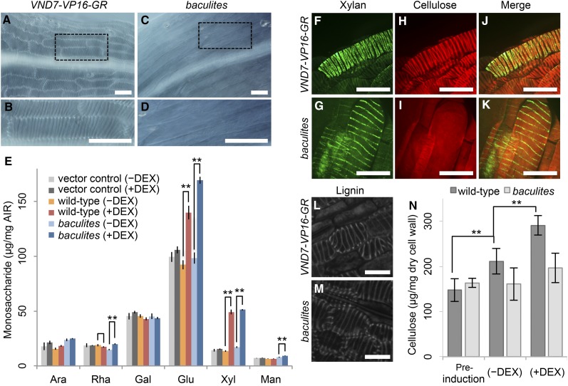
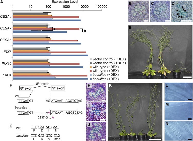
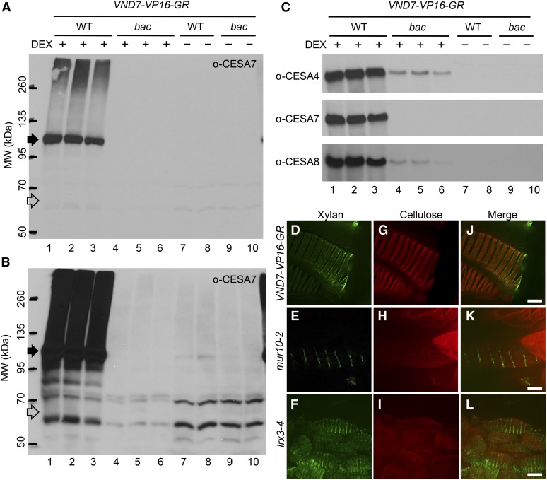
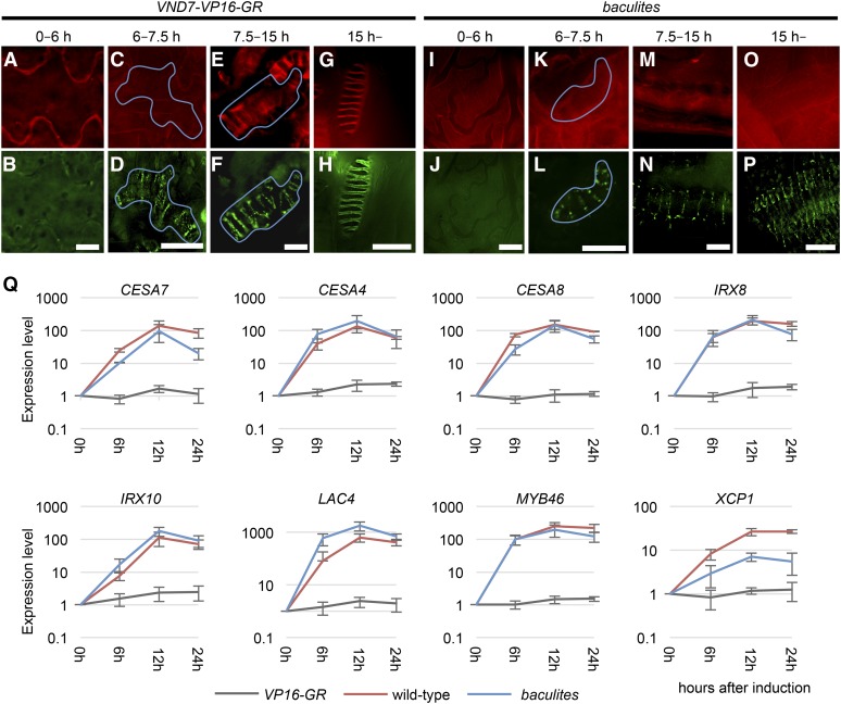
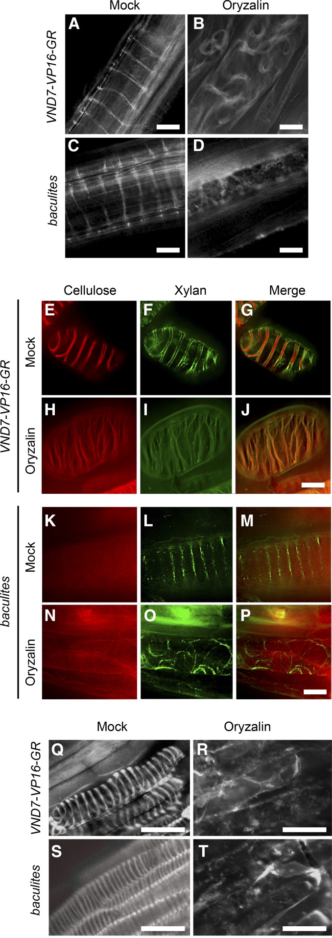
The secondary cell wall (SCW) of xylem vessel cells provides rigidity and strength that enables efficient water conduction throughout the plant. To gain insight into SCW deposition, we mutagenized Arabidopsis thaliana VASCULAR-RELATED NAC-DOMAIN7-inducible plant lines, in which ectopic protoxylem vessel cell differentiation is synchronously induced. The baculites mutant was isolated based on the absence of helical SCW patterns in ectopically-induced protoxylem vessel cells, and mature baculites plants exhibited an irregular xylem (irx) mutant phenotype in mature plants. A single nucleic acid substitution in the CELLULOSE SYNTHASE SUBUNIT 7 (CESA7) gene in baculites was identified: while the mutation was predicted to produce a C-terminal truncated protein, immunoblot analysis revealed that cesa7bac mutation results in loss of production of CESA7 proteins, indicating that baculites is a novel cesa7 loss-of-function mutant. In cesa7bac , despite a lack of patterned cellulose deposition, the helically-patterned deposition of other SCW components, such as the hemicellulose xylan and the phenolic polymer lignin, was not affected. Similar phenotypes were found in another point mutation mutant cesa7mur10-2 , and an established knock-out mutant, cesa7irx3-4 Taken together, we propose that the spatio-temporal deposition of different SCW components, such as xylan and lignin, is not dependent on cellulose patterning.
© 2018 American Society of Plant Biologists. All rights reserved.
Figures
Comment in
-
Microtubules Direct Lignin and Xylan Deposition in a Cellulose-Independent Manner.Plant Cell. 2018 Nov;30(11):2644-2645. doi: 10.1105/tpc.18.00820. Epub 2018 Oct 29. Plant Cell. 2018. PMID: 30373759 Free PMC article. No abstract available.
-
Polymers out of sync.Nat Plants. 2018 Dec;4(12):980. doi: 10.1038/s41477-018-0331-6. Nat Plants. 2018. PMID: 30518835 No abstract available.
Similar articles
-
A review of xylan and lignin biosynthesis: foundation for studying Arabidopsis irregular xylem mutants with pleiotropic phenotypes.Crit Rev Biochem Mol Biol. 2014 May-Jun;49(3):212-41. doi: 10.3109/10409238.2014.889651. Epub 2014 Feb 24. Crit Rev Biochem Mol Biol. 2014. PMID: 24564339 Review.
-
The structure and interaction of polymers affects secondary cell wall banding patterns in Arabidopsis.Plant Cell. 2024 Oct 3;36(10):4309-4322. doi: 10.1093/plcell/koae233. Plant Cell. 2024. PMID: 39163271 Free PMC article.
-
The Carbon Flow Shifts from Primary to Secondary Metabolism during Xylem Vessel Cell Differentiation in Arabidopsis thaliana.Plant Cell Physiol. 2023 Dec 21;64(12):1563-1575. doi: 10.1093/pcp/pcad130. Plant Cell Physiol. 2023. PMID: 37875012 Free PMC article.
-
A R2R3-MYB transcription factor that is specifically expressed in cotton (Gossypium hirsutum) fibers affects secondary cell wall biosynthesis and deposition in transgenic Arabidopsis.Physiol Plant. 2015 Jul;154(3):420-32. doi: 10.1111/ppl.12317. Epub 2015 Jan 26. Physiol Plant. 2015. PMID: 25534543
-
The cell biology of secondary cell wall biosynthesis.Ann Bot. 2018 May 11;121(6):1107-1125. doi: 10.1093/aob/mcy005. Ann Bot. 2018. PMID: 29415210 Free PMC article. Review.
Cited by
-
Hevea brasiliensis coniferaldehyde-5-hydroxylase (HbCAld5H) regulates xylogenesis, structure and lignin chemistry of xylem cell wall in Nicotiana tabacum.Plant Cell Rep. 2021 Jan;40(1):127-142. doi: 10.1007/s00299-020-02619-8. Epub 2020 Oct 17. Plant Cell Rep. 2021. PMID: 33068174 Free PMC article.
-
Transport of UDP-rhamnose by URGT2, URGT4, and URGT6 modulates rhamnogalacturonan-I length.Plant Physiol. 2021 Apr 2;185(3):914-933. doi: 10.1093/plphys/kiaa070. Plant Physiol. 2021. PMID: 33793913 Free PMC article.
-
Overexpression of PnMYB2 from Panax notoginseng induces cellulose and lignin biosynthesis during cell wall formation.Planta. 2022 Apr 21;255(5):107. doi: 10.1007/s00425-022-03891-6. Planta. 2022. PMID: 35445881
-
Developmental landscape and asymmetric gene expression in the leaf vasculature of Brassica rapa revealed by single-cell transcriptome.Hortic Res. 2025 Feb 26;12(6):uhaf060. doi: 10.1093/hr/uhaf060. eCollection 2025 Jun. Hortic Res. 2025. PMID: 40271455 Free PMC article.
-
Plant biomechanics and resilience to environmental changes are controlled by specific lignin chemistries in each vascular cell type and morphotype.Plant Cell. 2022 Sep 21;34(12):4877-96. doi: 10.1093/plcell/koac284. Online ahead of print. Plant Cell. 2022. PMID: 36215679 Free PMC article.
References
-
- Atalla R.H., Hackney J.M., Uhlin I., Thompson N.S. (1993). Hemicelluloses as structure regulators in the aggregation of native cellulose. Int. J. Biol. Macromol. 15: 109–112. - PubMed
-
- Avci U., Earl Petzold H., Ismail I.O., Beers E.P., Haigler C.H. (2008). Cysteine proteases XCP1 and XCP2 aid micro-autolysis within the intact central vacuole during xylogenesis in Arabidopsis roots. Plant J. 56: 303–315. - PubMed
-
- Awano T., Takabe K., Fujita M. (1998). Localization of glucuronoxylans in Japanese beech visualized by immunogold labelling. Protoplasma 202: 213–222.
-
- Balakshin M., Capanema E., Gracz H., Chang H.M., Jameel H. (2011). Quantification of lignin-carbohydrate linkages with high-resolution NMR spectroscopy. Planta 233: 1097–1110. - PubMed
Publication types
MeSH terms
Substances
LinkOut - more resources
Full Text Sources
Other Literature Sources
Molecular Biology Databases
