. 2018 Nov;39(11):2095-2102.
doi: 10.3174/ajnr.A5854. Epub 2018 Oct 18.

Spinal Epidural Arteriovenous Fistula with Perimedullary Venous Reflux: Clinical and Neuroradiologic Features of an Underestimated Vascular Disorder

Affiliations

Spinal Epidural Arteriovenous Fistula with Perimedullary Venous Reflux: Clinical and Neuroradiologic Features of an Underestimated Vascular Disorder

M Mull et al. AJNR Am J Neuroradiol. 2018 Nov.

Abstract

Background and purpose: The purpose of this study was to discuss the clinical and radiologic characteristics of spinal epidural arteriovenous fistulas (SEAVF) and demonstrate their specific angiomorphology in a single-center series.

Materials and methods: Thirteen consecutive patients were diagnosed with SEAVF at RWTH Aachen University Hospital between 2006 and 2018 and were included in this study. All patients had MR imaging and DSA before treatment; 10 of these 13 patients received contrast-enhanced MRA (CE-MRA).

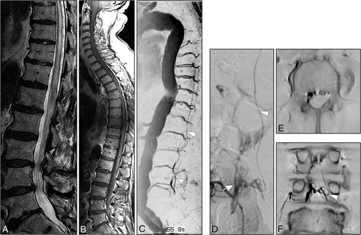
Results: The mean patient age was 72 ± 8 years. Paraparesis was present in 12 (92%) patients. Sphincter dysfunction and sensory symptoms were observed in 7 (54%) and 6 (46%) patients, respectively. The mean duration of symptoms was 6 ± 8 months. Congestive myelopathy on MR imaging was present in all patients. Prominent arterialized perimedullary veins were demonstrated in only 3 cases. CE-MRA revealed arterialized perimedullary veins and an arterialized epidural pouch in 9/10 (90%) patients, mostly located ventrolaterally. DSA demonstrated a multisegmental extension of the arterialized ventrolateral epidural pouch in 6 (46%) cases. An intradural radicular drainage vein was localized distant from the original fistula point in 3 (23%) patients.

Conclusions: Congestive myelopathy with an acute/subacute clinical course was the dominant finding in spinal epidural arteriovenous fistulas. CE-MRA is a powerful diagnostic tool for identifying arterialized perimedullary veins as well as an arterialized epidural pouch. While arterialized perimedullary veins frequently present with only mild enlargement and elongation in spinal epidural arteriovenous fistulas, the arterialized epidural pouch is frequently located ventrolaterally and may extend over several vertebral levels. DSA remains the criterion standard to precisely visualize a spinal epidural arteriovenous fistula and its intradural radicular drainage vein, which may be located distant from the fistulous point.


Figures

Fig 1.
Fig 1.
A, Sagittal T2- weighted images (3T; T2-TSE; slice thickness, 3 mm) reveal extensive congestive myelopathy (white arrowhead). B–D, Spinal CE-MRA (3T; time-resolved imaging with strochastic trajectories (TWIST); sagittal MIP; coronal and axial MPR) shows arterialized pouch in the lumbar ventrolateral epidural space (white arrows) in association with arterialized perimedullary veins in the thoracic region (white arrowheads) suspicious for a SEAVF in the lumbar region. E, DSA in lateral projection shows a SEAVF (white arrow) supplied via branches of the left L2 segmental artery (black arrowhead) and drained via the respective intradural radicular vein (white arrowheads). Note the extraspinal venous outlet (asterisk).
Fig 2.
Fig 2.
A–B, Sagittal T2- and contrast-enhanced T1-weighted images (3T; T2-TSE; T1-TSE; slice thickness, 3 mm) show extensive congestive thoracic myelopathy. C, Spinal CE-MRA (sagittal MIP) reveals an abnormal arterialized epidural pouch in the lumbar region (white arrow) in addition to thoracic arterialized perimedullary veins (white arrowhead). D, DSA (posteroanterior projection) exams identify the fistula in the epidural space on the vertebral level of L4 (white arrow), supplied via the right L4 segmental artery and drained by the contralateral L4 intradural radicular vein (white arrowhead). E and F, Axial and coronal MPR of DynaCT, 2 mm, 8 seconds rotation: Note the multisegmental and bilateral extension of the arterialized epidural pouch and the left sided origin of the intradural radicular drainage vein crossing the dura at the contralateral neural foramen (white arrowhead).
Fig 3.
Fig 3.
A, Spinal CE-MRA (1.5 T, sagittal MIP) reveals an extensive pathological arterialization of a ventrolateral epidural venous pouch extending over four vertebral levels (white arrow). B–C, Further reconstructions of the source MRA images (coronal and axial MPR) demonstrate precisely the epidural pouch (white arrow) and show the filling of the intradural radicular drainage vein (white arrowhead). D–E, DSA exams (posteroanterior projections) identify the multisegmental ventrolateral epidural pouch of the SEAVF (white arrow) with multiple left-sided arterial feeders supplied by the thoracic segmental arteries T 10 and T 11. Note the distant origin of the intradural radicular drainage vein (asterisk).

Similar articles

Cited by

References

    1. Oldfield EH, Doppman JL. Spinal arteriovenous malformations. Clin Neurosurg 1988;34:161–83 - PubMed
    1. Clarke MJ, Patrick TA, White JB, et al. . Spinal extradural arteriovenous malformations with parenchymal drainage: venous drainage variability and implications in clinical manifestations. Neurosurg Focus 2009;26:E5 10.3171/FOC.2009.26.1.E5 - DOI - PubMed
    1. Brinjikji W, Yin R, Nasr DM, et al. . Spinal epidural arteriovenous fistulas. J Neurointerv Surg 2016;8:1305–10 10.1136/neurintsurg-2015-012181 - DOI - PubMed
    1. Aminoff MJ, Logue V. The prognosis of patients with spinal vascular malformations. Brain 1974;97:211–18 10.1093/brain/97.1.211 - DOI - PubMed
    1. Jablawi F, Nikoubashman O, Schubert GA, et al. . Clinical and radiologic characteristics of deep lumbosacral dural arteriovenous fistulas. AJNR Am J Neuroradiol 2018;39:392–98 10.3174/ajnr.A5497 - DOI - PMC - PubMed

LinkOut - more resources