. 2019 Jan;25(1):90-104.
doi: 10.1261/rna.065466.117. Epub 2018 Oct 18.

UGA stop codon readthrough to translate intergenic region of Plautia stali intestine virus does not require RNA structures forming internal ribosomal entry site

Affiliations

UGA stop codon readthrough to translate intergenic region of Plautia stali intestine virus does not require RNA structures forming internal ribosomal entry site

Nobuhiko Kamoshita et al. RNA. 2019 Jan.

Abstract

The translation of capsid proteins of Plautia stali intestine virus (PSIV), encoded in its second open reading frame (ORF2), is directed by an internal ribosomal entry site (IRES) located in the intergenic region (IGR). Owing to the specific properties of PSIV IGR in terms of nucleotide length and frame organization, capsid proteins are also translated via stop codon readthrough in mammalian cultured cells as an extension of translation from the first ORF (ORF1) and IGR. To delineate stop codon readthrough in PSIV, we determined requirements of cis-acting elements through a molecular genetics approach applied in both cell-free translation systems and cultured cells. Mutants with deletions from the 3' end of IGR revealed that almost none of the sequence of IGR is necessary for readthrough, apart from the 5'-terminal codon CUA. Nucleotide replacement of this CUA trinucleotide or change of the termination codon from UGA severely impaired readthrough. Chemical mapping of the IGR region of the most active 3' deletion mutant indicated that this defined minimal element UGACUA, together with its downstream sequence, adopts a single-stranded conformation. Stimulatory activities of downstream RNA structures identified to date in gammaretrovirus, coltivirus, and alphavirus were not detected in the context of PSIV IGR, despite the presence of structures for IRES. To our knowledge, PSIV IGR is the first example of stop codon readthrough that is solely defined by the local hexamer sequence, even though the sequence is adjacent to an established region of RNA secondary/tertiary structures.

Keywords: dicistrovirus; internal ribosomal entry site; stop codon readthrough.


Figures

FIGURE 1.
FIGURE 1.
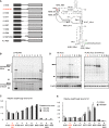
Structure of PSIV IGR and experimental system to analyze stop codon readthrough coincidental to internal initiation. (A) Schematic representation of genomic RNA of PSIV. Coding regions are boxed. Noncoding regions are shown with bold lines. The range of nucleotide sequence cloned into the expression vector is shown by the arrow. Nucleotide numbers are from registered sequence AB006531. Positions of the 3D replicase and VP2 capsid protein are shown along with the box. Dotted lines within the box represent expected cleavage sites of viral proteins. (B) Dicistronic expression unit of plasmid derived from pCI-neo with dicistronic mRNA transcribed, and with schematics of three polypeptides translated. Nucleotide and polypeptide regions for Renilla luciferase (RLuc) and firefly luciferase (FLuc) with a FLAG tag are shown with dark and light gray boxes, respectively. Promoters and polyadenylation site are depicted by triangles and a closed diamond, respectively. In the matrix for translated polypeptides, translation termination at UGA codon is shown by a filled square, while readthrough is shown by an open circle. Schematics of three translational products from full-length IGR, namely, readthrough polypeptides (RT), FLuc, and RLuc, are shown with numbers representing their expected molecular mass. (C) Primary nucleotide sequence of PSIV IGR presented with secondary and tertiary structural model (Kanamori and Nakashima 2001). Positions of stem–loop III (6005–6072) and three pseudoknots are shown. Termination codon of the first ORF (ORF1) and initiation codon of the second ORF (ORF2) are written in red and green, respectively. IGR, intergenic region; IRES, internal ribosomal entry site; N/A, not applicable; PK, pseudoknot; SL, stem–loop; UTR, untranslated region.
FIGURE 2.
FIGURE 2.
Effects of the viral sequence upstream of the UGA codon on the downstream translation of PSIV. (A) Dicistronic mRNAs with or without the viral ORF1 (open box) were used as a template for protein synthesis. Translation from dicistronic luciferase mRNA in which the wild-type full-length IGR sequence is preceded by an upstream viral sequence 5854–6003 (sample 1, 5854–6192) was compared with that of the standard sequence (sample 2, 6004–6192, written with red letters). Controls without stop codon readthrough (sample 3) or internal initiation (sample 4) were prepared by frameshift mutation after the stop codon or M1 mutation into pseudoknot I (PK I) that is essential for IRES activity, respectively. (B) Polypeptides labeled with L-[35S]-methionine in rabbit reticulocyte lysate (RRL) were separated by 8% SDS-PAGE. (C) According to the radioactivity quantified from the gel, the relative expression levels of stop codon readthrough and IRES to those of sample 2 were determined as described in the text and are shown with open and shaded bars, respectively. Mean and SD of three experiments. (D) Readthrough products RT and IRES-dependent FLuc, expressed after transfection into HEK293 cells, were detected with anti-FLAG antibody. Loading control of the blot assayed with anti-β-actin antibody is shown at the bottom. (E) According to the chemiluminescence of the immunoblot obtained using anti-FLAG antibody, the expression levels of RT and FLuc products relative to those of sample 2 are expressed with open and shaded bars, respectively. Mean from the three independent experiments with SD. In B and D, schematic representations of separated polypeptides and marker positions are shown alongside the gel. Mck, mock; X in sample 4, M1 mutation to inactivate IGR IRES. Other abbreviations are as shown in Figure 1. A sample name and lane numbers acting as references are colored red.
FIGURE 3.
FIGURE 3.
Effects of termination codon mutations on stop codon readthrough. (A) The UGA termination codon of PSIV IGR (sample 3) was changed to UAA (sample 1) or UAG (sample 2). To inactivate IRES, 165 nt of the IGR portion starting from PSIV nucleotide 6028A were deleted. In sample 4, the GCU codon following IGR was changed to UAG. The stop codon readthrough level of each sample was determined as the ratio of firefly to Renilla luciferase enzymatic activities after 48 h of transfection into indicated cells and is shown as the percentage relative to that of UGA–GCU (sample 3). Mean and SD from four different transfections are shown. (B) Polypeptides expressed in COS-1 cells after 60 h of transfection were immunoblotted with anti-RLuc or anti-FLAG antibody. Loading controls are shown as in Figure 2. Polypeptides expressed from IRES were run as a control in lane 5.
FIGURE 4.
FIGURE 4.
Effects of deletions from the 3′ end of IGR on UGA stop codon readthrough. (A) IGR cloned into the dicistronic unit was deleted from the 3′ end of full-length IGR (sample 1). Schematics of mutants (left) and positions of deletion alongside the nucleotide sequence in predicted structure (right). In the panel at right, numbers following an underscore symbol denote the lengths of IGR nucleotides remaining. (B) In RRL, polypeptides translated from dicistronic mRNAs with partial (lanes 3–7) to complete (lane 8) deletion of IGR were analyzed and compared with those from control mRNAs (lanes 2, 9, and 10). Shown below is the overexposed image of RT and FLuc products, with its corresponding part in the original image indicated with rectangles alongside. Potassium concentration was adjusted to 150 mM with KCl. Representative data among three reactions are shown. (C) Proportion of stop codon readthrough was determined from the radioactivity of each polypeptide and normalized to that of 6192M1 (sample 2). Values obtained at potassium concentrations of 100 and 150 mM are shown with open and dark gray bars, respectively. (D) Plasmids corresponding to A were transfected into COS-1 cells and expressed polypeptides were immunodetected in blots using anti-RLuc or anti-FLAG antibody. Loading controls against β-actin are shown below. (E) Relative expression levels determined from the chemiluminescence of readthrough polypeptides in COS-1 and HEK293 cells, normalized to that of 6192M1 mutant (lane 2), are shown with open and shaded bars, respectively. Mean from three different experiments.
FIGURE 5.
FIGURE 5.
Chemical mapping of RNA structures downstream from UGA1 codon in 3′ deletion mutants 6009 and 6072. IGR RNA structures of representative deletion mutants used in Figure 4 were chemically mapped with DMS (60 mM), CMCT (25 mM), or NAI (100 mM). (Left) Chemically modified ribonucleotides in mutant 6009 (A) or 6072 (B) were determined by the inhibition of reverse transcription from the downstream 32P-labeled primer hybridized to FLuc. Two different preparations of RNAs were modified in DMS and CMCT. The positions of modified nucleotides are indicated with lines at the side of the gel. Strong modifications observed in NAI are depicted with thick lines. (Right) In the schematics, the positions of landmark nucleotides are shown with dots and numbers. Modified nucleotides are indicated with bars (A) and lines (B). Codons 6004UGA, 6007CUA, and 6193GCU are color-coded with red, orange, and green, respectively. The positions of coding sequences for RLuc and Δcapsid–FLuc are boxed. In B (left), the positions of predicted paired regions in SLIII are indicated by arrows on the side of the gel. The positions of CMCT modification were determined from the short-exposure image (see Supplemental Fig. S4C).
FIGURE 6.
FIGURE 6.
Synonymous mutations for 6007–6009CUA. (A) Synonymous mutations coding for CUA/leucine were introduced into the GCU/alanine Δcapsid initiation codon of mutant 6006. Readthrough levels in HEK293 cells normalized to the CUA wild-type sequence, indicated with orange, are shown as mean ± SEM from five different experiments. (B) Polypeptides expressed in HEK293 cells were analyzed with immunoblots using anti-RLuc or anti-FLAG antibody. Loading controls obtained in immunoblots for β-actin are shown below.
FIGURE 7.
FIGURE 7.
Readthrough efficiency in cultured cells. (A) Expression of 108 kDa RT polypeptides from the UGA codon (sample 2, indicated in red) was compared with that of the corresponding products from GGA sense codon control (sample 1). IRES-dependent translation was inactivated by M1 mutation, shown with an X, in pseudoknot I. In the matrix, polypeptides expressed or not expressed are shown with a solid or dotted box, respectively. (B) FLuc activity 48 h after transfection was normalized to coexpressed fluorescence of EGFP used as an internal control. Relative FLuc activities normalized to that expressed from the UGA codon (sample 2) were determined from three independent transfections. Mean and SD in COS-1 and HEK293 cells are shown with open and shaded bars, respectively. N/A, not applicable.

Similar articles

References

    1. Abeyrathne PD, Koh CS, Grant T, Grigorieff N, Korostelev AA. 2016. Ensemble cryo-EM uncovers inchworm-like translocation of a viral IRES through the ribosome. Elife 5: e14874 10.7554/eLife.14874 - DOI - PMC - PubMed
    1. Au HHT, Elspass VM, Jan E. 2018. Functional insights into the adjacent stem-loop in honey bee dicistroviruses that promotes IRES-mediated translation and viral infection. J Virol 92: e01725-17 10.1128/JVI.01725-17 - DOI - PMC - PubMed
    1. Beier H, Grimm M. 2001. Misreading of termination codons in eukaryotes by natural nonsense suppressor tRNAs. Nucleic Acids Res 29: 4767–4782. 10.1093/nar/29.23.4767 - DOI - PMC - PubMed
    1. Bertram G, Innes S, Minella O, Richardson J, Stansfield I. 2001. Endless possibilities: translation termination and stop codon recognition. Microbiology 147: 255–269. 10.1099/00221287-147-2-255 - DOI - PubMed
    1. Bonning BC, Miller WA. 2010. Dicistroviruses. Annu Rev Entomol 55: 129–150. 10.1146/annurev-ento-112408-085457 - DOI - PubMed

Publication types

Supplementary concepts

LinkOut - more resources