. 2018 Oct 18;9(11):1066.
doi: 10.1038/s41419-018-1071-2.

YLT-11, a novel PLK4 inhibitor, inhibits human breast cancer growth via inducing maladjusted centriole duplication and mitotic defect

Affiliations

YLT-11, a novel PLK4 inhibitor, inhibits human breast cancer growth via inducing maladjusted centriole duplication and mitotic defect

Qian Lei et al. Cell Death Dis. .

Abstract

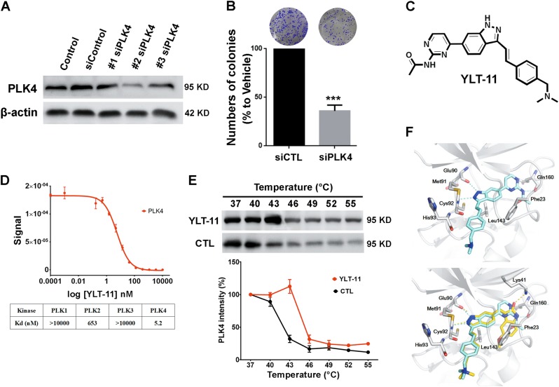
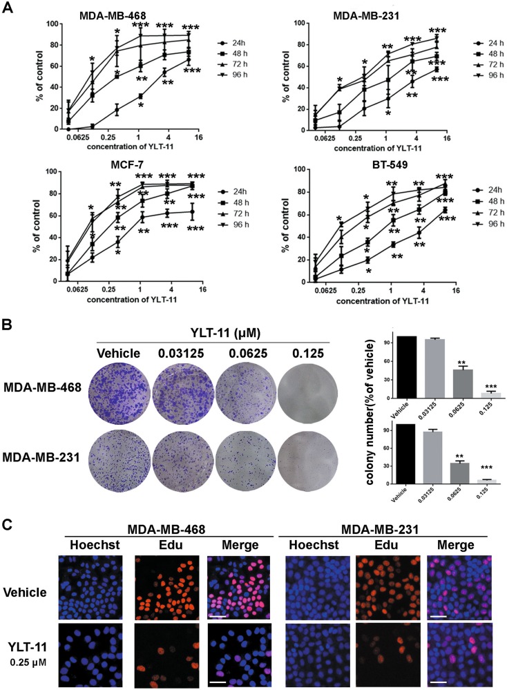
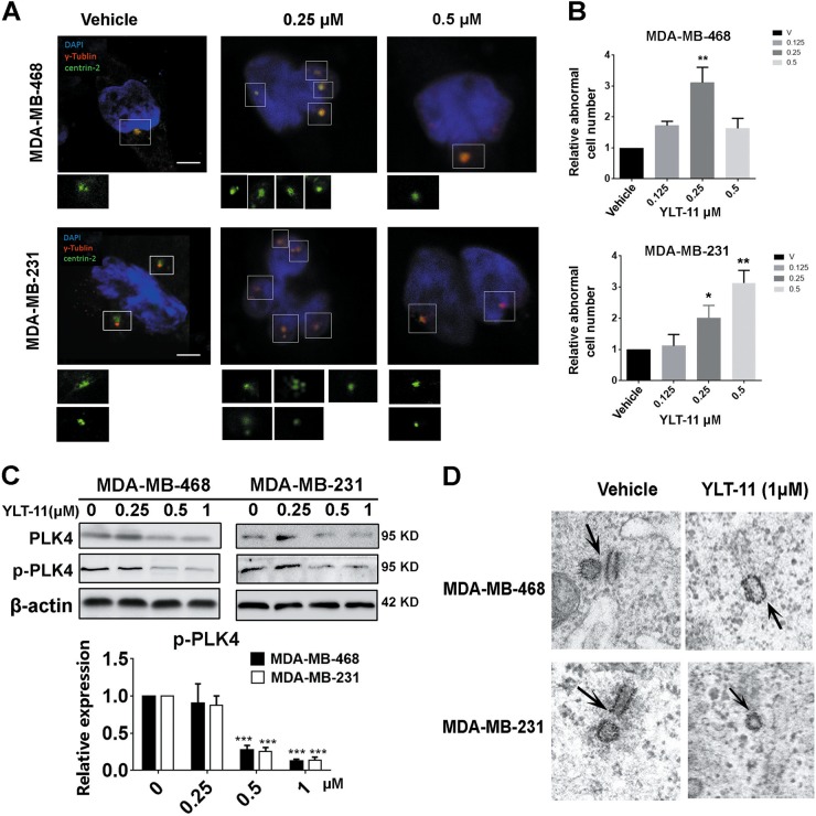
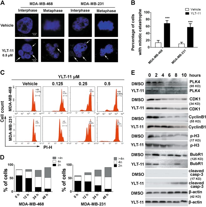
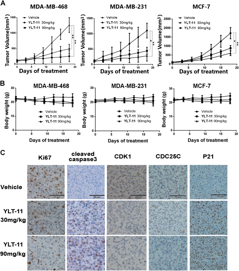
Polo-like kinase 4 (PLK4) is indispensable for precise control of centriole duplication. Abnormal expression of PLK4 has been reported in many human cancers, and inhibition of PLK4 activity results in their mitotic arrest and apoptosis. Therefore, PLK4 may be a valid therapeutic target for antitumor therapy. However, clinically available small-molecule inhibitors targeting PLK4 are deficient and their underlying mechanisms still remain not fully clear. Herein, the effects of YLT-11 on breast cancer cells and the associated mechanism were investigated. In vitro, YLT-11 exhibited significant antiproliferation activities against breast cancer cells. Meanwhile, cells treated with YLT-11 exhibited effects consistent with PLK4 kinase inhibition, including dysregulated centriole duplication and mitotic defects, sequentially making tumor cells more vulnerable to chemotherapy. Furthermore, YLT-11 could strongly regulate downstream factors of PLK4, which was involved in cell cycle regulation, ultimately inducing apoptosis of breast cancer cell. In vivo, oral administration of YLT-11 significantly suppressed the tumor growth in human breast cancer xenograft models at doses that are well tolerated. In summary, the preclinical data show that YLT-11 could be a promising candidate drug for breast tumor therapy.


Conflict of interest statement

Conflict of interest

The authors declare that they have no conflict of interest.

Disclaimer

We claim that none of the materials in the paper has been published or is under consideration for publication elsewhere, all authors are aware of the submission and agree to its publication.

Figures

Fig. 1
Fig. 1. The activity of PLK4 could be potently inhibited by YLT-11, a novel and specific PLK4 inhibitor.
a MDA-MB-231 transfected with PLK4 siRNA. The expression of PLK4 was determined by Western blotting. b The number of colonies in MDA-MB-231 transfected with PLK4 siRNA. Quantification was shown in the lower panel. Data are expressed as mean ± SD for three independent experiments. Columns, mean, bars, SD (***p < 0.001 vehicle control). c Chemical structure of YLT-11. d Binding assays for YLT-11–PLK4 interaction. Upper panel: Determination of quantitative binding constants of PLK4; lower panel: binding constants of PLK family. e Cellular thermal shift assay from 40 to 55 ℃ of MDA-MB-231 lysates with or without YLT-11 incubation. The image (upper panel) and quantification of the band intensities (lower panel) in immunoblotting. Graphic data were run in triplicate and shown as the mean ± SD. f YLT-11 is docked into the active site of PLK4, showing interactions between YLT-11 and PLK4 in the three-dimentional structure, and YLT-11 binds to PLK4 via a mode highly similar to that of the inhibitor 400631
Fig. 2
Fig. 2. Antiproliferative activities of YLT-11 against human breast cancer cell lines.
a Four different breast cancer cells were treated with increasing doses of YLT-11 for 24, 48, 72, and 96 h, respectively. Each point represents the mean ± SD for three independent experiments (*p < 0.05, **p < 0.01, ***p < 0.001 vs. vehicle control).  b Effects of YLT-11 on cell colony formation after treatement for 2 weeks. Quantification is shown in the right panel. Columns, means (n = 3); bars, standard deviation (*p < 0.05, **p < 0.01, ***p < 0.001). c YLT-11 inhibited MDA-MB-468 and MDA-MB-231 cell proliferation. The EdU incorporation assay was examined on cells after exposure to YLT-11 for 24 h. EdU-positive (marked by red fluorescent staining) and Hoechst 33342 staining (marked by blue fluorescent staining) cells represented the proliferating and total cells, respectively. Images shown are representatives of three independent experiments. Scale bars, ×20 for micrograph
Fig. 3
Fig. 3. YLT-11 induced aberrant centriole duplication in breast cancer cells.
a MDA-MB-468 and MDA-MB-231 were treated with YLT-11 with an increasing concentration for 24 h, and the centrioles and centrosomes were determined by immunostaining for centrin 2 (green fluorescent staining) and γ-tublin (red fluorescent staining), respectively. DNA was marked by DAPI. Insets show higher magnification views of the centrioles at the spindle pole (boxed regions). Scale bars, 5 μm; 1 μm for insets. b The relative number of cancer cells with abnormal centrioles after treatement with YLT-11 for 24 h, compared with the vehicle group, were counted. Columns, means (n = 3); bars, standard deviation (*p < 0.05, **p < 0.01). c Expression of p-PLK4 was determined by Western blotting. MDA-MB-468 and MDA-MB-231 were treated with DMSO or the indicated concentrations of YLT-11 (0.25, 0.5, and 1 μM) for 48 h, and the expressions of p-PLK4 and PLK4 were detected with a specific antibody. Each has the expression of β-actin as the internal control. Protein expression was quantified by densitometry analysis using ImageJ and normalized against β-actin expression. Columns, mean; bars, SD, *p < 0.05. d Electron micrographs showed the number of centrioles in cells treated with 1 μM YLT-11 for 48 h (right panels), whereas the DMSO group showed only two centrioles (right panel). Scale bar, 500 nm
Fig. 4
Fig. 4. YLT-11 induced mitotic defects and disturbed mitotic checkpoint function.
a MDA-MB-468 and MDA-MB-231 cells were treated with DMSO or 0.5 μM YLT-11 for 30 h and then stained with DAPI to detect the karyomorphism in different stages. Multinucleation, micronuclei, and chromosome mal-disjunction were marked by arrowheads. b Percentage of cells with mitotic catastrophe is quantified. Columns, means (n = 3); bars, standard deviation, (***p < 0.001). c Breast cancer cell lines were treated with an increasing concentration of YLT-11 for 30 h and stained with propidium iodide prior to analysis of DNA content by flow cytometry. Gating (>4N) indicated the percentage of aneuploid/polyploid cells. d Breast cancer cell lines were treated with YLT-11 (0.25 μM) for indicated times. Percentage of cancer cells in different stages. 2N, 4N, and >4N presented G1, G2/M, and aneuploid/polyploid cells, respectively. e Western analyses of key mitotic checkpoint protein. MDA-MB-468 cells were synchronized by nocodazole for 16 h and allowed to proceed in the presence or absence of 0.5 μM YLT-11. β-actin served as a loading control
Fig. 5
Fig. 5. YLT-11 induced apoptosis of breast cancer cell lines.
a Cell morphological alterations and nuclear changes of MDA-MB-468 and MDA-MB-231 cells were analyzed by staining with Hoechst 33342 (10 mg/mL) and visualized using a microscope after treatment with increasing doses of YLT-11 for 24 h. b MDA-MB-468 and MDA-MB-231 cells were treated with YLT-11 for 48 h and were detected by flow cytometry after Annexin V/PI staining. c The expressions of cleaved caspase-3 and cleaved PARP1 were determined via western blotting. Protein expressions were quantized by densitometry analysis using ImageJ (shown in the right panel). Data are expressed as mean ± SD for three independent experiments. Columns, mean; bars, SD (*p < 0.05, **p < 0.01, ***p < 0.001)
Fig. 6
Fig. 6. Effects of YLT-11 on the proliferation of xenografts in vivo.
a, b Tumor suppression of MDA-MB-468, MDA-MB-231, and MCF-7 tumor xenografts in mice treated with different concentrations of YLT-11. Tumor size and body weight were measured and calculated every 3 days and presented as mean ± SD (n = 5; *p < 0.05; **p < 0.01). c Tumor tissues from MDA-MB-231 xenografts treated with vehicle or YLT-11 were immunohistochemically analyzed with anti-Ki67, cleaved caspase-3, and CDC25C, CDK1, and P21
Fig. 7
Fig. 7. Preliminary safety evaluation of YLT-11 in BALB/c mice.
a The difference of body weights between two administrated groups (female and male mice) and two vehicle groups (female and male mice) were not significant. Data are expressed as mean ± SD (n = 5). b Hematological and serum biochemical values of mice at day 14 (n = 5) for both vehicle and treated groups. Units of the parameters are as follows: WBC white blood cell (109/L); RBC red blood cell (1012/L); HGB hemoglobin (g/L); ALB albumin and TP total protein (g/L); ALT alanine transarninase (U/L); AST aspartate aminotransferase (U/L); TBIL total bilirubin (μmol/L); ALP alkaline phosphatase (U/L); BUN blood urea nitrogen; and GLU glucose (mM). c YLT-11 did not cause obvious pathologic abnormalities in normal tissues. Paraformaldehyde-fixed organs (heart, liver, spleen, lungs, and kidneys) were processed for paraffin embedding and then stained by hematoxylin and eosin. Images shown are representatives from each group. Scale bars, ×20 for micrograph

Similar articles

Cited by

References

    1. DeSantis C, Ma J, Bryan L, Jemal A. Breast cancer statistics, 2013. Cancer J. Clin. 2014;64:52–62. doi: 10.3322/caac.21203. - DOI - PubMed
    1. Fan L, et al. Breast cancer in China. Lancet Oncol. 2014;15:e279–289. doi: 10.1016/S1470-2045(13)70567-9. - DOI - PubMed
    1. Torre LA, Siegel RL, Ward EM, Jemal A. Global cancer incidence and mortality rates and trends—an update. Cancer Epidemiol. Biomark. Prev. 2016;25:16–27. doi: 10.1158/1055-9965.EPI-15-0578. - DOI - PubMed
    1. Goldhirsch A, et al. Personalizing the treatment of women with early breast cancer: highlights of the St Gallen International Expert Consensus on the Primary Therapy of Early Breast Cancer 2013. Ann. Oncol. 2013;24:2206–2223. doi: 10.1093/annonc/mdt303. - DOI - PMC - PubMed
    1. Lin NU, et al. Clinicopathologic features, patterns of recurrence, and survival among women with triple-negative breast cancer in the National Comprehensive Cancer Network. Cancer. 2012;118:5463–5472. doi: 10.1002/cncr.27581. - DOI - PMC - PubMed

Publication types

MeSH terms