. 2018 Oct 18;9(1):4330.
doi: 10.1038/s41467-018-06797-8.

Structure of MHC class I-like MILL2 reveals heparan-sulfate binding and interdomain flexibility

Affiliations

Structure of MHC class I-like MILL2 reveals heparan-sulfate binding and interdomain flexibility

Mizuho Kajikawa et al. Nat Commun. .

Abstract

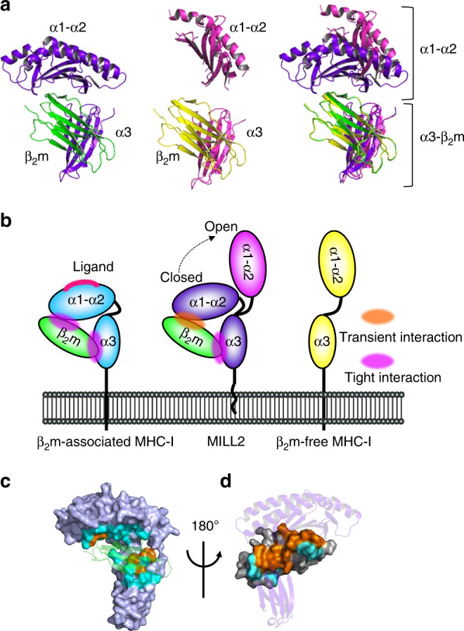
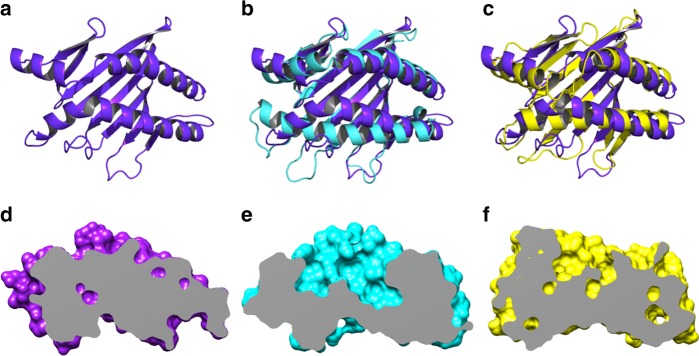
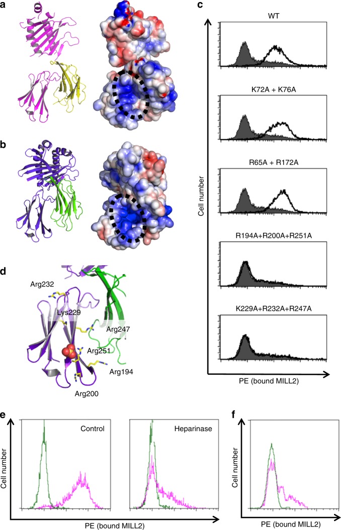
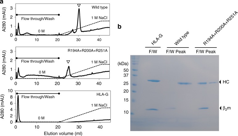
The MILL family, composed of MILL1 and MILL2, is a group of nonclassical MHC class I molecules that occur in some orders of mammals. It has been reported that mouse MILL2 is involved in wound healing; however, the molecular mechanisms remain unknown. Here, we determine the crystal structure of MILL2 at 2.15 Å resolution, revealing an organization similar to classical MHC class I. However, the α1-α2 domains are not tightly fixed on the α3-β2m domains, indicating unusual interdomain flexibility. The groove between the two helices in the α1-α2 domains is too narrow to permit ligand binding. Notably, an unusual basic patch on the α3 domain is involved in the binding to heparan sulfate which is essential for MILL2 interactions with fibroblasts. These findings suggest that MILL2 has a unique structural architecture and physiological role, with binding to heparan sulfate proteoglycans on fibroblasts possibly regulating cellular recruitment in biological events.


Conflict of interest statement

The authors declare no competing interests.

Figures

Fig. 1
Fig. 1
MILL2 crystals form both closed and open conformations. a The closed conformation (left), open conformation (center), and superimposition of these two conformations based on the position of β2m chain (right) are represented by ribbon diagrams. Purple, closed conformation of MILL2; magenta, open conformation of MILL2; green, β2m associated with the closed conformation of MILL2; yellow, β2m associated with the open conformation of MILL2. b Schematic model of the flexible membrane-distal α1-α2 domains (center). In typical β2m-associated MHC-I molecules, the α1–α2 domains have no flexibility because of tight interactions with β2m (left). By contrast, the α1–α2 domains of β2m–free MHC-I molecules (e.g. human MIC family) are located far from the membrane-proximal α3 domain. c Surface model of MILL2 showing contact residues with β2m. Orange, conserved contact residues with β2m on MILL2 and H-2Db; cyan, MILL2-specific contact residues with β2m. β2m is represented by a transparent ribbon diagram (green). d Surface model showing contact residues on β2m with MILL2. Orange, contact residues on β2m conserved between MILL2 and H-2Db; cyan, MILL2-specific contact residues on β2m. MILL2 heavy chains are represented by a ribbon diagram (purple)
Fig. 2
Fig. 2
The narrow space between the two helices in α1-α2 domains of MILL2. a Top view of the membrane-distal α1-α2 domains of MILL2 represented by a ribbon diagram (purple). b, c Superimposition of MILL2 α1-α2 domains with b HLA-B (PDB ID: 1E27) or c MICB (PDB ID: 1JE6), represented by ribbon diagrams. Purple, MILL2; cyan, HLA-B; yellow, MICB. df Side view of the α1-α2 domains of d MILL2, e HLA-B (PDB ID: 1E27), and f MICB (PDB ID: 1JE6) represented by cutaway model. Purple, MILL2; cyan, HLA-B; yellow, MICB
Fig. 3
Fig. 3
Putative heparan sulfate-binding site on the α3 domain of MILL2. a, b Side views of a the open and b the closed conformation of MILL2 shown by a ribbon diagram (left) and its electrostatic surface potential model (right). Magenta and yellow indicate open conformation of MILL2 and β2m, respectively. Purple and green indicate closed conformation of MILL2 and β2m, respectively. Red and blue indicate negatively and positively charged areas, respectively. The black dotted circles highlight the position of the basic patch. c Histograms show the binding levels of MILL2 and the respective alanine-substituted tetramers to NIH-3T3 cells. PE-conjugated MILL2 tetramer and tetramers incorporating alanine substitutions in the basic patch of the α1-α2 domains (K72A + K76A and R65A + K172A) or α3 domain (R194A + R200A + R251A, K229A + R232A + R247A) were generated. Staining with PE-conjugated streptavidin (shaded histograms) or PE-conjugated tetramer (open histograms) was measured by flow cytometry. d An SO4 ion is located near the basic patch on the α3 domain in MILL2 crystals. The α3 domain of MILL2 is shown as a ribbon diagram (purple) and sticks represent the side chains of residues in this patch. The SO4 ion is shown as a space-filling model. e Histograms show staining of NIH-3T3 cells with wild type MILL2 tetramers with or without heparinase treatment. Cells were preincubated with PBS (control) or PBS including heparinase (heparinase). After treatment, cells were stained with PE-conjugated streptavidin (green histogram) or PE-conjugated MILL2 tetramer (pink histogram) and binding was measured by flow cytometry. f Histogram shows heparin-competition for the binding of MILL2 tetramers to NIH-3T3 cells. PE-conjugated streptavidin (green histogram) or PE-conjugated MILL2 tetramer (pink histogram) staining was performed with heparin. Binding was measured by flow cytometry
Fig. 4
Fig. 4
MILL2 binds directly to heparin. a Heparin affinity chromatography. MILL2 (Wild type or R194A + R200A + R251A) or HLA-G was loaded on a HiTrap Heparin HP column equilibrated with 20 mM Tris-HCl buffer pH 8.0. Subsequently, column were washed with 20 column volumes of 20 mM Tris-HCl buffer pH 8.0 and bound proteins were eluted with 20 column volumes of a linear NaCl gradient (0 to 1 M NaCl in 20 mM Tris-HCl buffer pH 8.0). Open arrowheads indicate peaks eluted by the NaCl gradient. b SDS-PAGE analysis of collected flow-through/wash fraction (F/W) and eluted peak fraction (Peak). Arrows indicate heavy chain (HC) or β2m. An uncropped scanned image of this gel is shown in Supplementary Fig. 15

Similar articles

Cited by

References

    1. Tanaka K, Kasahara M. The MHC class I ligand-generating system: roles of immunoproteasomes and the interferon-gamma-inducible proteasome activator PA28. Immunol. Rev. 1998;163:161–176. doi: 10.1111/j.1600-065X.1998.tb01195.x. - DOI - PubMed
    1. Bjorkman PJ, et al. Structure of the human class I histocompatibility antigen, HLA-A2. Nature. 1987;329:506–512. doi: 10.1038/329506a0. - DOI - PubMed
    1. Rudolph MG, Wilson IA. The specificity of TCR/pMHC interaction. Curr. Opin. Immunol. 2002;14:52–65. doi: 10.1016/S0952-7915(01)00298-9. - DOI - PubMed
    1. Salter RD, et al. A binding site for the T-cell co-receptor CD8 on the alpha 3 domain of HLA-A2. Nature. 1990;345:41–46. doi: 10.1038/345041a0. - DOI - PubMed
    1. Wang R, Natarajan K, Margulies DH. Structural basis of the CD8 alpha beta/MHC class I interaction: focused recognition orients CD8 beta to a T cell proximal position. J. Immunol. 2009;183:2554–2564. doi: 10.4049/jimmunol.0901276. - DOI - PMC - PubMed

Publication types

MeSH terms