. 2018 Oct 18;8(1):15427.
doi: 10.1038/s41598-018-33723-1.

Crystallographic snapshots of ligand binding to hexameric purine nucleoside phosphorylase and kinetic studies give insight into the mechanism of catalysis

Affiliations

Crystallographic snapshots of ligand binding to hexameric purine nucleoside phosphorylase and kinetic studies give insight into the mechanism of catalysis

Zoran Štefanić et al. Sci Rep. .

Abstract

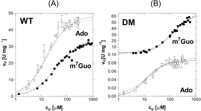
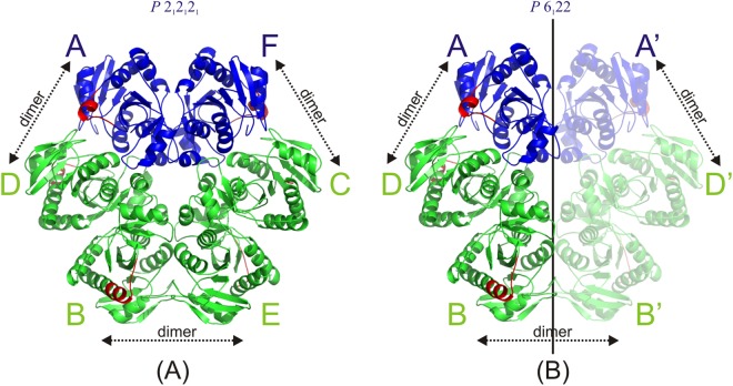
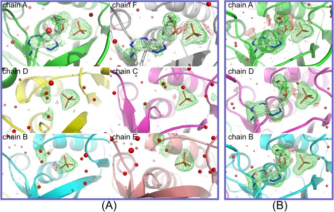
Purine nucleoside phosphorylase (PNP) catalyses the cleavage of the glycosidic bond of purine nucleosides using phosphate instead of water as a second substrate. PNP from Escherichia coli is a homohexamer, build as a trimer of dimers, and each subunit can be in two conformations, open or closed. This conformational change is induced by the presence of phosphate substrate, and very likely a required step for the catalysis. Closing one active site strongly affects the others, by a yet unclear mechanism and order of events. Kinetic and ligand binding studies show strong negative cooperativity between subunits. Here, for the first time, we managed to monitor the sequence of nucleoside binding to individual subunits in the crystal structures of the wild-type enzyme, showing that first the closed sites, not the open ones, are occupied by the nucleoside. However, two mutations within the active site, Asp204Ala/Arg217Ala, are enough not only to significantly reduce the effectiveness of the enzyme, but also reverse the sequence of the nucleoside binding. In the mutant the open sites, neighbours in a dimer of those in the closed conformation, are occupied as first. This demonstrates how important for the effective catalysis of Escherichia coli PNP is proper subunit cooperation.


Conflict of interest statement

The authors declare no competing interests.

Figures

Figure 1
Figure 1
Left panel: Open and closed conformation of E. coli PNP. Monomers (A) (green) and (B) (magenta) of the wild-type E. coli ternary complex with phosphate/sulphate and FA structure, WT-6(P/S)-6FA, are overlapped to show the difference between closed and open active site conformation. The positions of catalytic amino acids Asp204 and Arg217 mutated to Ala in the double mutant structures are shown. The extent of movement of the loop and segmentation of the helix H8 is indicated by arrows. Right panel: structure of PNP ligands used in this study, PNP inhibitor FA, the N9 deaza analogue of the natural enzyme substrate adenosine, and 7-methylguanosine, the substrate bearing the positive charge on the five-membered ring of purine as the result of methylation at position N7.
Figure 2
Figure 2
Activity of E. coli PNP and its DM mutant, with nucleoside, Ado (□) and m7Guo (■), as variable substrates, and a constant, saturating concentration of the second substrate, phosphate. The reactions were conducted at 25 °C in 50 mM Hepes buffer at pH 7.0 and followed by direct spectrophotometric assays. Fitting was performed with the equation (1), representing allosteric interaction between binding sites (solid lines) and with the Michaelis-Menten (MM) equation (dashed lines). For WT enzyme (A), MM equation described the data properly, while for DM (B) and data sets shown here F-test indicated that equation (1) was more appropriate (see also Materials and Methods). Error bars represent a standard deviation of the average rate from several independent measurements. For the DM and m7Guo as variable substrate averaging of rates lead to high errors of fitted parameters therefore fitting was performed separately for each experiment and one such experiment is depicted on this figure. Kinetic parameters obtained from the fitting of MM and (1) equations, for WT and DM, respectively, are shown in Table 1.
Figure 3
Figure 3
Deuterium uptake curve for unliganded protein (black trace), phosphate complex (red trace) and complex with phosphate and FA (blue trace). Each deuterium incorporation point is an average of 3 independent experiments. Analysis of the standard deviation for all triplicates provided a 98% confidence interval of about ±0.3 Da. Error bars were marked in the figure but are too small to be clearly visible. Deuterium incorporation graphs for (A) peptides 202–216 VSDHIRTHEQTTAAE and 217–224 RQTTFNDM of the wild-type E. coli PNP and (B) peptides 202–216 VSAHIRTHEQTTAAE and 217–224 AQTTFNDM of the E. coli PNP Asp204Ala/Arg217Ala mutant.
Figure 4
Figure 4
Schematic drawing of the hexamers of PNP in the two space groups. The colours indicate open (green) and closed (blue) conformations of monomers’ active sites. The amino acids of helix H8 (214–219) that move during the change of active sites’ conformation are shown in red. Arrows show which chains join together to form a dimer. (A) In the case of orthorhombic structures WT-6(P/S)-2FA, DM-6(P/S)-2FA and DM-6(P/S)-4FA, chains A and F are in closed conformation and chains B, C, D and E are in open conformation. (B) In the case of hexagonal structures WT-6(P/S)-6FA and DM-6(P/S)-6FA, only one half of the hexamer is crystallographically independent and the other half is generated by crystallographic two-fold axes (indicated by the paler half). In terms of open and closed conformations, in structures WT-6(P/S)-6FA and DM-6(P/S)-6FA, chain A (and by symmetry also A’) is in closed conformation, while B and D (and their symmetry equivalents B’ and D’) are in open conformation.
Figure 5
Figure 5
Electron density maps of the active site of the wild-type structures (A) WT-6(P/S)-2FA and (B) WT-6(P/S)-6FA given in approximately equal orientations. Maps shown are omit maps where the water and FA molecules in the active sites were omitted from the refinement (i.e. their occupancy was set to 0 and their coordinates were fixed). Maximum likelihood mFo-DFc difference maps contoured at levels 3σ and -3σ are given in green and red, respectively. (A) In the case of structure WT-6(P/S)-2FA, chains A and F are in closed conformation and the presence of difference electron density indicates the presence of FA molecules in the active site. The remaining four chains (B–E) are in open conformation, and no continuous density could be identified where FA molecules could be placed. (B) Higher concentration of FA in the structure WT-6(P/S)-6FA results in filling the active sites of all the chains. Here also only chains A and F are in closed conformations. Difference electron density around FA molecule in chain D is slightly less noticeable than in the other open site B, probably indicating a lower binding affinity.
Figure 6
Figure 6
Electron density maps of the active sites of the double mutant crystal structures DM-6(P/S)-2FA, DM-6(P/S)-4FA and DM-6(P/S)-6FA given in approximately equal orientations. Maps shown are omit maps where the water and FA molecules in the active sites were omitted from the refinement (i.e. their occupancy was set to 0 and their coordinates were fixed). Maximum likelihood mFo-DFc difference maps contoured at levels 3σ and -3σ are given in green and red, respectively. (A) In the structure DM-6(P/S)-2FA, the FA electron density is most pronounced in chains C and D and it clearly shows the presence of FA ligands. This is in marked contrast to chains A and F, which are in the usual closed conformation, but with electron density in the form of separate blobs, which are modelled as water molecules. Electron density is poorest in the open-open dimer B-E and is modelled as separate water molecules. (B) In the structure DM-6(P/S)-4FA, electron density around FA molecules is still most prominent in the open sites C and D, but this time also shows the presence of FA molecules in the corresponding closed chains A and F. Open-open dimer B-E remains occupied by water molecules only. (C) The concentration of FA molecules in the structure DM-6(P/S)-6FA is such to allow all active sites to be occupied and the electron density is clearly present in all six active sites of the hexamer (the other half of the molecule is symmetrically equivalent).

Similar articles

Cited by

References

    1. Bzowska A, Kulikowska E, Shugar D. Purine nucleoside phosphorylases: properties, functions, and clinical aspects. Pharmacol. Ther. 2000;88:349–425. doi: 10.1016/S0163-7258(00)00097-8. - DOI - PubMed
    1. Štefanić Z, et al. Still a Long Way to Fully Understanding the Molecular Mechanism of Escherichia coli Purine Nucleoside Phosphorylase. Croat. Chem. Acta. 2013;86:117–127. doi: 10.5562/cca2116. - DOI
    1. Almendros M, Berenguer J, Sinisterra J-V. Thermus thermophilus nucleoside phosphorylases active in the synthesis of nucleoside analogues. Appl. Environ. Microbiol. 2012;78:3128–35. doi: 10.1128/AEM.07605-11. - DOI - PMC - PubMed
    1. Kazazić S, et al. New Insights into Active Site Conformation Dynamics of E. coli PNP Revealed by Combined H/D Exchange Approach and Molecular Dynamics Simulations. J. Am. Soc. Mass Spectrom. 2016;27:73–82. doi: 10.1007/s13361-015-1239-2. - DOI - PubMed
    1. Koellner G, et al. Open and closed conformation of the E. coli purine nucleoside phosphorylase active center and implications for the catalytic mechanism. J. Mol. Biol. 2002;315:351–71. doi: 10.1006/jmbi.2001.5211. - DOI - PubMed

Publication types

MeSH terms

LinkOut - more resources