. 2018 Oct 18;8(1):15345.
doi: 10.1038/s41598-018-33738-8.

A Microfluidic Device for Simultaneous Extraction of Plasma, Red Blood Cells, and On-Chip White Blood Cell Trapping

Affiliations

A Microfluidic Device for Simultaneous Extraction of Plasma, Red Blood Cells, and On-Chip White Blood Cell Trapping

Da-Han Kuan et al. Sci Rep. .

Abstract

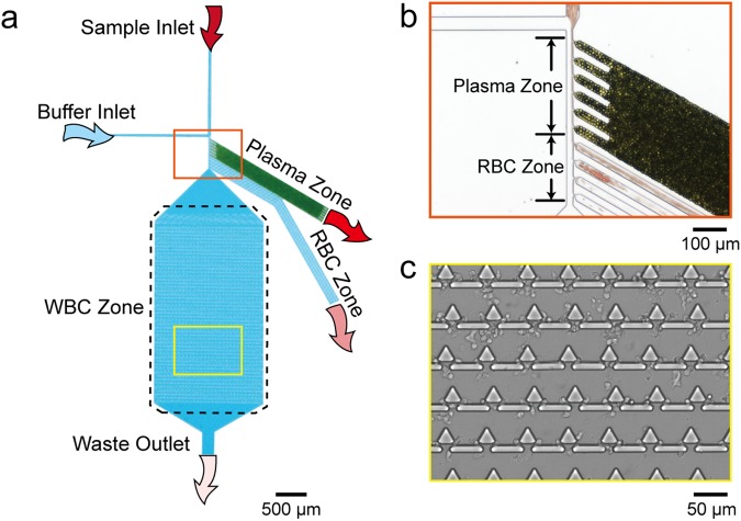
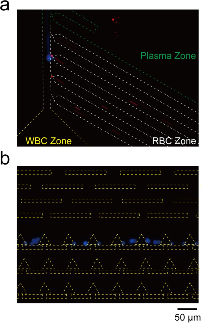
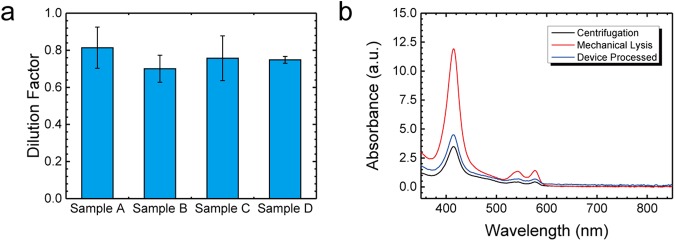
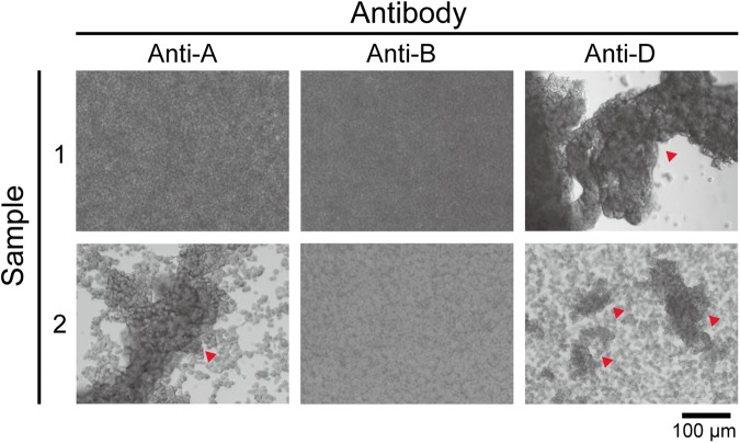
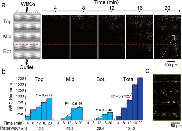
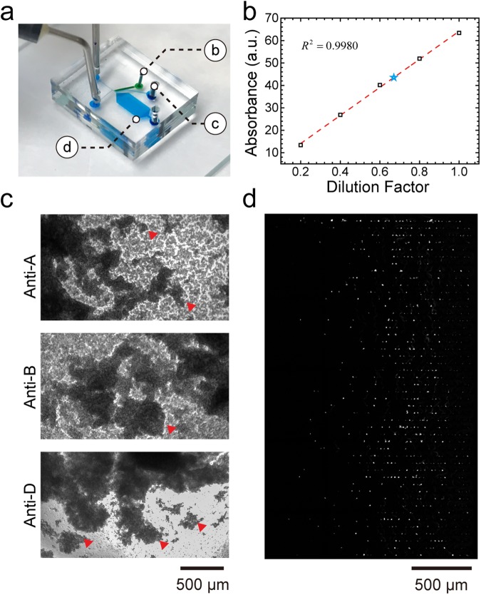
This study reports a microfluidic device for whole blood processing. The device uses the bifurcation law, cross-flow method, and hydrodynamic flow for simultaneous extraction of plasma, red blood cells, and on-chip white blood cell trapping. The results demonstrate successful plasma and red blood cell collection with a minimum dilution factor (0.76x) and low haemolysis effect. The extracted red blood cells can also be applied for blood type tests. Moreover, the device can trap up to ~1,800 white blood cells in 20 minutes. The three components can be collected simultaneously using only 6 μL of whole blood without any sample preparation processes. Based on these features, the microfluidic device enables low-cost, rapid, and efficient whole blood processing functionality that could potentially be applied for blood analysis in resource-limited environments or point-of-care settings.


Conflict of interest statement

The authors declare no competing interests.

Figures

Figure 1
Figure 1
(a) Schematic of the microfluidic device, which contains a whole blood sample inlet and a buffer inlet, respectively. After the whole blood sample flowed to the bifurcation region (orange zone), plasma and RBCs were extracted to plasma zone and RBC zone, respectively. WBCs flowed to the main channel and were trapped in the WBC zone. (b) Photograph of the plasma zone and RBC zone entrance. Green particles are packed beads, and red particles are RBCs. (c) Photograph of the WBC zone with trapped WBCs.
Figure 2
Figure 2
The fluorescence images of beads in the microfluidic device. Green, white and yellow dashed line represent to the plasma zone, RBC zone, and WBC zone, respectively. (a) Most 2-μm red fluorescent beads flow into the RBC zone; (b) 10-μm blue fluorescent beads flow into the WBC zone.
Figure 3
Figure 3
(a) Plot of the dilution factor test in 4 clinical samples. Each sample was processed by 3 individual devices. (b) The UV-VIS spectra of the plasma extracted from centrifugation (black line), the microfluidic device (blue line), and mechanically lysed whole blood (red line).
Figure 4
Figure 4
Blood type test images from the extracted RBC solutions of blood sample 1 and sample 2 under anti-A, anti-B, and anti-D treatment. The red triangles indicate blood agglutination.
Figure 5
Figure 5
WBC seeding pattern in the trapping units of the microfluidic device. (a) Schematic and time-lapse fluorescence images of WBC zone. (b) Trapped WBC numbers in top, middle, and bottom regions at different time points. The red dashed lines are the regression lines of each region, representing the WBC capture rate. (c) Enlarged image of the yellow box showing the WBC seeding profile.
Figure 6
Figure 6
Simultaneous extraction of plasma, RBCs and on-chip white blood cell trapping. (a) Image of the microfluidic device showing the position of each zone. (b) Standard curve of the absorbance intensity versus dilution factor of manually diluted blood (black rectangles and red dashed line). The blue star represents the absorbance and corresponding dilution factor (0.67x) of extracted plasma in this test. (c) Blood type test images from the extracted RBC solution. The red triangles represent blood agglutination. (d) WBC seeding pattern in the WBC zone.

References

    1. Christensen RD, Henry E, Jopling J, Wiedmeier SE. The CBC: Reference Ranges for Neonates. Seminars in Perinatology. 2009;33:3–11. doi: 10.1053/j.semperi.2008.10.010. - DOI - PubMed
    1. Spell DW, Jones DV, Harper WF, David Bessman J. The value of a complete blood count in predicting cancer of the colon. Cancer Detection and Prevention. 2004;28:37–42. doi: 10.1016/j.cdp.2003.10.002. - DOI - PubMed
    1. Mach AJ, Adeyiga OB, Di Carlo D. Microfluidic sample preparation for diagnostic cytopathology. Lab on a Chip. 2013;13:1011–1026. doi: 10.1039/c2lc41104k. - DOI - PMC - PubMed
    1. Hou HW, et al. Microfluidic Devices for Blood Fractionation. Micromachines. 2011;2:319. doi: 10.3390/mi2030319. - DOI
    1. Kersaudy-Kerhoas M, Sollier E. Micro-scale blood plasma separation: from acoustophoresis to egg-beaters. Lab on a Chip. 2013;13:3323–3346. doi: 10.1039/c3lc50432h. - DOI - PubMed

Publication types